
You are in the right place. Dr. Tom Biernacki, DPM, FACFAS — board-certified foot & ankle surgeon with 3,000+ surgeries — explains exactly what foot/ankle X-ray procedure means and what works. Call (810) 206-1402 for same-day appointment at Howell or Bloomfield Hills.
Quick answer: Xray Foot Ankle What To Expect is a common foot/ankle topic that affects many patients. Effective treatment starts with a targeted diagnosis, conservative-first treatment, and escalation only when needed. We treat this regularly at our Howell and Bloomfield Hills practices. Call (810) 206-1402.
Medically reviewed by Dr. Tom Biernacki, DPM
Board-certified podiatric surgeon | Balance Foot & Ankle
Last reviewed: May 2026
The most important clinical decision with Xray Foot Ankle What To Expect isn't which treatment to start with — it's which subtype or underlying cause you actually have. Our podiatrists regularly see patients who've been treated for months for the wrong diagnosis. The correct identification changes the entire treatment path. Call (810) 206-1402 — Dr. Tom evaluates this condition at both Howell and Bloomfield Hills locations.
The most important clinical decision with Xray Foot Ankle What To Expect isn’t which treatment to start with — it’s identifying the correct subtype. That changes everything. Call (810) 206-1402.
Table of Contents
- What to Expect During Your Foot X-Ray
- What Your Podiatrist Looks for on X-Ray
- Standard X-Ray Views Explained
- What X-Rays Can’t Show
- Frequently Asked Questions
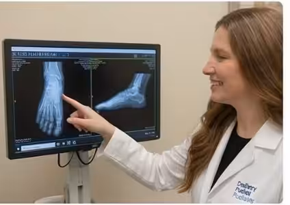
Getting an X-ray at your first podiatry visit can feel intimidating — especially if you’re wondering what the images will reveal and what happens next. In our Howell and Bloomfield Hills clinics, we take in-office digital X-rays for most new patients with foot or ankle pain, and walking you through the findings is one of the most important parts of your appointment. Here’s exactly what we’re looking at and why.


What to Expect During Your Foot or Ankle X-Ray
Foot and ankle X-rays are quick, painless, and performed right in our office — no hospital trip needed. Most patients are surprised by how fast the process is. Here’s exactly what happens from the moment we decide imaging is needed:
- You’ll remove your shoes and socks. Jewelry around the ankle should also come off — metal artifacts block the image.
- Weight-bearing views are standard. For most foot and ankle conditions, we want to see the bones under the load of your body weight. You’ll stand on the X-ray plate while the machine captures the image — this takes seconds.
- Multiple angles are captured. Typically 3 views per foot are taken (anteroposterior, lateral, oblique), sometimes more depending on the suspected pathology.
- Radiation exposure is minimal. A standard 3-view foot X-ray delivers approximately 0.001 mSv — equivalent to about 3 hours of natural background radiation. It’s extremely low risk.
- Results are immediate. Our digital X-ray system processes images in seconds. Your podiatrist reviews them with you during the same appointment.
Key takeaway: In-office weight-bearing X-rays are more diagnostically useful than non-weight-bearing images taken in a radiology suite, because deformities and joint space narrowing appear differently under load.
What Your Podiatrist Looks for on a Foot X-Ray
When I review a foot or ankle X-ray, I’m systematically evaluating bone integrity, joint spaces, alignment angles, and soft tissue shadows in a specific sequence. Here are the key findings we assess:
- Fractures and stress fractures. Acute fractures usually show a clear break line. Stress fractures are subtler — early stress fractures may not show on plain X-ray at all, which is why we sometimes follow up with MRI if the clinical picture doesn’t match the X-ray.
- Arthritis and joint space narrowing. Healthy joints have uniform space between the bones. Osteoarthritis narrows that space and often produces osteophytes (bone spurs) at joint margins. We grade severity from mild to end-stage.
- Bunion (hallux valgus) angle. We measure the hallux abductus angle and the intermetatarsal angle — the exact degrees guide whether conservative care or surgery is most appropriate.
- Flatfoot deformity. Weight-bearing X-rays reveal talar-first-metatarsal alignment (Meary’s angle), calcaneal pitch, and arch height — all critical for orthotic and surgical planning.
- Bone spurs. Heel spurs, dorsal foot spurs, and ankle bone spurs are clearly visible and help correlate with pain location.
- Bone density changes. Areas of increased or decreased density can suggest bone tumors, cysts, or avascular necrosis that require further workup.
- Foreign bodies. Metal, glass, and some types of gravel are visible on X-ray — important if a patient reports stepping on something.
Standard X-Ray Views and What Each Shows
Each X-ray angle reveals different aspects of foot anatomy. Understanding which view is ordered and why helps patients follow along when we review images together in the exam room.
- Anteroposterior (AP) view — top-down. Shows the forefoot and midfoot from above. Best for assessing metatarsal alignment, toe deformities, Lisfranc joint integrity, and bunion angles.
- Lateral view — side profile. Shows the entire foot from the side. Critical for assessing arch height, calcaneal pitch, ankle joint alignment, Achilles calcification, and flatfoot deformity severity.
- Oblique view — 45° angle. Reveals the midfoot joints (cuboid, navicular, cuneiforms) and the 3rd–5th metatarsal bases — an area easily missed on the other two views.
- Ankle AP and mortise views. For ankle injuries, the mortise view (15° internal rotation) shows the medial clear space and talar dome — essential for ruling out ankle fracture with syndesmotic injury.
- Sesamoid view. A specialized axial view of the two small bones under the big toe joint, ordered when sesamoiditis or sesamoid fracture is suspected.
Key takeaway: The oblique view catches fractures and arthritis in the midfoot that are completely invisible on AP and lateral views alone. Never skip it for midfoot pain — a Lisfranc injury missed on initial X-ray is a career-ending mistake in musculoskeletal medicine.
What X-Rays Can’t Show — When We Order MRI or CT
X-rays are excellent at showing bone but provide almost no information about soft tissue structures. Tendons, ligaments, cartilage, nerve entrapments, and early stress reactions in bone are all invisible or near-invisible on plain X-ray. The most important conditions we can miss on X-ray include:
- Ligament tears (ankle ligaments, Lisfranc ligament complex) — require MRI
- Tendon tears (Achilles, peroneal, posterior tibial tendon) — require MRI or ultrasound
- Early stress fractures before cortical disruption — require MRI (most sensitive) or bone scan
- Cartilage damage and osteochondral defects in the ankle — require MRI
- Plantar fascia thickness and tears — best assessed with diagnostic ultrasound in the office
- Complex fracture configuration before surgery — CT scan gives 3D bone detail for surgical planning
⚠️ When to Seek Imaging Urgently
- Acute injury with inability to bear weight at all
- Visible deformity or bony prominence after trauma
- Foot or ankle pain after a fall from height or car accident
- Open wound near a joint after an injury
- Severe swelling that developed within 1 hour of injury
Ready to Get Relief?
Same-day appointments available in Howell & Bloomfield Hills, MI
4.9★ | 1,123 Reviews | 3,000+ Surgeries
Or call: (810) 206-1402
In-Office Treatment at Balance Foot & Ankle
If home treatment isn’t providing relief for your foot and ankle conditions, our podiatry team at Balance Foot & Ankle can help with same-day evaluations and advanced in-office care.
Same-day appointments available. (810) 206-1402
Doctor Hoy’s Natural Pain Relief Gel
Natural topical pain relief I use in our clinic. Arnica + camphor formula — apply directly to the area 3–4x daily. ($20–25)
Shop Doctor Hoy’s →Frequently Asked Questions
Do I need a referral for a foot X-ray?
No. As a board-certified podiatrist, Dr. Biernacki can order and perform X-rays in our office without a referral from your primary care physician. In-office digital X-rays are included as part of your podiatric evaluation and are typically covered by most insurance plans as a diagnostic service. We bill your insurance directly.
Is a foot X-ray safe during pregnancy?
Foot X-rays use extremely low radiation — approximately 0.001 mSv — and the beam is directed at your foot, far from your abdomen. The American College of Radiology considers foot X-rays during pregnancy to carry negligible fetal risk. That said, we always discuss your pregnancy status before imaging and use a lead apron as standard precaution. For pregnant patients with suspected soft tissue injuries, we frequently use diagnostic ultrasound as a radiation-free alternative.
Can an X-ray detect plantar fasciitis?
Not directly. The plantar fascia itself is a soft tissue structure invisible on X-ray. However, X-rays help rule out other causes of heel pain (stress fractures, calcaneal cysts, tumors) and may show a heel spur — a bony projection on the bottom of the calcaneus often associated with plantar fasciitis. The presence or absence of a heel spur doesn’t diagnose plantar fasciitis, and we typically use clinical examination and diagnostic ultrasound for definitive diagnosis.
How soon do I get my X-ray results?
Immediately. Our in-office digital X-ray system produces images in seconds, and Dr. Biernacki reviews the images with you during the same appointment. This is one of the advantages of seeing a podiatrist with in-office imaging — you leave your first appointment with a diagnosis and treatment plan, not waiting days for a radiology report.
The Bottom Line
Foot and ankle X-rays are one of our most valuable diagnostic tools — fast, safe, and immediately informative. Whether you’ve suffered an acute injury, have persistent pain we need to explain, or you’re preparing for surgery and we need precise measurements, X-rays give us the structural map we need. If you’re dealing with foot or ankle pain in the Howell or Bloomfield Hills area, call us at (810) 206-1402 or book online for same-day imaging and diagnosis.
Sources
- American College of Radiology. “ACR Appropriateness Criteria: Acute Trauma to the Foot.” 2022.
- Beaman DN et al. “Radiographic analysis of foot and ankle articular surfaces.” Foot Ankle Int. 2006.
- Nork SE et al. “Lisfranc injuries: diagnosis and treatment.” J Am Acad Orthop Surg. 2003.
Frequently Asked Questions
When should I see a podiatrist?
If symptoms persist past 2 weeks, affect your normal activity, or are accompanied by red-flag symptoms (warmth, redness, swelling, inability to bear weight).
What does treatment cost?
Most diagnostic visits and conservative treatments are covered by Medicare and major insurers. Out-of-pocket costs vary by your specific plan.
How quickly can I get an appointment?
Most non-urgent cases see us within 5 business days. Urgent cases (sudden pain, possible fracture) typically same or next business day.
What is Foot pain?
Foot pain is a common foot/ankle condition that affects mobility and quality of life. Understanding the underlying cause is the first step in successful treatment. Our podiatrists at Balance Foot & Ankle perform a hands-on biomechanical exam, review your activity history, and use diagnostic imaging when appropriate to identify the root cause—not just treat the symptom. Many patients have been told to “rest and ice” without a deeper diagnostic workup; our approach is different.
Symptoms and warning signs
Common signs of foot pain include pain that worsens with activity, morning stiffness, swelling, tenderness when palpated, and difficulty bearing weight. If you experience sudden severe pain, inability to walk, visible deformity, numbness or color change, contact our office the same day or visit urgent care—these can signal a more serious injury such as a fracture, tendon rupture, or vascular compromise. Diabetics with any foot wound should seek same-day care.
Conservative treatment options
Most cases of foot pain respond to non-surgical care: structured rest, supportive footwear changes, custom orthotics, targeted stretching and strengthening protocols, anti-inflammatory medications when medically appropriate, and in-office procedures such as ultrasound-guided injections. We also offer advanced therapies including MLS laser therapy, EPAT/shockwave, regenerative injections, and image-guided procedures. Treatment is sequenced from least invasive to most invasive, and we explain the rationale at every step.
When is surgery considered?
Surgery is reserved for cases that fail 3-6 months of well-structured conservative care, when there is structural pathology (severe deformity, complete tear, advanced arthritis), or when imaging shows damage that will not heal without intervention. Our surgeons have performed 3,000+ foot and ankle procedures and prioritize minimally-invasive techniques whenever appropriate. We discuss recovery timelines, return-to-activity milestones, and realistic outcome expectations before any procedure is scheduled.
Recovery timeline and prevention
Recovery from foot pain varies based on severity and chosen treatment path. Conservative cases often improve within 4-8 weeks with consistent adherence to the protocol. Post-procedural recovery may range from a few days (in-office procedures) to several months (reconstructive surgery). Long-term prevention involves footwear assessment, activity modification, structured strengthening, and regular check-ins with your podiatrist if you have a history of recurrence. We provide written home-exercise plans and digital follow-up support.
Ready to feel better?
Same-week appointments available in Howell and Bloomfield Hills, Michigan.
Book Your VisitReady to fix this for good?
Reading goes so far. The fastest path is a 30-minute office visit. Same-day Howell or Bloomfield Hills. Call (810) 206-1402.
Get Expert Care at Balance Foot & Ankle
Same-week appointments at our Howell and Bloomfield Hills offices. Board-certified podiatric surgeons. Most insurance accepted.
Dr. Tom Biernacki, DPM is a board-certified foot & ankle surgeon (ABFAS & ABPM) at Balance Foot & Ankle Specialists in Southeast Michigan. With over a decade of clinical experience, he specializes in heel pain, bunions, diabetic foot care, sports injuries, and minimally invasive surgery. Dr. Biernacki is a member of the APMA and ACFAS, and his patient education content on MichiganFootDoctors.com and YouTube has made him one of the most-followed foot & ankle educators on YouTube.