

Quick answer: Ankle Cartilage Repair Surgery Microfracture Oats is a common foot/ankle topic that affects many patients. The 2026 evidence-based approach combines proper diagnosis, conservative-first treatment, and escalation only when needed. We treat this regularly at our Howell and Bloomfield Hills practices. Call (810) 206-1402.
Medically reviewed by Dr. Tom Biernacki, DPM — Board-Certified Podiatric Surgeon — Balance Foot & Ankle, Howell & Bloomfield Hills, MI. Last updated April 2026.
Medically Reviewed by Dr. Tom Biernacki, DPM — Board-Certified Podiatrist, Balance Foot & Ankle Specialists, Michigan. Last updated April 2026.
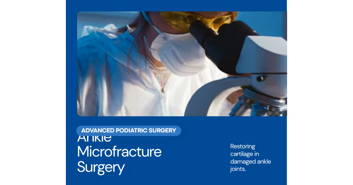
Surgical Cartilage Restoration for Ankle Osteochondral Defects
Articular cartilage — the smooth, glistening tissue that covers joint surfaces — has extremely limited natural healing capacity. Unlike bone, which can regenerate through a reliable healing response, cartilage lacks blood vessels and depends on nutrient diffusion from joint fluid for survival. When cartilage is damaged in the ankle by trauma, repetitive stress, or osteochondral lesion, the body cannot spontaneously regenerate the same high-quality hyaline cartilage that was lost. Without treatment, cartilage defects progress and accelerate arthritis development.
Surgical cartilage restoration aims to either stimulate new cartilage tissue formation or transplant healthy cartilage to fill the defect. At Balance Foot & Ankle, our foot and ankle surgeons are experienced in all major cartilage restoration techniques and match the procedure to each patient defect characteristics, age, activity demands, and prior treatment history.
Preoperative Planning
Careful preoperative planning determines which cartilage restoration technique is most appropriate. Key factors include defect size (smaller defects respond well to bone marrow stimulation; larger defects require cell-based or osteochondral graft techniques), lesion depth (purely cartilaginous versus those extending into subchondral bone), cyst formation beneath the lesion, defect location on the talar dome (centrally located versus shoulder lesions have different access and outcome profiles), patient age and biological healing potential, activity demands, and prior treatment history.
MRI characterizes cartilage integrity and the extent of subchondral involvement. Weight-bearing CT scan provides precise three-dimensional size measurement and identifies subchondral cysts that may require bone grafting concurrent with cartilage repair. These imaging findings directly guide the surgical approach.
Arthroscopic Debridement and Microfracture
For lesions smaller than approximately 1.5 cm in diameter without significant cyst formation, arthroscopic microfracture is the standard first-line treatment. The procedure is performed entirely through small arthroscopic portals without opening the ankle joint.
The unstable cartilage is debrided using arthroscopic shavers and curettes until a stable cartilage edge surrounds the defect. The underlying calcified cartilage layer is removed with a curette, preparing the bone surface for new tissue attachment. An arthroscopic awl is used to create multiple perforations through the subchondral bone plate at 3 to 4 millimeter intervals. These perforations access the subchondral bone marrow, releasing mesenchymal stem cells, growth factors, and blood that fill the defect with a superclot.
Over 6 to 8 weeks, the superclot matures into a fibrocartilage tissue — mechanically functional but composed of type I collagen rather than the type II collagen of native hyaline cartilage. Fibrocartilage is less stiff and wears more quickly than hyaline cartilage, explaining why microfracture outcomes tend to deteriorate for larger defects over time.
Recovery requires 4 to 6 weeks of non-weight bearing to protect the maturing clot, followed by progressive weight bearing and physical therapy. Return to full athletic activity typically requires 4 to 6 months.
Osteochondral Autograft Transfer System (OATS)
OATS transfers one or more cylindrical plugs of bone and articular cartilage from a non-weight-bearing area of the knee (typically the lateral trochlea or intercondylar notch) to the ankle defect. The transplanted cartilage is true hyaline cartilage — the same tissue type as the native talar dome — providing superior mechanical properties compared to the fibrocartilage from microfracture.
OATS is indicated for lesions of 1.5 to 2.5 cm in diameter where microfracture would likely yield inferior results, for cystic lesions requiring bone grafting, and for revision of failed prior bone marrow stimulation. The procedure may be performed arthroscopically or through a small open approach (miniarthrotomy) depending on defect location and size.
The harvest from the knee creates a donor site defect that fills with fibrocartilage over time. Donor site pain and stiffness affect some patients temporarily but significant long-term donor site morbidity is uncommon. Careful graft sizing and seating is critical — proud grafts (those that sit too high in the recipient site) accelerate wear of the opposing tibial cartilage.
Recovery after OATS is similar to microfracture in timeline, with 4 to 6 weeks of non-weight bearing protecting the graft incorporation period followed by progressive rehabilitation. Full return to sport requires 6 to 9 months.
BioCartilage Augmentation
BioCartilage (Arthrex) is a particulated extracellular cartilage matrix derived from cadaveric cartilage and dehydrated into a powder or putty form. Mixed with platelet-rich plasma (PRP) and packed into the microfracture defect, it provides a three-dimensional scaffold that promotes more organized fibrocartilage tissue formation compared to microfracture alone.
BioCartilage augments — rather than replaces — the microfracture technique, providing a single-stage procedure with improved tissue quality compared to standard microfracture. It is appropriate for lesions in the 1.0 to 2.0 cm range where microfracture alone would be expected to yield suboptimal results but OATS is not yet required. Intermediate-term results are promising, though long-term comparative data with OATS and cell-based techniques is still accumulating.
Autologous Chondrocyte Implantation (ACI and MACI)
For large defects (greater than 2.5 cm), failed prior cartilage procedures, or patients for whom maximum cartilage quality is the priority, cell-based techniques including autologous chondrocyte implantation (ACI) and matrix-induced ACI (MACI) provide the most biologically advanced restoration possible.
ACI/MACI is a two-stage procedure. In Stage 1, a small cartilage biopsy is harvested arthroscopically from a non-weight-bearing knee area and sent to a laboratory where the chondrocytes are grown in culture for 4 to 6 weeks, expanding the cell population dramatically. In Stage 2, the cultured cells are implanted into the ankle defect — either under a periosteal patch (ACI) or seeded onto a collagen scaffold that is sutured into the defect (MACI).
The implanted cells mature over 12 to 18 months into hyaline-like cartilage. Long-term outcomes for ACI/MACI in the ankle are superior to microfracture for large defects, though the two-stage nature, specialized laboratory processing, and higher cost limit its use to appropriately selected cases.
Choosing the Right Cartilage Repair Approach
The optimal approach is determined by defect characteristics, not by patient or surgeon preference alone. Defects under 1.0 cm in a young, active patient: microfracture. Defects 1.0 to 1.5 cm with BioCartilage augmentation as a single-stage enhancement. Defects 1.5 to 2.5 cm with cystic changes: OATS. Defects greater than 2.5 cm or failed prior procedures: MACI or ACI. Concurrent alignment problems must be corrected simultaneously to prevent overloading the repair site.
Contact Balance Foot & Ankle for hands-on exam plus imaging when needed of persistent ankle pain, especially following prior ankle injuries. Our foot and ankle surgeons provide expert assessment and individualized treatment planning, including all modern cartilage restoration techniques, for patients throughout Southeast Michigan.
Ready to Relieve Your Foot Pain?
Board-certified podiatrists serving Southeast Michigan. Same-week appointments available.
Ankle Cartilage Repair Surgery at Balance Foot & Ankle
Ankle cartilage damage (osteochondral lesions) can cause chronic pain and instability if untreated. Dr. Tom Biernacki at Balance Foot & Ankle performs microfracture, OAT grafting, and other cartilage restoration procedures at our Howell and Bloomfield Hills offices.
Learn About Our Ankle Surgery Options | Book Your Appointment | Call (810) 206-1402
Clinical References
- Zengerink M, et al. “Treatment of osteochondral lesions of the talus: a systematic review.” Knee Surgery, Sports Traumatology, Arthroscopy. 2010;18(2):238-246.
- Chuckpaiwong B, et al. “Microfracture for osteochondral lesions of the ankle.” Arthroscopy. 2008;24(1):106-112.
- Savage-Elliott I, et al. “Osteochondral lesions of the talus: a current concepts review.” Foot and Ankle Specialist. 2014;7(5):414-422.
Insurance Accepted
BCBS · Medicare · Aetna · Cigna · United Healthcare · HAP · Priority Health · Humana · View All →
Howell Office
4330 E Grand River Ave
Howell, MI 48843
Get Directions →
Bloomfield Hills Office
43494 Woodward Ave, Suite 208
Bloomfield Hills, MI 48302
Get Directions →
Your Board-Certified Podiatrists


Ready to Get Back on Your Feet?
Same-week appointments available at both locations.
Book Your AppointmentMore Podiatrist-Recommended Arthritis Essentials
Cushioned Running Shoe

Hoka Clifton 10 — max cushioning reduces joint impact for arthritic feet.
Wide Walking Shoe
New Balance 990v6 — wide toe box accommodates arthritic first-MTP (hallux rigidus).

As an Amazon Associate, Balance Foot & Ankle earns from qualifying purchases. Product recommendations are based on clinical experience; prices and availability shown above update live from Amazon.

When to See a Podiatrist
Foot and ankle arthritis progresses silently — cartilage doesn’t regrow, but joint fusion, cheilectomy, and biologic injections can restore function at every stage. Balance Foot & Ankle offers the full arthritis spectrum: bracing, injections, and reconstructive surgery. Start with a consult so we can image the joint and give you a realistic 5-year outlook.
Call Balance Foot & Ankle: (810) 206-1402 · Book online · Offices in Howell & Bloomfield Hills
When Shoes Aren’t Enough — Dr. Tom’s Top 9 Orthotics
About 30% of patients I see for foot pain need MORE than a great shoe — they need a structured insole. Below: my complete 2026 orthotic ranking with pros, cons, and the specific patient I’d give each one to.
★ DR. TOM’S COMPLETE 2026 ORTHOTIC RANKING
9 Best Prefab Orthotics by Use Case
PowerStep, Currex, Spenco, Vionic, and PowerStep Pinnacle — every orthotic I’ve fitted to thousands of patients across both Michigan offices. Each card includes pros, cons, and the specific patient I’d give it to. Real Amazon ratings, review counts, and prices below.
Best All-Purpose Orthotic for Most Patients
Semi-rigid arch shell + dual-layer cushion + deep heel cup. The orthotic I’ve fitted to more patients than any other for 15 years. APMA-accepted. Trim-to-fit design works in athletic shoes, casual shoes, and most work boots.
✓ Pros
- Semi-rigid arch shell provides true biomechanical correction
- Deep heel cup centers the heel and reduces lateral instability
- Dual-layer cushion (top + bottom) lasts 9-12 months daily wear
- Available in 8 sizes for precise fit
- APMA-accepted and clinically validated
- Lower price than CURREX RunPro for equivalent function
✗ Cons
- Too thick for most dress shoes (use ProTech Slim instead)
- Some break-in period required (3-7 days for arch tolerance)
- Not enough correction for severe pes planus or rigid pes cavus
Dr. Tom’s Recommendation: If a patient has run-of-the-mill plantar fasciitis, mild flat feet, or arch fatigue, this is the first orthotic I try. Better value than PowerStep Pinnacle for 90% of patients, which is why I swapped it into our clinic kits three years ago. Sub-$50 typically.
Maximum Motion Control · Flat Feet & Severe Over-Pronation
PowerStep’s most aggressive stability orthotic. Adds a 2°-7° medial heel post on top of the standard PowerStep platform — designed specifically for flat-footed patients and severe pronators who need real corrective force.

✓ Pros
- 2°-7° medial heel post adds aggressive pronation control
- Same trusted PowerStep arch shell, more correction
- Built specifically for flat-foot biomechanics
- Excellent for posterior tibial tendon dysfunction (PTTD)
- Removable top cover for cleaning
✗ Cons
- Too aggressive for neutral-arch patients
- Needs longer break-in (10-14 days) due to stronger correction
- Adds 2-3 mm of stack height — won’t fit slim dress shoes
Dr. Tom’s Recommendation: When a patient comes in with significant flat feet AND symptoms (heel pain, arch pain, knee pain), the Original PowerStep isn’t aggressive enough. The Maxx is what gets prescribed. About 25% of my flat-footed patients end up here.
Low-Profile · Fits Dress Shoes & Narrow Casuals
3 mm slim profile with podiatrist-designed tri-planar arch technology. Engineered specifically to fit inside dress shoes, oxfords, loafers, and women’s flats without crowding the toe box. Vionic was founded by an Australian podiatrist.
✓ Pros
- 3 mm slim profile (vs 7-10 mm for standard orthotics)
- Tri-planar arch technology adds support without bulk
- Built-in deep heel cup despite slim design
- Fits dress shoes WITHOUT having to remove the factory insole
- Trim-to-fit · APMA-accepted
✗ Cons
- Less arch support than full-volume orthotics
- Top cover wears faster than thicker alternatives
- Not enough correction for severe foot deformities
Dr. Tom’s Recommendation: My default when a patient says ‘I need orthotics but I have to wear dress shoes for work.’ Slim enough to fit in oxfords and pumps without the heel sliding out. The single highest-impact change you can make for office workers with foot pain.
Built-In Metatarsal Pad · Morton’s Neuroma · Ball-of-Foot Pain
Standard Pinnacle orthotic with a built-in metatarsal pad positioned proximal to the metatarsal heads — the exact location that offloads neuromas and metatarsalgia. No need for separate met pads or pad placement guesswork.

✓ Pros
- Built-in met pad eliminates DIY pad placement errors
- Specifically designed for Morton’s neuroma + metatarsalgia
- Same trusted PowerStep arch + heel cup platform
- Top cover protects sensitive forefoot skin
- Faster relief than orthotics + add-on met pads
✗ Cons
- Met pad position is fixed (can’t fine-tune individual placement)
- Some patients with very small or very large feet need custom
- Slightly thicker than the standard Pinnacle
Dr. Tom’s Recommendation: If a patient has Morton’s neuroma, sesamoiditis, or generalized ball-of-foot pain (metatarsalgia), this saves a clinic visit and a prescription. The built-in pad placement is anatomically correct for 80% of feet. Way better than DIY met pads.
Adaptive Dynamic Arch · Athletic & Daily Wear
Currex’s flagship adaptive arch technology — the orthotic flexes with your gait instead of fighting it. Different stiffness zones along the length give you targeted support at the heel, midfoot, and forefoot. Available in three arch heights (low/medium/high).

✓ Pros
- Dynamic flex zones adapt to natural gait cycle
- Three arch heights ensure precise fit
- Lighter than rigid orthotics (no ‘heavy foot’ feel)
- Excellent for runners and athletic walkers
- European podiatric design (German engineering)
✗ Cons
- More expensive than PowerStep Original ($55-65 typically)
- Less aggressive correction than Pinnacle Maxx for severe cases
- Three arch heights means you must self-select correctly
Dr. Tom’s Recommendation: I started recommending Currex three years ago for runners who said PowerStep felt ‘too rigid.’ The dynamic flex zones respect natural gait. Best for active patients who walk 8K+ steps daily and don’t need maximum motion control.
Running-Specific · Heel Strike + Forefoot Strike Compatible
Currex’s purpose-built running orthotic. The midfoot flex zone is positioned for runner’s gait mechanics, with a flared heel cushion for heel strikers and a forefoot rocker for midfoot/forefoot strikers. Tested on 1000+ runners during product development.

✓ Pros
- Designed by German biomechanics lab specifically for runners
- Dynamic arch flexes with running gait (not static like PowerStep)
- Three arch heights (low/medium/high)
- Reduces overuse injury risk in mid-distance runners
- Lightweight (no impact on cadence)
✗ Cons
- Premium price ($60-75)
- Not aggressive enough for severe over-pronators (use Pinnacle Maxx)
- Runner-specific design = less ideal for daily walking shoes
Dr. Tom’s Recommendation: If a patient runs 20+ miles per week and has plantar fasciitis or shin splints, this is the orthotic I prescribe. The dynamic flex zones respect running biomechanics in a way that no rigid PowerStep can match. Pricier but worth it for serious runners.
Cavus Foot & High-Arch Patients
Polyurethane base with a deeper heel cup and higher arch profile than PowerStep — built for cavus (high-arched) feet that need maximum cushion and support. The 5-zone cushioning system addresses the unique pressure points of high-arch feet.

✓ Pros
- Deeper heel cup centers the heel for cavus foot stability
- Higher arch profile fills the void under high arches
- 5-zone cushioning addresses cavus foot pressure points
- Polyurethane base lasts 12+ months
- Available in Wide width
✗ Cons
- Too tall/aggressive for normal or low arches
- Won’t fit slim dress shoes
- Pricier than PowerStep Original
- Some patients find the arch height uncomfortable initially
Dr. Tom’s Recommendation: Cavus foot patients are often misdiagnosed and given low-arch orthotics — that makes everything worse. Spenco’s Total Support has the arch profile that high-arch feet actually need. About 15% of my patients have cavus feet; this is what they wear.
Cushion Layer · Standing All Day · Gel Pressure Relief
NOT a true biomechanical orthotic — this is a cushion insole. But for patients who want gel pressure relief instead of arch correction (or to add ON TOP of factory insoles in work boots), this is the best gel option on Amazon.

✓ Pros
- Genuine gel cushioning (not foam pretending to be gel)
- Targeted gel waves under heel and ball of foot
- Trim-to-fit · works in most shoe types
- Sub-$15 price (most affordable option in this list)
- Massaging texture is genuinely soothing
✗ Cons
- ZERO arch support — this is cushion only
- Won’t fix plantar fasciitis or flat-foot issues
- Compresses faster than PowerStep (4-6 months)
- Top cover wears through in high-mileage applications
Dr. Tom’s Recommendation: I recommend these to patients who tell me ‘I just want my feet to stop hurting at the end of my shift’ and who don’t have a biomechanical issue. Construction workers, factory workers, retail. Pure cushion does the job for them.
Tight-Fitting Shoes · Cycling Shoes · Hockey Skates
PowerStep Pinnacle’s slim version of their famous Green insole. The trademark stabilizer cap is preserved but the overall thickness is reduced — works in cycling shoes, hockey skates, ski boots, and other tight-fitting footwear that the standard CURREX RunPro can’t fit into.

✓ Pros
- Stabilizer cap centers the heel (PowerStep Pinnacle’s signature feature)
- Slim profile fits tight athletic footwear
- Lasts 12+ months daily wear
- Excellent for cycling shoes specifically
- Built-in odor-control treatment
✗ Cons
- Premium price ($45-55)
- Less cushion than PowerStep equivalents
- Not as aggressive correction as Pinnacle Maxx for flat feet
- The signature ‘heel cup feel’ takes 1-2 weeks to adapt to
Dr. Tom’s Recommendation: If you’re a cyclist with foot numbness, hot spots, or knee pain — this is the orthotic. The stabilizer cap solves cycling-specific biomechanical issues that no other orthotic addresses. Worth the premium for athletes.
None of these solving your foot pain?
Some patients (about 30%) need custom-molded prescription orthotics. We make 3D-scanned custom orthotics in our Howell and Bloomfield Hills offices — specifically built for your foot mechanics.
Schedule a Custom Orthotic Fitting →FSA/HSA eligible · Most insurance accepted · (810) 206-1402
🦶 Dr. Tom’s Recommended Products
These are the at-home products I recommend most often to patients at Balance Foot & Ankle in Howell, MI.
The OTC orthotic I recommend most in our clinic. Medical-grade arch support at a fraction of custom orthotic cost.
View on Amazon →
Natural topical pain relief I use in our clinic. Arnica + menthol formula — apply directly to the area 3-4x daily. FSA-eligible.
View on Amazon →
FTC Disclosure: As an Amazon Associate and Foundation Wellness affiliate, we earn from qualifying purchases. This never affects our clinical recommendations.
American Academy of Orthopaedic Surgeons: Osteochondral Lesions / Cartilage Repair
In-Office Treatment at Balance Foot & Ankle
If home treatment isn’t providing relief for your foot and ankle injuries, our podiatry team at Balance Foot & Ankle can help with same-day evaluations and advanced in-office care.
Same-day appointments available. (810) 206-1402
Get Expert Care at Balance Foot & Ankle
Same-week appointments at our Howell and Bloomfield Hills offices. Board-certified podiatric surgeons. Most insurance accepted.
Dr. Tom Biernacki, DPM is a board-certified foot & ankle surgeon (ABFAS & ABPM) at Balance Foot & Ankle Specialists in Southeast Michigan. With over a decade of clinical experience, he specializes in heel pain, bunions, diabetic foot care, sports injuries, and minimally invasive surgery. Dr. Biernacki is a member of the APMA and ACFAS, and his patient education content on MichiganFootDoctors.com and YouTube has made him one of the most-followed foot & ankle educators on YouTube.