

Quick answer: Need a podiatrist’s opinion on this? Call Balance Foot & Ankle at (810) 206-1402 for same-week appointments in Howell or Bloomfield Hills, Michigan. Most insurance accepted, including Medicare.

Medically Reviewed by Dr. Tom Biernacki, DPM, FACFAS — Board-certified podiatric surgeon | Balance Foot & Ankle | Last updated: May 2026
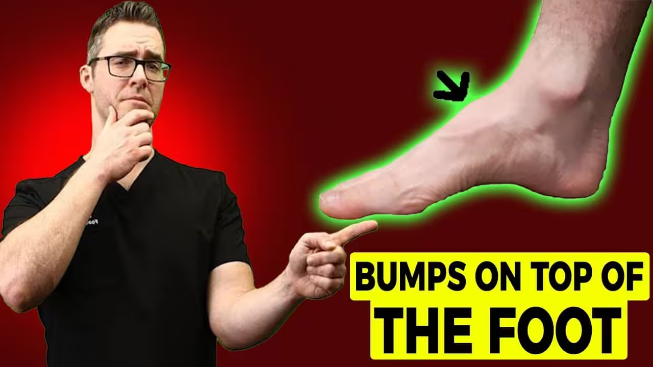
Quick Answer: Chipped Bone in the Foot
A chipped bone in the foot (avulsion fracture) occurs when ligament or tendon tension pulls a small bone fragment free at its attachment point — most commonly at the base of the 5th metatarsal, the navicular, or the cuboid. These injuries cause localized pain, swelling, and bruising after a twist or impact. Most foot avulsion fractures heal in 4–8 weeks with a walking boot. However, the 5th metatarsal base fracture has a tendency toward non-union and requires careful management to avoid refracture.
The foot contains 26 bones and 33 joints, any of which can sustain a chipped or avulsion fracture under sufficient force. While ankle avulsion fractures get more attention, foot avulsions are equally common and equally misdiagnosed — often labeled as “soft tissue injuries” until persistent pain drives a patient back for additional imaging weeks later.
In our Howell and Bloomfield Hills clinics, we evaluate foot chipped-bone injuries every week. The evaluation starts with identifying the exact pain location, mechanism of injury, and whether the Ottawa Foot Rules indicate imaging.
⚠️ When to see a podiatrist:
- Cannot bear weight after foot injury
- Visible deformity or significant swelling
- Multiple areas of tenderness suggesting more complex fracture
- No improvement in pain after 2 weeks of rest and protection
⭐ 4.4★ | 5K+ Sold
Protects chipped foot bones during recovery — reduces pressure on the fracture site while allowing controlled walking.
PowerStep Pinnacle Arch Support Insoles
⭐ 4.7★ | 50K+ Sold
Provides the arch support needed to offload chipped foot bones during recovery and prevent re-injury.
Ready to Get Relief? Same-Day Appointments Available
Board-certified podiatric surgeon | Howell & Bloomfield Hills, MI | Most Insurance Accepted
4.9★ | 1,123 Reviews | 3,000+ Surgeries
Or call: (810) 206-1402
Most Common Sites for Chipped Bones in the Foot
| Location | How It Chips | Key Risk | Healing Time |
|---|---|---|---|
| 5th metatarsal base | Peroneus brevis avulsion | Non-union, zone 2 Jones fracture | 4–8 weeks |
| Navicular | Tibialis posterior avulsion | Flat foot deformity if missed | 6–10 weeks |
| Cuboid | Lateral ligament tension | Frequently missed on plain X-ray | 4–6 weeks |
| Styloid process (5th met) | Plantaris/peroneus chip | Confused with Jones fracture | 4–6 weeks |
| Sesamoid bones | Direct impact, hyperextension | Big toe push-off pain lasting months | 6–12 weeks |
The 5th Metatarsal Base Chip: Critical Distinctions
The most important clinical distinction with foot bone chips is differentiating a true avulsion fracture at the styloid process of the 5th metatarsal (Zone 1 — heals well) from a Jones fracture at the metaphyseal-diaphyseal junction (Zone 2 — high non-union risk) or a diaphyseal stress fracture (Zone 3 — often requires surgery). These three fracture types occur within centimeters of each other and look nearly identical clinically. X-ray with proper foot positioning — and sometimes CT — is needed to classify them correctly, because Zone 1 can be booted while Zone 2–3 may require surgical fixation with an intramedullary screw.
Watch: Navicular — Foot Bone Pain Explained
Dr. Tom covers mid-foot bone injuries including the navicular, which is a common site for chipping and stress fractures:

Book a same-day foot fracture evaluation → | (810) 206-1402
Frequently Asked Questions
How do you know if you chipped a bone in your foot?
Point tenderness directly over a specific bone (rather than a ligament), swelling, and bruising after a foot injury are the primary clinical signs. The Ottawa Foot Rules indicate X-ray when there is tenderness at the navicular or at the base of the 5th metatarsal. Many foot chips are missed on initial imaging — if pain persists at a specific bone location beyond 2–3 weeks despite normal X-rays, request an MRI or bone scan to detect occult fractures invisible on plain films.
Can you walk on a chipped bone in your foot?
For small, non-displaced chips at non-weight-bearing surfaces, limited walking in a stiff-soled walking boot may be tolerated. However, the 5th metatarsal base should be protected in a boot regardless of pain level — continued forefoot loading stresses the fracture site with every step. Sesamoid chips and navicular avulsions also benefit from boot immobilization even when weight-bearing is possible, because repetitive micromotion prevents healing.
How long does a chipped foot bone take to heal?
Most foot avulsion fractures heal in 4–8 weeks. Navicular avulsions and cuboid chips typically heal in 4–6 weeks in a boot. The 5th metatarsal styloid avulsion heals in 4–6 weeks with conservative management; Jones fractures take 6–8 weeks minimum, with higher non-union rates. Sesamoid fractures heal slowly — often 8–12 weeks — and occasionally require surgical removal if they become a persistent source of pain.
What happens if a chipped foot bone doesn’t heal?
Non-union of a foot bone chip results in a fibrous pseudoarthrosis — a false joint filled with scar tissue rather than bone. This typically causes chronic, activity-related pain that worsens with walking and is relieved by rest. Treatment options range from bone stimulator therapy to surgical excision or fixation with bone graft. Jones fracture non-union specifically may require intramedullary screw fixation with bone graft stimulation.
Do I need surgery for a chipped bone in my foot?
Most foot avulsion fractures do not require surgery. Surgical indications include: Jones fracture in an athlete (for faster return to sport), any fracture with >2mm displacement involving a joint surface, documented non-union after 12+ weeks of conservative care, or sesamoid fractures causing intractable pain after conservative exhaustion. A podiatrist can classify the fracture type and recommend the most appropriate management pathway.
Foot Pain After a Twist or Impact? Get Imaged Today.
Dr. Tom Biernacki, DPM provides same-day foot fracture evaluation and X-rays at both locations.
Related: Chipped Ankle Bone | Chipped Shin Bone | Signs of a Broken Ankle | Protruding Bone Outside Foot
Podiatrist-Recommended Products for Foot Fracture Recovery
- Doctor Hoy’s Natural Pain Relief Gel — topical pain relief for fracture site soreness during the healing and return-to-activity phases
- Plantar Fasciitis Compression Socks — graduated compression reduces swelling around the fracture site during recovery
- PowerStep Maxx — maximum-support insole for return to weight-bearing after bone healing is confirmed
These are the same products Dr. Biernacki recommends in clinic. Available through our partner Foundation Wellness.
In-Office Treatment at Balance Foot & Ankle
If home treatment isn’t providing relief for your foot fracture, our podiatry team at Balance Foot & Ankle can help with same-day evaluations and advanced in-office care.
When should I see a podiatrist?
If symptoms persist past 2 weeks, affect your normal activity, or are accompanied by red-flag symptoms (warmth, redness, swelling, inability to bear weight).
What does treatment cost?
Most diagnostic visits and conservative treatments are covered by Medicare and major insurers. Out-of-pocket costs vary by your specific plan.
How quickly can I get an appointment?
Most non-urgent cases see us within 5 business days. Urgent cases (sudden pain, possible fracture) typically same or next business day.
What is Foot pain?
Foot pain is a common foot/ankle condition that affects mobility and quality of life. Understanding the underlying cause is the first step in successful treatment. Our podiatrists at Balance Foot & Ankle perform a hands-on biomechanical exam, review your activity history, and use diagnostic imaging when appropriate to identify the root cause—not just treat the symptom. Many patients have been told to “rest and ice” without a deeper diagnostic workup; our approach is different.
Symptoms and warning signs
Common signs of foot pain include pain that worsens with activity, morning stiffness, swelling, tenderness when palpated, and difficulty bearing weight. If you experience sudden severe pain, inability to walk, visible deformity, numbness or color change, contact our office the same day or visit urgent care—these can signal a more serious injury such as a fracture, tendon rupture, or vascular compromise. Diabetics with any foot wound should seek same-day care.
Conservative treatment options
Most cases of foot pain respond to non-surgical care: structured rest, supportive footwear changes, custom orthotics, targeted stretching and strengthening protocols, anti-inflammatory medications when medically appropriate, and in-office procedures such as ultrasound-guided injections. We also offer advanced therapies including MLS laser therapy, EPAT/shockwave, regenerative injections, and image-guided procedures. Treatment is sequenced from least invasive to most invasive, and we explain the rationale at every step.
When is surgery considered?
Surgery is reserved for cases that fail 3-6 months of well-structured conservative care, when there is structural pathology (severe deformity, complete tear, advanced arthritis), or when imaging shows damage that will not heal without intervention. Our surgeons have performed 3,000+ foot and ankle procedures and prioritize minimally-invasive techniques whenever appropriate. We discuss recovery timelines, return-to-activity milestones, and realistic outcome expectations before any procedure is scheduled.
Recovery timeline and prevention
Recovery from foot pain varies based on severity and chosen treatment path. Conservative cases often improve within 4-8 weeks with consistent adherence to the protocol. Post-procedural recovery may range from a few days (in-office procedures) to several months (reconstructive surgery). Long-term prevention involves footwear assessment, activity modification, structured strengthening, and regular check-ins with your podiatrist if you have a history of recurrence. We provide written home-exercise plans and digital follow-up support.
Ready to feel better?
Same-week appointments available in Howell and Bloomfield Hills, Michigan.
Book Your VisitGet Expert Care at Balance Foot & Ankle
Same-week appointments at our Howell and Bloomfield Hills offices. Board-certified podiatric surgeons. Most insurance accepted.
Dr. Tom Biernacki, DPM is a board-certified foot & ankle surgeon (ABFAS & ABPM) at Balance Foot & Ankle Specialists in Southeast Michigan. With over a decade of clinical experience, he specializes in heel pain, bunions, diabetic foot care, sports injuries, and minimally invasive surgery. Dr. Biernacki is a member of the APMA and ACFAS, and his patient education content on MichiganFootDoctors.com and YouTube has made him one of the most-followed foot & ankle educators on YouTube.