
Medically reviewed by Dr. Tom Biernacki, DPM
Board-certified podiatric surgeon | Balance Foot & Ankle, Howell & Bloomfield Hills, MI
Last reviewed: May 2026

| Bursa Location | Condition | Cause | Key Sign | Primary Treatment |
|---|---|---|---|---|
| Retrocalcaneal (posterior heel) | Retrocalcaneal bursitis | Achilles impingement, pump bump (Haglund’s) | Tenderness between Achilles + calcaneus | Heel lift, open-back shoe, cortisone injection |
| Subcutaneous (over Achilles) | Superficial Achilles bursitis | Shoe counter friction | Fluid-filled sac behind heel | Padding, shoe modification, aspiration |
| 1st MTP joint (medial) | Bunion bursa (adventitial) | Shoe pressure on bunion prominence | Redness + swelling over medial bunion | Wide-toe shoe, padding, injection |
| 5th MTP joint (lateral) | Tailor’s bunion / bunionette bursa | Narrow shoe lateral pressure | Pain + bursa over 5th MT head | Wide shoe, padding, injection ± surgery |
| Plantar heel | Plantar heel bursitis | Fat pad atrophy, calcaneal spur | Plantar heel pain, soft swelling | Heel cushion, cortisone injection |
| 2nd–4th MTP (intermetatarsal) | Intermetatarsal bursitis | Metatarsal overload, narrow shoes | Pain between toes, positive Mulder’s click | Metatarsal pad, wide shoe, injection |
| Treatment | Best For | Expected Relief | Notes |
|---|---|---|---|
| Shoe Modification (wide box, open back) | All foot bursitis | Immediate reduction in friction | First and most important step |
| Padding / Donut Pads | Bunion bursa, heel bursa | Days to weeks | Offloads pressure directly over bursa |
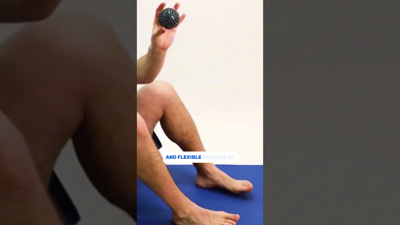
| Ice Therapy | Acute inflammation | Immediate (symptomatic) | 20 min on/off; reduces swelling |
| NSAIDs (oral or topical) | Mild-moderate bursitis | 3–7 days | Short courses; topical diclofenac for focal areas |
| Corticosteroid Injection | Refractory bursitis, retrocalcaneal | Days to weeks | Max 2 injections; avoid near Achilles (rupture risk) |
| Aspiration | Tense, fluid-filled bursa | Immediate volume reduction | Culture if septic bursitis suspected |
| Surgical Bursectomy | Chronic, recurrent, failed injections | Permanent resolution | Often combined with bony prominence removal (e.g., Haglund’s) |
Quick answer: Foot Bursitis Michigan Podiatrist is a common foot/ankle topic that affects many patients. The 2026 evidence-based approach combines proper diagnosis, conservative-first treatment, and escalation only when needed. We treat this regularly at our Howell and Bloomfield Hills practices. Call (810) 206-1402.
Foot pain isn’t resolving?
Same-week appointments at Howell & Bloomfield Hills
Medically Reviewed | Dr. Tom Biernacki, DPM | Board-Certified Podiatric Surgeon | Balance Foot & Ankle, Michigan


Understanding Foot Bursae and Bursitis
A bursa is a small closed sac lined with synovial membrane that acts as a fluid-filled cushion between structures subject to friction — tendons, bones, and overlying skin. Bursae are found throughout the body at pressure points and are normally thin, almost imperceptible structures. When irritated by friction, pressure, or inflammatory disease, the bursal lining secretes excess fluid, the sac expands, and local inflammation creates pain and swelling — this is bursitis.
Several anatomically distinct bursae exist in the foot and ankle, each with a characteristic clinical presentation. Dr. Biernacki evaluates foot bursitis systematically, using ultrasound when needed to confirm the diagnosis and guide injection therapy.
Types of Foot Bursitis
Retrocalcaneal Bursitis
The retrocalcaneal bursa lies between the superior calcaneal surface and the Achilles tendon. It is the most clinically significant foot bursa and is frequently involved in Haglund’s deformity (posterior heel exostosis). When the posterior heel bone is prominent, it repetitively compresses the bursa with each step, creating chronic bursal inflammation.
Presentation: pain at the back of the heel, anterior to the Achilles tendon. Tenderness on both sides of the Achilles near its insertion. Worse with shoes that have a firm heel counter. Frequently mistaken for Achilles tendinopathy, which may coexist. Ultrasound reveals fluid-filled bursal distension between the tendon and calcaneus.
Subcutaneous (Superficial Achilles) Bursitis
Between the Achilles tendon and overlying skin lies the subcutaneous calcaneal bursa. Shoe pressure — particularly from stiff heel counters of dress shoes, pumps, or new athletic footwear — creates friction over this bursa. The result is a soft, tender, fluctuant bump at the posterior heel that may evolve into a visible “pump bump” in chronic cases.
Presentation: visible swelling and tenderness at the skin surface overlying the Achilles insertion. Less deep than retrocalcaneal bursitis — palpable as a superficial soft mass. Resolves predictably with footwear modification.
Intermetatarsal Bursitis
Small bursae lie between the metatarsal heads in the plantar forefoot, adjacent to the interdigital nerve branches. Chronic compression from narrow-toe-box shoes or metatarsal head overload causes these bursae to enlarge, creating pain and occasionally a palpable mass in the ball of the foot. Intermetatarsal bursitis is an important clinical mimic of Morton’s neuroma — both cause third interspace forefoot pain that may radiate into the toes, and both respond to similar conservative measures. MRI or ultrasound distinguishes enlarged bursae from true neuromas.
Presentation: pain and burning at the ball of the foot, typically in the second or third web space. Worsened with shoes, better barefoot. Lateral compression of the forefoot (Mulder’s sign) may produce a click in Morton’s neuroma but not in pure bursitis.
First Metatarsophalangeal (Bunion) Bursitis
A subcutaneous bursa overlies the medial first metatarsal head in most people. In bunion deformity, the medially prominent metatarsal head repetitively contacts shoe upper, creating chronic bursal inflammation — the tender, red swelling at the bunion that patients commonly report. This “red bunion” appearance reflects bursitis as much as the underlying bone deformity.
Presentation: redness, warmth, swelling, and tenderness directly at the bunion prominence. Markedly worse in narrow shoes. Occasionally infected in patients who manipulate or pop the bursa. Soft tissue and bone deformity evaluation required to assess for concurrent hallux valgus correction needs.
Plantar Heel Bursitis
A bursa between the plantar fascia insertion and the calcaneal fat pad can develop with chronic plantar heel pressure. Less common than plantar fasciitis and usually coexisting with it. Presents as focal posterior heel tenderness on weight-bearing, indistinguishable clinically from plantar fasciitis without imaging. Ultrasound distinguishes bursal fluid accumulation from plantar fascia thickening.
Diagnosis
Clinical examination — location of tenderness, character of swelling, shoe-related symptom pattern — guides initial diagnosis. Confirmatory imaging when needed:
- Ultrasound — real-time visualization of bursal fluid, distension, and location. Available at Balance Foot & Ankle. Guides aspiration and injection placement.
- MRI — confirms bursitis and evaluates coexisting pathology (Achilles tendon, plantar fascia, interdigital nerve).
- X-ray — identifies Haglund’s exostosis, Achilles calcification, and sesamoid pathology that may be contributing to bursitis.
Treatment
Footwear Modification
The most important non-surgical intervention for all forms of foot bursitis. Soft heel counters eliminate retrocalcaneal and Achilles bursa pressure. Wide toe boxes reduce intermetatarsal and bunion bursa irritation. Open-heel sandals or heel-cut shoe modifications provide immediate relief for posterior heel bursitis.
Padding and Offloading
U-shaped horseshoe pads around the posterior heel unload retrocalcaneal and Achilles bursitis. Metatarsal pads proximal to the metatarsal heads reduce intermetatarsal compression. Bunion shields protect the medial first metatarsal head.
Ice and Anti-Inflammatory Therapy
Ice application, NSAIDs, and topical anti-inflammatory gels reduce acute bursal inflammation during flare-ups.
Aspiration and Corticosteroid Injection
Ultrasound-guided aspiration removes fluid from the distended bursa, providing immediate decompression. Corticosteroid injection into the bursa reduces inflammation and prevents re-accumulation. Effective for refractory bursitis not responding to conservative measures. Repeated injections are used cautiously near the Achilles tendon due to tendon weakening risk.
Surgical Bursectomy
Surgical excision of the chronically inflamed bursa is rarely required but is appropriate when conservative care including injection has failed repeatedly. In retrocalcaneal bursitis associated with Haglund’s deformity, surgical removal of the exostosis with bursectomy addresses both the structural cause and the inflammatory result simultaneously.
Dr. Tom’s Product Recommendations
Heel That Pain Plantar Fasciitis Heel Seats
⭐ Highly Rated
Viscoelastic heel cups with a posterior heel cutout design that unloads the Achilles insertion and retrocalcaneal region. Useful for retrocalcaneal and Achilles bursitis management.
Dr. Tom says: “My Achilles heel bursitis flared every fall when I started wearing my new boots — these heel cups took the pressure right off.”
Retrocalcaneal bursitis, Achilles insertion pain, posterior heel pressure offloading
Posterior heel cutout may not fit all shoe types; check fit before extended use
Disclosure: We earn a commission at no extra cost to you.
Bunion Bootie Splint Sleeve
⭐ Highly Rated
Gel-lined silicone sleeve with bunion pad that cushions the medial first metatarsal head and reduces shoe friction against the bunion bursa.
Dr. Tom says: “My bunion bursa would flare whenever I wore dress shoes. This sleeve protects it just enough to get through a long workday.”
Bunion bursitis, medial first metatarsal head protection
Does not correct underlying bunion deformity; consult podiatrist for progressive or surgical bunion management
Disclosure: We earn a commission at no extra cost to you.
✅ Pros / Benefits
- Foot bursitis typically responds well to targeted conservative care — footwear modification, padding, and injection produce resolution in most cases
- Ultrasound-guided injection confirms bursal location and delivers treatment precisely — available at Balance Foot & Ankle
- Bursitis is often a secondary finding alongside structural problems (Haglund’s, bunions) — addressing the structural cause prevents recurrence
❌ Cons / Risks
- Retrocalcaneal bursitis associated with Haglund’s deformity may recur without addressing the underlying bone prominence
- Intermetatarsal bursitis can be clinically indistinguishable from Morton’s neuroma without ultrasound or MRI — treatment protocols differ significantly
Dr. Tom Biernacki’s Recommendation
Bursitis is one of those satisfying diagnoses because it almost always responds to treatment — and often very quickly once the offending pressure is relieved. The posterior heel bursas in particular respond dramatically to footwear modification and a well-placed injection. The key diagnostic challenge is distinguishing intermetatarsal bursitis from Morton’s neuroma, because both cause that aching ball-of-foot pain that radiates into the toes. Ultrasound tells us within minutes which we’re dealing with and allows us to inject accurately. Getting the diagnosis right means the treatment works.
— Dr. Tom Biernacki, DPM | Board-Certified Podiatric Surgeon | Balance Foot & Ankle
Frequently Asked Questions
What does foot bursitis feel like?
Localized, aching pain with swelling and tenderness at the specific bursa site. Retrocalcaneal bursitis: deep posterior heel pain, worse with heel counter of shoe. Bunion bursitis: red, warm swelling at the bunion. Intermetatarsal bursitis: ball-of-foot aching between the metatarsal heads, occasionally with toe tingling. Symptoms are typically worse with certain shoes and better barefoot or in open footwear.
How long does foot bursitis take to heal?
With footwear modification and conservative care, mild bursitis improves in 2–4 weeks. More established bursitis with significant fluid accumulation may require 6–8 weeks of treatment, or aspiration with injection for faster resolution. Structural bursitis (from Haglund’s, bunions) tends to recur unless the underlying cause is addressed.
Can you drain a bursa in the foot?
Yes. Ultrasound-guided aspiration of an enlarged bursa can be performed in the office at Balance Foot & Ankle. Aspiration removes the fluid, providing immediate decompression and pain relief. Corticosteroid injection into the bursa is typically performed at the same time to prevent rapid re-accumulation.
Is foot bursitis serious?
Foot bursitis is generally not serious and responds well to conservative treatment. In immunocompromised patients or diabetics, infected bursitis (septic bursitis) is a more serious complication requiring urgent drainage and antibiotics. A red, warm, tender posterior heel with fever or systemic symptoms requires immediate evaluation.
Michigan Foot Pain? See Dr. Biernacki In Person
4.9★ rated | 1,123 Reviews | 3,000+ Surgeries
Same-week appointments · Howell & Bloomfield Hills
📞 (810) 206-1402 Book Online →Frequently Asked Questions
When should I see a podiatrist?
If symptoms persist past 2 weeks, affect your normal activity, or are accompanied by red-flag symptoms (warmth, redness, swelling, inability to bear weight).
What does treatment cost?
Most diagnostic visits and conservative treatments are covered by Medicare and major insurers. Out-of-pocket costs vary by your specific plan.
How quickly can I get an appointment?
Most non-urgent cases see us within 5 business days. Urgent cases (sudden pain, possible fracture) typically same or next business day.
Visit Balance Foot & Ankle — Same-Day Appointments Available
Our podiatry team serves patients throughout Michigan including Howell, Brighton, and Bloomfield Hills. Whether you’re dealing with heel pain, ingrown toenails, or a foot injury, we have same-day appointment availability.
American Podiatric Medical Association: Find a Podiatrist
Ready to Get Relief?
Same-day appointments available in Howell & Bloomfield Hills, MI
4.9★ | 1,123 Reviews | 3,000+ Surgeries
Or call: (810) 206-1402
Dr. Tom Biernacki, DPM is a board-certified foot & ankle surgeon (ABFAS & ABPM) at Balance Foot & Ankle Specialists in Southeast Michigan. With over a decade of clinical experience, he specializes in heel pain, bunions, diabetic foot care, sports injuries, and minimally invasive surgery. Dr. Biernacki is a member of the APMA and ACFAS, and his patient education content on MichiganFootDoctors.com and YouTube has made him one of the most-followed foot & ankle educators on YouTube.