

The most important clinical decision with Jones Fracture Treatment isn’t which treatment to start with — it’s identifying the correct subtype. That changes everything. Call (810) 206-1402.
Jones Fracture Treatment: Zone Classification Determines Surgery vs. Boot
Not all 5th metatarsal fractures are equal — and the most dangerous treatment error is applying the wrong protocol based on the wrong diagnosis. A Zone 1 avulsion fracture (the most common) heals reliably in a walking boot. A true Jones fracture (Zone 2) has a 20-30% non-union rate without surgery. Zone 3 stress fracture in an athlete almost always requires surgical fixation. The zone of the fracture — determined by precise X-ray measurement — determines everything.
| Fracture Zone | Location | Mechanism | Non-Union Risk | Standard Treatment | Return to Sport |
|---|---|---|---|---|---|
| Zone 1 — Avulsion fracture (pseudo-Jones) | Tip or base of the 5th metatarsal styloid process; fracture line is at or proximal to the 4th-5th intermetatarsal articulation | Acute ankle inversion; peroneus brevis tendon or plantar fascia pulls bone fragment off the styloid; most common lateral foot fracture | LOW — 95%+ union rate; abundant blood supply at the styloid; reliable healing | Walking boot or hard-soled shoe × 4-6 weeks; weight-bearing as tolerated from day 1; crutches for comfort only; gradual return to activity at 6-8 weeks | 4-8 weeks; no surgical restriction; sport return when pain-free and full ROM |
| Zone 2 — True Jones fracture | Metaphyseal-diaphyseal junction (the “watershed” zone); fracture line at or just distal to the 4th-5th intermetatarsal articulation; within 1.5cm of the base | Acute: varus force to the midfoot; also occurs with lateral ankle sprain; the fracture line is transverse or slightly oblique at the junction | HIGH — 20-30% non-union rate with conservative treatment due to poor blood supply at this zone; “watershed area” between two arterial territories | Non-athletes/low-demand: non-weight-bearing cast × 6-8 weeks, then boot; 60-75% union at 3 months. Athletes/high-demand: surgical fixation (intramedullary screw) recommended — reduces time to return and non-union rate to <5% | Conservative: 10-16 weeks. Surgical: 6-8 weeks; significantly faster return to competitive sport |
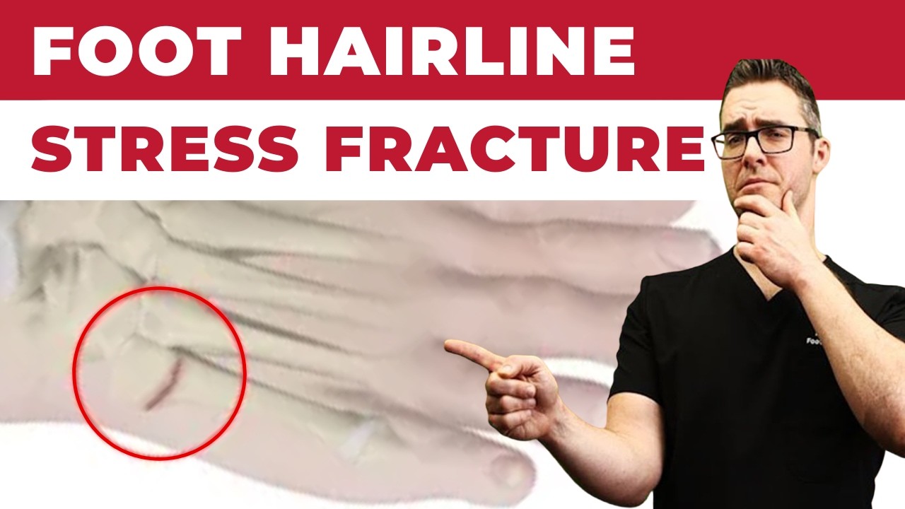
| Zone 3 — Diaphyseal stress fracture | Proximal diaphysis (shaft), distal to Zone 2; chronic/repetitive stress pattern; may have periosteal reaction, intramedullary sclerosis, or cortical hypertrophy on X-ray | Repetitive loading (running, basketball, football); NOT acute trauma; insidious onset with progressive lateral foot pain; history of prior Jones fracture increases risk | VERY HIGH — highest non-union risk of all zones; diaphysis has poorest blood supply; intramedullary sclerosis on X-ray = established chronic stress change indicating highest risk | Athletes: surgical fixation (intramedullary screw, larger diameter than Zone 2 screw); conservative treatment (cast + NWB) appropriate only for non-athletes or those refusing surgery; high re-fracture rate with conservative treatment in athletes | Surgical: 8-12 weeks; requires documented bone healing on CT scan before return to sport; CT preferred over X-ray for confirming union |
Jones Fracture Surgery vs. Conservative Treatment: Who Gets Which?
| Factor | Conservative Treatment (Boot/Cast) | Surgical Fixation (Intramedullary Screw) |
|---|---|---|
| Fracture zone | Zone 1 (avulsion) — almost always conservative; Zone 2 in non-athletes (60-75% union conservative) | Zone 2 in athletes — standard of care; Zone 3 in any active patient — strongly recommended |
| Patient activity level | Sedentary or low-demand patient; willing to accept 10-16 week recovery timeline; no competitive sport requirements | Competitive athlete (any level); patient who cannot tolerate 10-16 weeks non-weight-bearing; physically demanding occupation requiring faster return |
| X-ray characteristics | Acute fracture with no prior stress changes; clean fracture line without intramedullary sclerosis; first-time fracture | Intramedullary sclerosis present (chronic stress changes — highest non-union risk); delayed union at 8-12 weeks on conservative treatment; prior Jones fracture on same foot |
| Fracture displacement | Non-displaced (<2mm step-off) — adequate alignment for conservative healing | Displaced (>2mm) — requires anatomic reduction; surgical fixation required |
| Timeline to return | 10-16 weeks (Zone 2 conservative); 4-8 weeks (Zone 1) | 6-8 weeks (Zone 2 surgical); 8-12 weeks (Zone 3 surgical) |
| Re-fracture risk | Higher — 25-50% re-fracture rate in athletes returning to sport after conservative treatment of Zone 2 | Lower — <5% re-fracture with intramedullary screw fixation; screw provides structural protection during healing |
| Cost and invasiveness | Non-invasive; boot + follow-up X-rays; lower initial cost | Outpatient surgery; general or regional anesthesia; screw may remain permanently or be removed if symptomatic; higher initial cost, lower total cost if conservative fails |
Quick answer: Treatment for jones fracture treatment follows a stepwise approach: 1) conservative care first (rest, ice, supportive footwear, OTC anti-inflammatories), 2) physical therapy and targeted exercises, 3) in-office treatments (injections, custom orthotics) if conservative fails at 4-6 weeks, 4) surgery for refractory cases. Most patients resolve at step 1 or 2. Call (810) 206-1402.
Foot pain isn't resolving?
Same-week appointments at podiatrist in Howell & podiatrist in Bloomfield Hills
Board-Certified Podiatric Foot & Ankle Surgeon · Last reviewed: May 5, 2026
Medically Reviewed by Dr. Tom Biernacki, DPM — Board-Certified Podiatric Surgeon | 3,000+ surgeries | Balance Foot & Ankle, Howell & Bloomfield Hills MI
Quick Answer: Jones Fracture Treatment
A Jones fracture is treated with 6–8 weeks non-weight-bearing in a cast or boot for most patients. Athletes and active individuals are often better served by surgical fixation with an intramedullary screw, which cuts healing time to 6–8 weeks and dramatically reduces the ~30% non-union risk seen with conservative care. Early treatment is critical — delayed union is the most common complication.
Table of Contents
If you twisted your ankle or rolled your foot outward and now have sharp pain on the outer edge of your foot — and an X-ray shows a fracture at the base of your 5th metatarsal — you may be wondering what happens next. The answer depends almost entirely on exactly where that fracture is. A Jones fracture is one of the most commonly mismanaged foot fractures because it looks similar to two other fractures but has a dramatically different healing profile. In our clinic, we evaluate 5th metatarsal fractures carefully before making any treatment recommendation, because getting this wrong means months of unnecessary pain or a fracture that never heals.
What Is a Jones Fracture
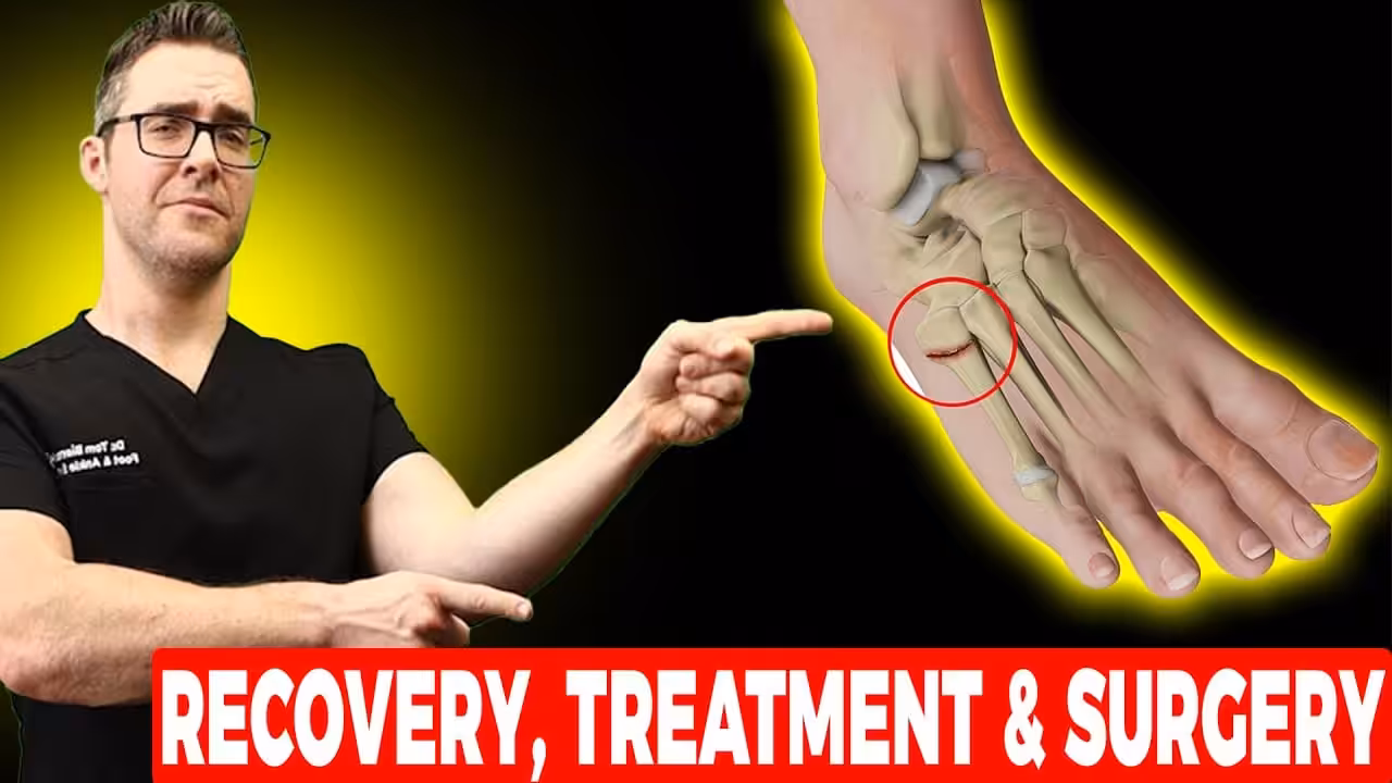
A Jones fracture is a break at the metaphyseal-diaphyseal junction of the 5th metatarsal — the bone on the outer edge of your foot that connects to your little toe. Specifically, it occurs in Zone II, approximately 1.5 to 2 centimeters from the base of the bone. This location is critical: it sits at a watershed area where two blood supplies meet poorly, making it one of the slowest-healing fractures in the foot.
Named after orthopedic surgeon Sir Robert Jones (who fractured his own foot in 1902), this injury accounts for roughly 20–25% of all 5th metatarsal fractures but causes a disproportionate amount of treatment complications. In our clinic, we see Jones fractures most often in athletes — basketball players landing from a jump, soccer players planting and cutting, and runners with excessive supination (underpronation) who load the lateral foot heavily.
The anatomy matters because the 5th metatarsal Zone II receives blood supply from two directions — a metaphyseal branch proximally and a nutrient artery diaphyseally — and the junction between them is a relative avascular zone. When fractured here, the bone struggles to mount the robust healing response seen in other foot fractures, making non-union a genuine and well-documented risk.
Symptoms and Diagnosis of a Jones Fracture
Most patients with a Jones fracture report a sudden, sharp pop or snap on the outer foot followed by immediate pain and difficulty bearing weight. Unlike some fractures that hurt but allow some walking, Jones fractures typically produce enough pain that putting full weight on the foot is either impossible or severely limited from the moment of injury.
Common symptoms include: immediate lateral foot pain at or just above the base of the 5th metatarsal, rapid swelling along the outer foot border, bruising (ecchymosis) appearing within 24–48 hours under and around the little toe, pain that worsens with any inversion or eversion of the ankle, and tenderness directly over the outer metatarsal shaft when pressed.
Diagnosis: Standard weight-bearing X-rays of the foot confirm the fracture in most cases. The AP and oblique views best visualize the 5th metatarsal. In subtle cases or when stress fracture is suspected, MRI provides earlier detection and reveals bone marrow edema before cortical changes appear on X-ray. CT scan is useful when surgical planning requires precise fracture gap measurement or when evaluating for non-union. In our clinic, we always obtain weight-bearing films — non-weight-bearing X-rays can miss subtle displacement and underestimate fracture severity.
5th Metatarsal Fracture Classification: Lawrence-Botte Zones
Not all 5th metatarsal fractures are Jones fractures. The Lawrence-Botte classification divides the proximal 5th metatarsal into three zones, and treatment differs significantly between them. Getting the zone right is the single most important step in fracture management.
| Zone | Location | Common Name | Healing Rate | Treatment |
|---|---|---|---|---|
| Zone I | Tuberosity (base) | Dancer’s fracture / avulsion | Excellent — 6–8 weeks | Walking boot or hard-sole shoe; rarely surgical |
| Zone II | Metaphyseal-diaphyseal junction | Jones fracture | Moderate — non-union risk 30%+ | NWB cast vs. intramedullary screw |
| Zone III | Diaphysis (shaft) | Diaphyseal stress fracture | Poorest — high refracture risk | Surgery strongly preferred; prolonged NWB if conservative |
This is why precise localization on X-ray matters so much. A Zone I tuberosity avulsion fracture can be treated in a walking boot and heals reliably — telling a patient with a Zone II Jones fracture to “walk it off” in a boot is a significant clinical error that dramatically increases non-union risk.
Conservative (Non-Surgical) Jones Fracture Treatment
Conservative management of a Jones fracture centers on strict non-weight-bearing immobilization in a short-leg cast for 6–8 weeks, followed by gradual progressive weight-bearing as bone healing is confirmed radiographically. This approach is appropriate for lower-demand patients, sedentary individuals, and patients who prefer to avoid surgery.
Protocol: A fiberglass or plaster short-leg non-weight-bearing cast is applied immediately. Crutches are required for the full immobilization phase. X-rays are repeated at 6 weeks to assess healing — callus bridging across the fracture line is the target sign. If callus is present and the patient is low-demand, transition to a walking boot for 2–4 more weeks. Full return to activity follows once the fracture demonstrates complete bridging on X-ray and the patient is pain-free with normal gait.
The evidence problem: Studies consistently show that conservative Jones fracture treatment carries a non-union rate of 20–30% in the general population and higher in athletes. A landmark study in the American Journal of Sports Medicine found that athletes treated conservatively had a return-to-sport rate of only 72% compared to 95% for surgically treated athletes, with significantly longer time to return. In our clinic, we have an honest conversation with every patient: if you’re an athlete, weekend warrior, or person whose livelihood requires standing and walking, the evidence strongly favors surgical fixation.
Vitamin D and calcium optimization: Before any Jones fracture treatment, we check serum 25-OH vitamin D levels. Deficiency (below 30 ng/mL) is a modifiable risk factor for delayed union and should be corrected with supplementation during the healing phase. Calcium intake is reviewed and optimized to 1,000–1,200 mg daily.
Surgical Jones Fracture Treatment: Intramedullary Screw Fixation
Surgical fixation of a Jones fracture using an intramedullary screw is the treatment of choice for athletes, physically active individuals, patients with recurrent fracture, and anyone who has failed conservative management. The procedure provides immediate mechanical stability, compresses the fracture gap, and allows much earlier return to weight-bearing and activity.
The procedure: Performed under regional anesthesia (ankle block) as an outpatient surgery taking 30–45 minutes. A guidewire is inserted into the proximal 5th metatarsal under fluoroscopic guidance, followed by a solid or cannulated intramedullary screw (typically 4.5–6.5mm diameter). The screw spans the fracture, providing compression and rotational stability. No large incisions — the entry point is a small 1cm poke hole.
Post-operative protocol: Most patients are non-weight-bearing for 2 weeks in a surgical boot, then advance to weight-bearing in the boot at 2–4 weeks once initial healing is confirmed. Return to jogging typically occurs at 6–8 weeks; return to full sports at 8–12 weeks. Screw removal is optional and only indicated if the screw causes prominent hardware irritation — the majority of patients keep their screw permanently without issue.
Surgical outcomes: Union rates with intramedullary screw fixation exceed 95% for acute Jones fractures. Refracture rates are lower than conservative treatment, and hardware prominence requiring removal occurs in roughly 10–15% of cases. In competitive athletes, surgical fixation has become the standard of care at the Division I collegiate and professional levels.
Jones Fracture Recovery Timeline
| Phase | Conservative | Surgical |
|---|---|---|
| Non-weight-bearing | 6–8 weeks | 2 weeks |
| Walking boot | 2–4 weeks | 2–4 weeks |
| Return to regular shoes | 10–14 weeks | 6–8 weeks |
| Return to low-impact activity | 12–16 weeks | 8–10 weeks |
| Return to full sport | 16–24 weeks (if heals) | 10–14 weeks |
One of the most important things we tell patients: X-ray healing and clinical healing are not the same. Just because a patient feels better does not mean the fracture is consolidated. We always confirm bridging callus on repeat X-ray before advancing activity, regardless of how good the patient feels. Premature return to weight-bearing is the leading cause of refracture and delayed union.
Differential Diagnosis: Conditions That Look Like a Jones Fracture
| Condition | Key Distinguishing Feature | Imaging |
|---|---|---|
| Zone I avulsion fracture (Dancer’s) | Fracture at very tip of tuberosity; peroneus brevis tendon pull mechanism | X-ray: transverse fracture at base tip |
| Zone III diaphyseal stress fracture | Insidious onset; no acute trauma; pain with running over weeks | MRI: periosteal edema; X-ray may be normal early |
| Peroneus brevis tendon tear | Longitudinal tear along peroneal groove; pain behind lateral malleolus | MRI: split tear of peroneus brevis |
| Cuboid stress fracture | Pain more midlateral; dorsal tenderness over cuboid | MRI confirms; X-ray negative early |
| Os vesalianum / os peroneum | Accessory ossicle; rounded edges vs. acute fracture margins; no tenderness | X-ray: smooth corticated margins; bilateral views helpful |
Red Flags: When a Jones Fracture Needs Immediate Attention
⚠ Warning Signs Requiring Urgent Evaluation
- Inability to bear any weight on the foot after a foot/ankle injury — always warrants same-day imaging
- Visible deformity of the outer foot edge — suggests displaced fracture requiring urgent reduction
- Pain that returns or worsens after initial improvement — classic sign of delayed union or refracture
- Fracture pain in a diabetic patient — Charcot arthropathy must be excluded immediately
- Fracture history with recurrence on the same foot — metabolic workup (Vitamin D, PTH, DEXA) required before treatment
The Most Common Mistake with Jones Fractures
The most common mistake we see is treating a Jones fracture (Zone II) like a dancer’s fracture (Zone I). A patient presents with a 5th metatarsal fracture on X-ray, the provider says “walking boot, return in 4 weeks,” and three months later the patient is back in the office with a painful non-union. The key distinction — Zone I vs. Zone II — requires careful measurement on the oblique X-ray view. If the fracture line begins at or just distal to the articulation between the 4th and 5th metatarsal bases, that is a Jones fracture requiring a much more aggressive treatment protocol. When in doubt, consult a podiatric surgeon before discharging in a walking boot.
Recommended Products for Jones Fracture Recovery
During the recovery and return-to-activity phase, the right support products reduce lateral foot stress and protect against refracture. These are the products we recommend most frequently in our clinic:
Doctor Hoy’s Natural Pain Relief Gel — Acute Pain Management
During the acute phase and early recovery, Doctor Hoy’s provides topical arnica and camphor-based pain relief without the systemic effects of oral NSAIDs. Apply to the lateral foot and ankle 2–3× daily to reduce inflammation and discomfort during immobilization.
View at Foundation Wellness — 30% off →
Not ideal for: open wounds, broken skin, or patients with camphor sensitivity.
PowerStep Pinnacle — Return-to-Activity Arch Support
Once cleared to return to regular footwear, PowerStep Pinnacle insoles provide critical arch support and lateral stability that reduces stress on the 5th metatarsal. Patients with cavus foot (high arch) are at elevated risk for Jones fracture recurrence — proper arch support is essential, not optional, during the return-to-activity phase.
View at Foundation Wellness — 30% off →
Not ideal for: patients with severe cavus foot deformity requiring custom orthotics — see us for a custom prescription.
In-Office Jones Fracture Treatment at Balance Foot & Ankle
At Balance Foot & Ankle, we manage Jones fractures using weight-bearing digital X-ray in both our Howell and Bloomfield Hills locations for same-day zone classification. When surgical fixation is indicated, Dr. Tom Biernacki DPM and our surgical team perform outpatient intramedullary screw fixation with same-week scheduling whenever possible. We do not rely on a one-size-fits-all conservative protocol — every Jones fracture patient receives a treatment plan based on zone, activity level, occupation, and bone health.
If you’ve been told you have a 5th metatarsal fracture and aren’t sure which zone, call us for a second opinion. Getting this right in the first 2 weeks dramatically changes your outcome. Learn more about our fracture and surgical treatment options.
Diagnosed with a Jones Fracture?
Same-day appointments available. Dr. Tom Biernacki DPM · 3,000+ surgeries · 4.9 stars · 1,123 reviews
Book Your Appointment →Or call: (810) 206-1402 · Howell & Bloomfield Hills, MI
Frequently Asked Questions About Jones Fracture Treatment
How long does a Jones fracture take to heal?
A Jones fracture takes 8–16 weeks to heal with conservative treatment and 8–12 weeks with surgical fixation. Conservative healing is confirmed by callus bridging on X-ray at 6–8 weeks — if no callus is present, the fracture is at risk for delayed union or non-union and surgical consultation should be obtained. Surgical patients are often jogging by 6–8 weeks post-op.
Can you walk on a Jones fracture?
You should not walk on a Jones fracture during the acute treatment phase. Premature weight-bearing is the primary cause of non-union and refracture. After surgical fixation, controlled weight-bearing in a boot typically begins at 2–3 weeks. After conservative casting, weight-bearing is delayed 6–8 weeks until callus is confirmed on X-ray.
Is surgery necessary for a Jones fracture?
Surgery is not mandatory for all Jones fractures, but it significantly improves outcomes for athletes and active individuals. Studies show that operative fixation reduces non-union risk from 20–30% to under 5% and cuts return-to-sport time by 6–8 weeks compared to conservative casting. For sedentary or low-demand patients, a non-weight-bearing cast remains a reasonable option.
What is the difference between a Jones fracture and a dancer’s fracture?
A dancer’s fracture (Zone I avulsion) occurs at the very tip of the 5th metatarsal base where the peroneus brevis tendon inserts. It has an excellent prognosis and typically heals in 4–6 weeks in a walking boot. A Jones fracture (Zone II) occurs 1.5–2cm further down the shaft at a poorly vascularized junction and carries a 20–30% non-union risk — requiring much more aggressive management.
When should I see a podiatrist for a Jones fracture?
See a podiatrist immediately — ideally same day — if you have lateral foot pain after twisting your ankle or foot. Accurate zone classification within the first 1–2 weeks is essential to avoid undertreating a Jones fracture. Call Balance Foot & Ankle at (810) 206-1402 for same-day evaluation in Howell or Bloomfield Hills, MI.
Does insurance cover Jones fracture surgery?
Yes — Jones fracture surgical fixation is a standard covered procedure under Medicare and most commercial insurance plans. Intramedullary screw fixation (CPT 28476) is covered when medically necessary. Our team verifies your coverage before the procedure and provides a pre-authorization estimate so you know your out-of-pocket costs in advance.
Sources
1. Lawrence SJ, Botte MJ. “Jones’ fractures and related fractures of the proximal fifth metatarsal.” Foot & Ankle International. 1993;14(6):358–365.
2. Mologne TS, et al. “Early screw fixation versus casting in the treatment of acute Jones fractures.” American Journal of Sports Medicine. 2005;33(7):970–975.
3. Japjec M, et al. “Treatment of Jones fractures: screw fixation vs cast immobilization.” Injury. 2021;52(Suppl 5):S11–S16.
4. Porter DA, et al. “Fifth metatarsal Jones fractures in the athlete.” Foot & Ankle International. 2009;30(6):566–579.
Related Conditions & Resources
For more on related conditions and treatments:
- Foot stress fracture treatment
- Broken toe symptoms: how to tell if fractured
- Metatarsalgia: ball of foot pain causes
- Stone bruise on foot: causes & treatment
- Peroneal tendinopathy treatment
- Howell podiatrist office
- Bloomfield Hills podiatrist office
Need to see a podiatrist? Call (810) 206-1402 or book online. Same-week availability.
Frequently Asked Questions
What is a Jones fracture?
A specific type of fracture at the base of the 5th metatarsal (outer foot bone), in the proximal diaphysis (~1.5cm from the bone’s tip). Notorious for poor healing because of limited blood supply. Often confused with the more common avulsion fracture (which heals well). Misdiagnosis is common — accurate location is critical.
How long does a Jones fracture take to heal?
Conservative (boot/cast non-weight-bearing): 8-16 weeks for non-displaced fractures, with high non-union rate (15-30%). Surgical (intramedullary screw fixation): 6-8 weeks back to running, lower non-union rate (~5%). Athletes typically have surgery to ensure faster, more reliable healing.
Should a Jones fracture be treated with surgery?
Athletes and active adults: surgery is increasingly recommended for fast, reliable healing (intramedullary screw fixation in 30-minute outpatient procedure). Sedentary patients or older adults: conservative treatment (boot + non-weight-bearing) often sufficient if compliant. Decision depends on activity level + healing potential + patient preference.
Can I walk on a Jones fracture?
With non-surgical treatment: NO weight-bearing for 6-8 weeks until imaging shows healing. Walking on a Jones fracture causes non-union (the fracture doesn’t heal) — leading to chronic pain and eventually surgery anyway. Athletes who need to return to sport quickly should consider primary surgery to enable earlier weight-bearing.
How do I know if I have a Jones fracture?
Symptoms: pain on the outer foot (specifically just below the tip of the 5th metatarsal), bruising, swelling, painful walking. X-ray confirms — both the diagnosis AND the specific location matter (Jones fracture = proximal diaphysis; avulsion fracture = at the very tip). Get evaluated immediately for any outer-foot injury.
Podiatrist-Recommended Products for Jones Fracture Recovery
- Doctor Hoy’s Natural Pain Relief Gel — topical pain relief for lateral foot soreness during Jones fracture non-surgical recovery
- DASS Medical Compression Socks — graduated compression reduces swelling at the 5th metatarsal base fracture site
- PowerStep Maxx — maximum lateral support insole for return-to-activity after Jones fracture healing
These are the same products Dr. Biernacki recommends in clinic. Available through our partner Foundation Wellness.
Frequently Asked Questions
What is the difference between a Jones fracture and a 5th metatarsal avulsion fracture?
These are distinct injuries at different zones of the 5th metatarsal base: Zone 1 (avulsion fracture) — the peroneus brevis tendon pulls off a flake of bone at the styloid process, very common after ankle sprain, heals readily in a boot in 4–6 weeks. Zone 2 (Jones fracture, true) — fracture at the metaphyseal-diaphyseal junction, watershed blood supply zone, high non-union rate (25–35%), typically requires NWB × 6–8 weeks or surgical fixation. Zone 3 (stress fracture) — diaphyseal fracture from repetitive loading, worst blood supply, highest non-union risk. Accurate zone identification on X-ray determines treatment.
Do Jones fractures require surgery?
The decision for surgery on a Jones fracture depends on: (1) patient activity level — competitive athletes almost always benefit from intramedullary screw fixation for faster and more reliable return-to-sport; (2) fracture zone — Zone 2 true Jones fractures in active patients typically get screw fixation; (3) fracture displacement — any displacement >2mm suggests instability, favoring surgery; (4) prior failed conservative treatment — non-union after proper NWB immobilization is a clear surgical indication. For Zone 1 avulsion fractures, surgery is almost never needed. Conservative care is appropriate for non-displaced Zone 2 fractures in sedentary patients.
How long is recovery from a Jones fracture?
Conservative (NWB boot) treatment of a Jones fracture: 6–8 weeks non-weight-bearing, followed by 4–6 weeks in a walking boot, total recovery 12–16 weeks before return to sport — with a meaningful non-union risk. Surgical fixation with an intramedullary screw: 6–8 weeks protected weight-bearing, return to sport at 8–12 weeks. Both approaches require CT or MRI confirmation of healing before return to activity. Repeat fracture (refracture) occurs in 5–10% of surgically treated Jones fractures in athletes, typically from premature return.
What shoe should I wear after a Jones fracture?
During acute Jones fracture treatment: a non-weight-bearing CAM boot (for conservative treatment) or post-surgical boot. After healing is confirmed: stiff-soled shoes with a carbon fiber or rigid rocker sole to reduce lateral column stress during push-off. Avoid flexible shoes, minimalist shoes, and barefoot walking for 6+ months after Jones fracture healing — the bone remodeling period continues beyond the point of clinical healing. A lateral wedge insole (3–5mm) may reduce peroneus brevis tension and lateral column load during the return phase.
What is Stress fracture?
Stress fracture is a common foot/ankle condition that affects mobility and quality of life. Understanding the underlying cause is the first step in successful treatment. Our podiatrists at Balance Foot & Ankle perform a hands-on biomechanical exam, review your activity history, and use diagnostic imaging when appropriate to identify the root cause—not just treat the symptom. Many patients have been told to “rest and ice” without a deeper diagnostic workup; our approach is different.
Symptoms and warning signs
Common signs of stress fracture include pain that worsens with activity, morning stiffness, swelling, tenderness when palpated, and difficulty bearing weight. If you experience sudden severe pain, inability to walk, visible deformity, numbness or color change, contact our office the same day or visit urgent care—these can signal a more serious injury such as a fracture, tendon rupture, or vascular compromise. Diabetics with any foot wound should seek same-day care.
Conservative treatment options
Most cases of stress fracture respond to non-surgical care: structured rest, supportive footwear changes, custom orthotics, targeted stretching and strengthening protocols, anti-inflammatory medications when medically appropriate, and in-office procedures such as ultrasound-guided injections. We also offer advanced therapies including MLS laser therapy, EPAT/shockwave, regenerative injections, and image-guided procedures. Treatment is sequenced from least invasive to most invasive, and we explain the rationale at every step.
When is surgery considered?
Surgery is reserved for cases that fail 3-6 months of well-structured conservative care, when there is structural pathology (severe deformity, complete tear, advanced arthritis), or when imaging shows damage that will not heal without intervention. Our surgeons have performed 3,000+ foot and ankle procedures and prioritize minimally-invasive techniques whenever appropriate. We discuss recovery timelines, return-to-activity milestones, and realistic outcome expectations before any procedure is scheduled.
Recovery timeline and prevention
Recovery from stress fracture varies based on severity and chosen treatment path. Conservative cases often improve within 4-8 weeks with consistent adherence to the protocol. Post-procedural recovery may range from a few days (in-office procedures) to several months (reconstructive surgery). Long-term prevention involves footwear assessment, activity modification, structured strengthening, and regular check-ins with your podiatrist if you have a history of recurrence. We provide written home-exercise plans and digital follow-up support.
Ready to feel better?
Same-week appointments available in Howell and Bloomfield Hills, Michigan.
Book Your VisitGet Expert Care at Balance Foot & Ankle
Same-week appointments at our Howell and Bloomfield Hills offices. Board-certified podiatric surgeons. Most insurance accepted.
Same-Week Appointments in Howell & Bloomfield Hills
Three board-certified podiatric surgeons. 1,123+ five-star reviews. Most insurance accepted.
Dr. Tom Biernacki, DPM is a board-certified foot & ankle surgeon (ABFAS & ABPM) at Balance Foot & Ankle Specialists in Southeast Michigan. With over a decade of clinical experience, he specializes in heel pain, bunions, diabetic foot care, sports injuries, and minimally invasive surgery. Dr. Biernacki is a member of the APMA and ACFAS, and his patient education content on MichiganFootDoctors.com and YouTube has made him one of the most-followed foot & ankle educators on YouTube.