
Medically reviewed by Dr. Tom Biernacki, DPM, FACFAS
Board-certified podiatric surgeon | Balance Foot & Ankle | Last reviewed: June 2026
Most patients underestimate how much the post-operative phase determines bunion surgery outcomes — not the surgery itself. Our podiatric surgeons identify the single recovery variable that separates patients who return to full activity on schedule from those who experience setbacks. Call (810) 206-1402 — expert podiatric care across Michigan.

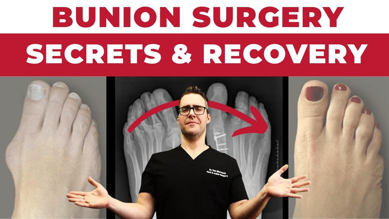
Watch Dr. Tom Biernacki DPM explain bunion surgery options and how to recover fast — MichiganFootDoctors YouTube
Bunion Surgery Options: Austin, Lapidus, and MICA — Which Is Right for You?
The vast majority of bunionectomies in the United States in 2026 are performed using a handful of techniques — the Austin/chevron osteotomy, the Lapidus procedure, minimally invasive MICA, and (for select larger deformities) the scarf osteotomy. The selection between them is determined by the severity of the deformity (IMA angle), the stability of the first tarsometatarsal joint (TMT joint), and patient-specific factors. This guide explains the key differences.
| Procedure | Best For | IMA Angle | Recurrence Risk | Recovery | Key Advantage | Key Limitation |
|---|---|---|---|---|---|---|
| Austin/Chevron Osteotomy | Mild to moderate bunion deformity with stable 1st TMT joint; no hypermobility; IMA correction up to 15°; most common bunionectomy for mild-moderate cases | IMA 9-15° (mild-moderate); HVA up to 30° | Low — under 5% with correct patient selection; higher if underlying TMT joint hypermobility is missed, because the osteotomy does not correct it | Surgical shoe 4-6 weeks; wide shoe at 6-8 weeks; running at 4-5 months; 8-12 weeks to return to standing work | Reliable, well-studied technique; immediate partial weight-bearing in most protocols; lower complication rate than TMT fusion; fastest recovery among the three options | Cannot fully address severe deformity (IMA >15°); higher recurrence than Lapidus if TMT hypermobility is the underlying cause; limited correction potential |
| Lapidus Procedure (1st TMT arthrodesis) | Moderate to severe bunion with TMT joint hypermobility (the anatomical cause of most bunion deformities); IMA correction >15°; recurrent bunion; flatfoot with bunion; most durable correction available | IMA 14-25° (moderate-severe); indicated regardless of IMA angle when TMT hypermobility is documented | Lowest — under 2%; addresses the underlying TMT hypermobility rather than just the metatarsal head deformity | Protected weight-bearing in a boot (fixation-dependent); boot to wide shoe at 8-12 weeks; running at 5-6 months; longest recovery of the three options; X-ray at 6 weeks to confirm fusion before advancing | Highest correction potential; addresses the anatomical cause (TMT hypermobility); most durable outcome; can correct concurrent flatfoot deformity; preferred for high-demand patients and moderate-severe deformity | Longest recovery of the three; fusion-related risks (non-union in 2-4%); requires strict boot and weight-bearing compliance; may slightly reduce 1st ray motion long-term |
| MICA (Minimally Invasive Chevron and Akin) | Mild to moderate bunion in appropriate candidates; surgeon experience required; emerging technique with growing evidence base; small incision approach to traditional chevron osteotomy | IMA 9-15° (mild-moderate); appropriate case selection critical | Similar to Austin/Chevron — under 5% in appropriately selected patients; same limitation regarding TMT hypermobility | Immediate full weight-bearing in surgical shoe in most protocols; return to regular shoes 4-6 weeks; faster functional recovery than open Austin; 2-4 months to running | Fastest functional recovery; minimal scarring; outpatient under ankle block; immediate weight-bearing in many protocols; growing popularity with experienced surgeons | Steep surgeon learning curve; outcomes highly technique-dependent; not appropriate for severe deformity; limited availability outside specialized centers; less long-term data than Austin or Lapidus |
Bunion Surgery: When Is It Indicated and What Results Should You Expect?
| Consideration | Surgery Indicated | Surgery Not Indicated / Premature |
|---|---|---|
| Pain level | Daily pain affecting quality of life; pain limiting walking, work, or activity; pain with normal shoes requiring constant accommodation (wide shoes, stretching, padding); pain at rest in severe cases | Purely cosmetic concern without significant pain; mild occasional pain managed with shoe modification; fear of future pain without current significant symptoms — surgery should not be prophylactic |
| Conservative treatment trial | At least 6 months of adequate conservative care has been tried without sufficient relief: wide toe-box shoes, bunion splints, custom orthotics, anti-inflammatory medication, and activity modification | Patient has not tried basic conservative measures (wider shoes, orthotics, padding) — surgery before conservative care failure is generally not indicated except in severe deformity cases |
| Radiographic severity | HVA >20° with significant deformity; IMA >13°; subluxation of the sesamoids; arthritic changes at the 1st MTP joint; progressive deformity documented on serial X-rays | Mild deformity (HVA <20°, IMA <13°) without functional limitation; deformity visible but not causing significant pain or shoe fitting problems |
| What to expect from surgery | 90-95% patient satisfaction at 5 years; significant pain reduction in 85-90% of patients; improved shoe fitting; return to most normal activities; cosmetic improvement (though not always “perfect” toe alignment) | Surgery does not guarantee return to all previous shoes; high heels and narrow dress shoes may still be limited post-surgery; bunion may partially recur over time (under 2-5% with modern procedure selection); return to running takes 4-6 months; visible scar is permanent |
| Surgeon experience matters | Surgeon should perform >50 bunionectomies per year; experience with the specific procedure planned; access to weight-bearing foot X-rays; pre-op CT if complex deformity suspected | General orthopedic surgeons with limited foot surgery volume; surgeons proposing the same procedure regardless of deformity type or TMT stability; practices without pre-operative weight-bearing X-ray evaluation |
What Is Hallux Valgus? (The Actual Pathomechanics)
A bunion — technically hallux valgus — is a progressive, triplanar deformity of the first ray, not simply a bump on the side of the foot. The visible prominence is a consequence of the underlying bone misalignment, not the primary pathology.
The deformity has two measurable components quantified on weight-bearing X-ray:
- Hallux Valgus Angle (HVA): The angle between the long axis of the first metatarsal and the long axis of the proximal phalanx of the hallux. Normal: under 15°. Mild deformity: 15–20°. Moderate: 20–40°. Severe: over 40°. This angle quantifies how far the big toe has deviated toward the second toe.
- Intermetatarsal Angle (IMA): The angle between the first and second metatarsal shafts. Normal: under 9°. Mild: 9–11°. Moderate: 11–16°. Severe: over 16°. This angle measures the divergence of the first metatarsal from the second — the primary skeletal deformity driving the bunion. When the IMA is elevated, the first metatarsal head has moved medially, leaving the sesamoids (which are held in place by their attachments) in relatively lateral position. The lateral sesamoid becomes a fulcrum that progressively pulls the hallux into valgus with each step.
The sequence: first metatarsal drifts medially (elevated IMA) → sesamoids sublux laterally → abductor hallucis tendon, now lateral to the axis of motion, loses its ability to abduct the hallux and instead contributes to further valgus drift → medial joint capsule stretches and attenuates → hallux deviates toward the second toe (elevated HVA). A positive feedback loop: the deforming forces strengthen as the deformity progresses.
The triggering factor in most cases is an unstable or hypermobile first tarsometatarsal (TMT) joint — also called the Lapidus instability. When the first TMT joint is excessively mobile, the first metatarsal cannot maintain its position under load, and the IMA increases progressively with each mile walked. This is why the Lapidus procedure — fusing the unstable first TMT joint — is the only procedure that addresses the actual cause of deformity rather than just correcting its angular effect.
Footwear contributes but is not the sole cause. Approximately 23% of people worldwide have hallux valgus, including populations that never wear shoes. However, constrictive footwear dramatically accelerates progression in genetically predisposed individuals. The inheritance pattern is autosomal dominant with variable penetrance — a positive family history is the strongest individual risk factor.
Who Gets Bunions?
- Prevalence: 23% of adults 18–65; 35% of adults over 65. Most common foot deformity requiring treatment in adults
- Sex: Women 2–4× more affected — partly footwear, partly hormone-mediated ligamentous laxity
- Genetics: Family history is the strongest risk factor; up to 90% of patients with severe hallux valgus have a first-degree relative with the same deformity
- Foot type: Hypermobile first ray, flatfoot (pes planus), and short first metatarsal all predispose to bunion formation
- Systemic: Rheumatoid arthritis, Marfan syndrome, Ehlers-Danlos syndrome (ligamentous laxity), and connective tissue disorders accelerate progression
- Age: Adolescent bunions (before growth plate closure) are a distinct entity requiring conservative management until skeletal maturity
Symptoms of Hallux Valgus
- Medial eminence pain: the hallmark — pain, redness, and swelling over the prominent medial first metatarsal head, typically worsened by shoe pressure and relieved when barefoot
- First MTP joint pain with activity: aching, stiffness, and pain with walking — distinct from the bump pain; indicates early arthritic change within the joint
- Second toe symptoms: as the hallux drifts laterally, it crowds and eventually underlies or overrides the second toe, causing second MTP joint pain and plantar metatarsalgia
- Sesamoiditis: inflammation of the sesamoid bones beneath the first MTP joint, causing plantar forefoot pain with each push-off phase
- Bursitis: a fluid-filled bursa develops over the medial prominence; inflamed bursae are warm, tender, and occasionally visibly swollen
- Difficulty with footwear: inability to wear shoes without rubbing and pain, even in wide shoes, as deformity progresses
Diagnosing Hallux Valgus
Weight-bearing X-rays are essential — non-weight-bearing X-rays underestimate deformity severity because the deforming forces (ground reaction force, body weight, muscle loading) are absent. Standard views: weight-bearing AP (anteroposterior), lateral, and sesamoid axial. On the AP view we measure HVA, IMA, and the distal metatarsal articular angle (DMAA), and assess sesamoid position on the Tibial Sesamoid Position (TSP) 1–7 scale. We also assess first TMT joint congruency for Lapidus planning.
Physical exam: First TMT joint mobility test (grasping the first metatarsal head and moving it plantarly/dorsally relative to the second — excessive motion >9mm indicates Lapidus instability), hallux range of motion, sesamoid position on palpation, and second toe assessment for compensatory hammertoe formation.
Advanced imaging (CT, MRI) is rarely needed for primary bunion planning but is used for revision cases or when underlying arthritis, osteonecrosis, or soft-tissue pathology is suspected.
Bunion vs. Similar Conditions
- Hallux rigidus: Arthritis of the first MTP joint that causes a dorsal osteophyte (“dorsal bunion”) rather than medial deviation. The toe does not deviate — the foot loses dorsiflexion range. Pain is at the top of the joint with motion, not medially with shoe pressure. These two conditions can coexist in older patients.
- Gout: Acute attacks of monosodium urate crystal deposition at the first MTP joint cause sudden, severe pain, redness, and swelling that mimic an inflamed bunion. Key difference: gout pain is acute (onset hours), intensely severe, and not localized to the medial eminence — the entire joint is affected. X-ray eventually shows punched-out erosions, not the angular deformity of hallux valgus.
- Sesamoiditis: Inflammation or stress fracture of the sesamoid bones beneath the first MTP head causes plantar forefoot pain with weight-bearing — particularly during push-off. No medial deviation of the hallux; tenderness is plantar (under the ball of the foot), not medial.
- Juvenile hallux valgus: Bunions developing before growth plate closure (typically ages 10–17) are a distinct entity — more likely to have a DMAA deformity (abnormal metatarsal head articular surface angle) and require different surgical planning. Conservative management is strongly preferred until skeletal maturity.
Types of Bunion Surgery: Procedure Selection by Deformity Severity
Procedure selection is driven by IMA, HVA, first TMT joint stability, and cartilage condition — not patient preference or surgeon default. Over 100 procedures have been described; 85–90% of cases are handled by four:
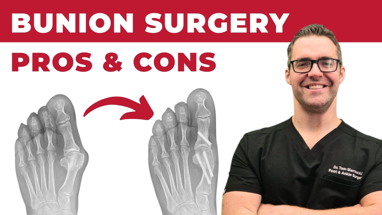
Dr. Tom Biernacki DPM compares the pros and cons of each bunion surgery type — MichiganFootDoctors YouTube
Austin/Chevron Osteotomy — Mild to Moderate (IMA up to 15°, HVA under 30°)
A V-shaped (chevron) cut in the first metatarsal head allows the capital fragment to be shifted laterally, reducing the IMA and the medial prominence. Fixed with one or two headless compression screws. The most commonly performed bunion procedure globally. Highly reliable with under 5% recurrence for correctly selected cases. Recovery: surgical shoe 4–6 weeks; regular wide shoe at 6–8 weeks. Not appropriate for: severe deformity (IMA >15°) or hypermobile first TMT joint (high recurrence).
Lapidus Bunionectomy — Moderate to Severe with Hypermobility (any IMA with unstable TMT joint)
The Lapidus procedure fuses the first tarsometatarsal joint, eliminating the hypermobility that drives recurrence in most patients. This is the procedure of choice when the first TMT joint demonstrates >9mm of mobility on clinical testing — because any osteotomy correction will recur if the unstable joint allowing the metatarsal to drift is left intact. Modern Lapidus with rigid internal fixation (locking plate systems) allows early protected weight-bearing in a boot, substantially shortening the traditional non-weight-bearing period. Recovery: protected weight-bearing in boot 6 weeks, transition to wide shoe at 8–12 weeks. Recurrence: under 2% — the lowest of any bunion procedure.
Minimally Invasive Bunion Surgery (MIS/MICA)
Third-generation minimally invasive techniques use 3–5mm percutaneous incisions and specialized rotary burrs under live fluoroscopic guidance to perform a chevron-equivalent osteotomy without a traditional open incision. Validated in Level I RCTs against open chevron osteotomy with equivalent angular correction, faster swelling resolution, and superior early cosmetic appearance. Best for: mild to moderate bunions (IMA up to 15°) in patients prioritizing cosmetic appearance or fastest return to shoe wear. Contraindicated for: severe deformity requiring large correction, hypermobile TMT joint, or revision cases.
Scarf Osteotomy — Moderate to Severe (IMA 13–18°, stable TMT joint)
A long, Z-shaped cut along the entire metatarsal shaft allows greater translation and correction than a chevron osteotomy allows. More technically demanding than chevron and preferred in some European training centers for larger deformities with a stable TMT joint. Recovery: surgical shoe 6–8 weeks, wide shoe at 8–10 weeks. Provides excellent correction for deformities too severe for standard chevron that do not have first TMT hypermobility.
Key takeaway: The most common reason bunions recur after surgery is performing an osteotomy (bone cut) in a patient who has first TMT joint hypermobility — the underlying cause of the deformity is left intact, and the metatarsal drifts back. This is why first TMT joint testing on clinical exam is a non-negotiable step in bunion surgical planning, not an optional assessment.
Bunion Surgery Recovery: Week-by-Week Timeline
Austin/Chevron and MIS Recovery
- Day of surgery: Walk same day in a surgical boot or post-op shoe. Foot is numb from the regional anesthetic block for 6–12 hours. Pain is typically 4–6/10 for the first 3–5 days, well controlled with oral medication.
- Week 1: Rest, elevate, ice for swelling. Surgical dressing changes at first post-op visit (3–5 days). Most patients transition to OTC pain relievers by day 5–7.
- Weeks 2–4: Continued walking in the surgical shoe. Swelling peaks around 2–3 weeks. X-rays at 4 weeks confirm bone position.
- Weeks 4–6: Transition to a wider athletic shoe if X-rays confirm healing. Most patients are in sneakers by 6–8 weeks.
- Months 3–4: Return to athletic activity. Full swelling resolution takes 3–6 months — normal and expected.
Lapidus Fusion Recovery
- Weeks 1–6: Protected weight-bearing in a boot (immediate or delayed depending on fixation construct and surgeon protocol). Crutches or knee scooter may assist during this phase.
- Weeks 6–8: Progressive weight-bearing in a boot as fusion consolidates on X-ray.
- Weeks 8–12: Transition to wide, supportive shoe. Physical therapy for range of motion and calf strengthening.
- Months 4–6: Return to athletic activity. Fusion maturation occurs at 4–6 months.
Bunion Surgery Recovery Essentials
Three recovery aids I most often recommend to my post-bunionectomy patients — always follow your surgeon’s specific protocol on when to start each one:
Bunion Bootie Day Sleeve — thin enough to wear inside a shoe during the weeks 8–16 shoe transition. Check Price on Amazon →
ViveSole Toe Separator Spacers — gentle realignment support once your surgeon clears toe spacers (typically weeks 4–12). Check Price on Amazon →
Copper Compression Bunion Night Splint — nighttime support during later-stage soft-tissue remodeling. Check Price on Amazon →
See the full bunion surgery recovery product guide →
As an Amazon Associate, Balance Foot & Ankle earns from qualifying purchases.
Conservative Care Before Surgery

Surgery is appropriate only after a genuine trial of conservative management — typically 6+ months. Conservative options reduce symptoms but do not correct the deformity:
- Wide, round toe-box shoes: The most effective conservative measure. Eliminates pressure on the medial eminence and reduces bursitis and corns. Athletic shoes with wide-last construction (HOKA, New Balance wide, Brooks wide) are most tolerable.
- Bunion splints/spacers: Silicone toe spacers between the hallux and second toe provide minimal deformity correction but can reduce the pressure-related pain of the toes crowding each other. Night splints have not been shown to correct bunion angle in clinical trials.
- Custom orthotics: Address the pronation and first ray hypermobility contributing to progression. Cannot reverse established deformity but can slow progression and reduce joint loading.
- Anti-inflammatory treatment: Oral NSAIDs, topical diclofenac, or ultrasound-guided corticosteroid injection into the first MTP or bursa for acute inflammatory flares.
- Padding: Donut-shaped bunion pads offload pressure directly over the medial eminence — effective for pain management, not deformity correction.
Doctor Hoy’s Natural Pain Relief Gel
Natural topical pain relief I use in our clinic. Arnica + camphor formula — apply directly to the area 3–4x daily. ($20–25)
Shop Doctor Hoy’s →Most Common Mistakes
- Choosing the wrong procedure for the deformity: An Austin osteotomy on a patient with first TMT hypermobility will recur — the deforming force (metatarsal hypermobility) was not addressed. Procedure selection must follow clinical testing and X-ray measurement, not size of the bump alone. This is the leading cause of bunion recurrence requiring revision surgery.
- Operating too early (adolescent patients): Performing definitive bunion surgery before growth plate closure in adolescents risks physeal injury and deformity recurrence as the foot continues to grow and mature. In patients under 16–18, conservative management until skeletal maturity is the standard of care except in extreme cases.
Red Flags — When to See a Podiatrist Immediately
- Sudden onset severe pain at the first MTP with acute redness and warmth — may indicate acute gout, septic joint, or pathologic fracture through the deformed joint; requires urgent evaluation and joint aspiration to rule out infection
- Second toe beginning to dislocate or cross over the hallux — indicates plantar plate failure at the second MTP joint, a structural injury requiring prompt evaluation to prevent permanent dislocation
- Open skin breakdown over the medial eminence in a diabetic patient — a wound over a bony prominence in a neuropathic patient risks deep infection and osteomyelitis; requires same-day podiatric evaluation
- Progressive bunion in a teenager causing functional limitation — adolescent bunions with significant IMA elevation and functional pain warrant early evaluation even if surgical treatment is deferred, because appropriate conservative management slows progression
- Numbness, paresthesias, or color change in the hallux or 2nd toe — vascular insufficiency to the great toe or nerve entrapment from the advancing deformity requires prompt vascular and neurologic assessment
Risks and Complications of Bunion Surgery
- Recurrence: Under 2% (Lapidus) to under 5% (Austin/chevron) with appropriate procedure selection. Soft-tissue-only procedures have 15–30% recurrence and are rarely indicated.
- Hardware irritation: Screws used to fix osteotomies can occasionally cause shoe-wear discomfort and may require removal as a minor outpatient procedure.
- Delayed healing or non-union: Most common in smokers, diabetics, and immunocompromised patients. Smoking cessation for 4+ weeks before and after surgery is a requirement, not a suggestion, in our practice.
- First MTP stiffness: Some loss of dorsiflexion is common post-operatively and improves with physical therapy; pre-operative joint mobility is the best predictor of post-operative mobility.
- Nerve sensitivity: Temporary numbness or hypersensitivity around the dorsomedial incision from dorsal cutaneous nerve irritation; typically resolves in 3–6 months.
- Infection: Under 1% with standard sterile technique and prophylactic antibiotics.
Frequently Asked Questions
How painful is bunion surgery recovery?
Most patients rate post-operative pain 4–6/10 for the first 3–5 days, well controlled with oral pain medication. The foot is numb for the first 6–12 hours from the local anesthetic block. By day 5–7, most patients transition to over-the-counter pain relievers. Swelling — not incision pain — is typically the primary symptom by week 2.
Will my bunion come back after surgery?
Recurrence depends heavily on procedure selection and underlying cause. Austin osteotomy: under 5% recurrence with appropriate footwear post-op. Lapidus fusion: under 2% because the unstable joint driving recurrence is eliminated. The highest recurrence rates are seen when the wrong procedure was selected for the deformity type — which is why preoperative X-ray measurement and first TMT joint testing are non-negotiable.
How long before I can wear regular shoes?
Austin/chevron and MIS: wide athletic shoe at 6–8 weeks. Dress shoes and narrow shoes at 3–4 months when swelling has substantially resolved. Lapidus: wide athletic shoe at 8–12 weeks; full range of footwear at 4–6 months. Residual foot swelling can persist 6–12 months — normal and not a sign of healing failure.
Is bunion surgery covered by insurance?
Most major insurance plans (including Medicare and Medicaid) cover bunion surgery for functional indications — documented pain limiting daily activity — with a diagnosis confirmed on X-ray and a documented conservative care trial of 6+ months. We handle pre-authorization and documentation at our clinic and verify coverage before scheduling.
Can both feet be done at the same time?
Bilateral simultaneous bunion surgery is possible but rarely recommended because it creates significant mobility challenges during recovery. We typically recommend spacing bilateral corrections 3–4 months apart. Patients who are wheelchair-mobile or have full-time caregiving may be candidates for simultaneous correction in select cases.
How successful is bunion surgery?
Modern bunion surgery achieves 90–95% patient satisfaction at 5 years with significant pain improvement and functional restoration. Recurrence is lowest with the Lapidus procedure (under 2%) because it eliminates the unstable joint that drives the deformity; Austin/chevron and MICA are under 5% with correct patient selection. Post-operative footwear compliance (wide toe box, low heel) is also an important factor in preventing recurrence.
Where can I get bunion surgery near me in Michigan?
Balance Foot & Ankle performs bunion surgery — including minimally invasive MICA correction — from two Michigan offices: 4330 E Grand River Ave, Howell, MI 48843 (Livingston County) and 43494 Woodward Ave #208, Bloomfield Hills, MI 48302 (Oakland County). Surgical consultations include weight-bearing X-rays at the first visit, and no referral is needed — call (810) 206-1402.
In-Office Bunion Evaluation at Balance Foot & Ankle
If you’ve been searching for bunion surgery near me, both offices serve the I-96 and Woodward corridors: Howell patients come from Brighton, Hartland, and Fowlerville; Bloomfield Hills patients from Birmingham, Troy, and West Bloomfield. Most patients are evaluated within the same week.
Every bunion consultation at our Howell and Bloomfield Hills clinics includes weight-bearing digital X-rays with HVA and IMA measurement, first TMT joint mobility testing, and a procedure-specific surgical plan when indicated. Dr. Tom Biernacki performs 100+ bunion corrections annually — volume that directly correlates with better outcomes and lower complication rates. Same-week new-patient appointments available.
Howell: 4330 E Grand River Ave, Howell MI 48843
Bloomfield Hills: 43494 Woodward Ave #208, Bloomfield Hills MI 48302
Phone: (810) 206-1402 | Book online →
Sources
- Nix S, et al. Prevalence of hallux valgus in the general population: a systematic review and meta-analysis. J Foot Ankle Res. 2010;3:21.
- Easley ME, Trnka HJ. Current concepts review: hallux valgus part II: operative treatment. Foot Ankle Int. 2007;28(6):748–758.
- Barg A, et al. Hallux valgus. Dtsch Arztebl Int. 2018;115(45):755–764.
- Kaufmann G, et al. Minimally invasive versus open chevron osteotomy for hallux valgus correction: prospective randomized trial. J Bone Joint Surg Am. 2019;101(15):1354–1361.
- Perera AM, et al. The number and diversity of published bunion corrections. Foot Ankle Int. 2011;32(4):426–427.
- American Academy of Orthopaedic Surgeons (AAOS): Bunions
Ready to Find Out if Bunion Surgery Is Right for You?
Weight-bearing X-rays & same-day surgical consultation — Howell & Bloomfield Hills, MI
4.9★ | 1,123 Reviews | 3,000+ Surgeries
Or call: (810) 206-1402
Dr. Tom Biernacki, DPM is a board-certified foot & ankle surgeon (ABFAS & ABPM) at Balance Foot & Ankle Specialists in Southeast Michigan. With over a decade of clinical experience, he specializes in heel pain, bunions, diabetic foot care, sports injuries, and minimally invasive surgery. Dr. Biernacki is a member of the APMA and ACFAS, and his patient education content on MichiganFootDoctors.com and YouTube has made him one of the most-followed foot & ankle educators on YouTube.