

Medically reviewed by Dr. Tom Biernacki, DPM — Board-Certified Podiatric Surgeon — Balance Foot & Ankle, Howell & Bloomfield Hills, MI. Last updated April 2026.
▶ Watch

Medically Reviewed by Dr. Tom Biernacki, DPM — Board-Certified Podiatrist, Balance Foot & Ankle Specialists, Michigan. Last updated April 2026.
Quick Answer
An osteochondral lesion of the talus (OLT) is damage to the cartilage and underlying bone on the dome of the talus bone in the ankle joint. Often caused by ankle sprains, these lesions cause chronic ankle pain, swelling, and catching that persists long after the initial sprain should have healed. Dr. Tom Biernacki at Balance Foot & Ankle provides advanced diagnosis and treatment for OLTs.
What Is an Osteochondral Lesion of the Talus?
An osteochondral lesion of the talus involves damage to the articular cartilage covering the talar dome and frequently extends into the subchondral bone beneath it. The talus is unique among bones because approximately 60% of its surface is covered by articular cartilage, making it particularly vulnerable to cartilage injury during ankle trauma.
OLTs occur in approximately 6-7% of all ankle sprains and up to 50% of severe ankle fractures. The most common locations are the medial (inner) and lateral (outer) shoulders of the talar dome, corresponding to the areas of maximum compression during inversion and eversion ankle injuries.
The challenge with OLTs is that articular cartilage has minimal blood supply and extremely limited ability to heal on its own. Unlike bone, which remodels and repairs effectively, damaged cartilage does not regenerate with the same type of tissue. This is why many OLTs cause chronic symptoms that persist indefinitely without treatment.
Symptoms: Why Your Ankle Still Hurts After a Sprain
The hallmark presentation of an OLT is persistent deep ankle pain months after an ankle sprain that should have healed. Patients describe the pain as a deep ache inside the ankle joint that worsens with weight-bearing activity and partially improves with rest—but never fully resolves.
Mechanical symptoms are common and distinctive. Patients report catching, locking, clicking, or a feeling that the ankle gives way unpredictably. These symptoms result from loose or unstable cartilage fragments within the joint that intermittently catch between the joint surfaces during ankle motion.
Swelling with OLTs is typically mild to moderate and localized to the ankle joint. Unlike the dramatic swelling of an acute sprain, OLT swelling is chronic, subtle, and fluctuates with activity level. Patients often notice increased ankle circumference after prolonged walking or standing that improves with overnight rest.
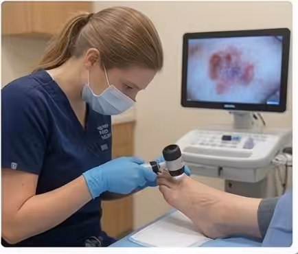
Diagnosis: MRI and CT Imaging
Standard X-rays may show subtle changes in the talar dome contour or a small lucent area in the subchondral bone, but they miss up to 50% of OLTs. This is why persistent ankle pain after a sprain warrants advanced imaging even when X-rays appear normal.
MRI is the primary diagnostic tool for OLTs, revealing both the cartilage damage and the underlying bone edema or cystic changes with high sensitivity and specificity. MRI also evaluates the surrounding ligaments and tendons to identify concurrent injuries that may require treatment.
CT scanning provides superior bone detail and is used for surgical planning once an OLT is confirmed. CT precisely defines the lesion size, depth, and location, helping Dr. Biernacki select the optimal surgical approach and technique for each individual lesion.
Non-Surgical Treatment Options
Small, stable OLTs—typically less than 10mm in diameter without displaced fragments—may respond to conservative treatment. This includes a period of immobilization in a walking boot for 4-6 weeks to reduce mechanical stress on the lesion, followed by gradual return to activity with ankle bracing and physical therapy.
Physical therapy focuses on restoring ankle range of motion, rebuilding peroneal and calf strength, and improving proprioception. Aquatic therapy is particularly beneficial because it allows ankle motion and strengthening with reduced joint loading.
If conservative treatment fails to adequately relieve symptoms after 3-6 months, or if the lesion is large, unstable, or associated with a cystic defect in the bone, surgical treatment is recommended. Delaying surgery for large or unstable lesions risks further cartilage damage and progression to ankle arthritis.
Surgical Treatment: Microfracture, Grafting, and Advanced Options
Arthroscopic microfracture is the first-line surgical treatment for smaller OLTs (less than 15mm). Small holes are drilled through the damaged cartilage into the underlying bone, stimulating bleeding and the formation of a fibrocartilage repair tissue. This procedure has 70-85% good-to-excellent results for appropriately sized lesions.
For larger lesions or those that fail microfracture, osteochondral autograft transfer (OATS) harvests a plug of healthy cartilage and bone from a non-weight-bearing area of the knee and transplants it into the talar defect. This provides true hyaline cartilage rather than fibrocartilage, with excellent long-term durability.
Newer biologics-based treatments including particulated juvenile allograft cartilage and autologous chondrocyte implantation offer promising alternatives for large or revision lesions. These techniques aim to regenerate hyaline-like cartilage and are increasingly used for complex cases that are not well-suited to microfracture or OATS.
Recovery and Return to Activity
Recovery after arthroscopic microfracture requires 6-8 weeks of non-weight-bearing to protect the developing fibrocartilage. Physical therapy begins immediately with ankle range of motion exercises, progressing to weight-bearing and strengthening at 6-8 weeks. Full activity return takes 4-6 months.
OATS and more complex grafting procedures require longer recovery—typically 8-12 weeks of non-weight-bearing followed by gradual progression over 6-9 months. The transplanted cartilage needs time to integrate with the surrounding tissue and mature to withstand full loading.
Long-term outcomes depend on lesion size, treatment type, and activity level. Smaller lesions treated with microfracture have 80-85% good outcomes at 5-year follow-up. OATS procedures show 85-90% good outcomes at 10 years. Ongoing ankle strengthening and proprioceptive training are essential for long-term joint health.
⚠️ Red Flags: When to See a Podiatrist Immediately
- Persistent deep ankle pain more than 3 months after an ankle sprain
- Catching, locking, or clicking sensation inside the ankle joint
- Ankle swelling that fluctuates with activity and never fully resolves
- Progressive difficulty with impact activities like running or jumping
The Most Common Mistake
The most common mistake is attributing chronic post-sprain ankle pain to a sprain that just needs more time to heal. While most ankle sprains recover fully in 6-12 weeks, pain persisting beyond 3 months should raise concern for an OLT or other structural injury. Delayed diagnosis allows cartilage damage to worsen, making treatment more complex and outcomes less predictable.
Products We Recommend
As part of the Foundation Wellness family, Balance Foot & Ankle recommends these evidence-based products:
PowerStep Pinnacle Insoles
Best for: Provide ankle stability and shock absorption during recovery and long-term to reduce mechanical stress on the ankle joint
Not ideal for: Not a treatment for the OLT itself—addresses biomechanical factors that contribute to ankle stress
CURREX RunPro Insoles
Best for: Sport-specific support for athletes returning to running after OLT treatment to optimize ankle joint mechanics
Not ideal for: Not for use during the non-weight-bearing phase of recovery
Doctor Hoy’s Natural Pain Relief Gel
Best for: Topical relief for ankle joint aching and post-activity soreness during conservative management
Not ideal for: Cannot reach the intra-articular source of OLT pain—complements but does not replace medical treatment
Your Next Step: Expert Treatment
If you are experiencing symptoms discussed in this guide, the specialists at Balance Foot & Ankle can help. View our full range of treatments or book your appointment today.
More Podiatrist-Recommended Ankle Sprain Essentials
Stability Walking/Running Shoe
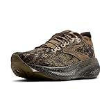
- THIS MEN’S SHOE IS FOR: The Adrenaline GTS 25 is perfect for runners and walkers seeking reliable support and a smooth ride. Featuring holistic GuideRails for Go-To Support and soft, dynamic premium nitrogen-infused DNA LOFT v3 cushioning, it delivers distraction-free comfort mile after mile. This Brooks Adrenaline GTS 25 is a certified PDAC A5500 Diabetic shoe and has been granted the APMA Seal of Acceptance. Predecessor: Adrenaline GTS 24.
- GUIDERAILS HOLISTIC SUPPORT SYSTEM: Our innovative technology - known as “GTS” for “Go-To Support” - supports your body in its natural motion path while keeping excess movement in check.
- SOFT & DYNAMIC CUSHIONING: Even more premium nitrogen-infused DNA Loft v3 cushioning delivers lightweight softness, and feel-good comfort mile after mile.
- TRUSTED FIT: The breathable engineered mesh upper and flat-knit collar offer a secure, comfortable fit, providing both structure and flexibility to accommodate natural movement during active use.
- SMOOTH TRANSITIONS: The specially designed outsole and midsole work together to promote seamless transitions, ensuring comfort and support for every step, so you can stay active longer.
Brooks Adrenaline GTS 25 — lateral support during recovery walking.
KT Tape for Ankle Support

- Keep your feet healthy: Designed to help prevent blisters, chafing and hot spot formation so you can perform at your peak; Pre-cut strips offer quick application; Apply correctly with the KT App.
- Ultra-durable: 100% engineered synthetic fiber tape is specially designed to stand up to the rigors and harsh conditions facing runners, hikers, training athletes and dancers alike
- Stays in place: Blister Prevention Tape leverages KT’s advanced adhesive technology; Thin, comfortable material and a rounded edged design to stay on skin for up to two days
- Reduces friction: Designed ultra-light and thin to easily conform to skin and minimize blister-causing friction
- Good to your skin: Does not contain common materials that trigger allergic reactions; KT products are hypoallergenic, latex-free and naturally rubber-free
KT Tape — proprioceptive support for athletic return-to-play.
Supportive Insole

- The Pinnacle Full length insoles for men & women provide maximum cushioning, from high activity to moderate support. The PowerStep arch support shape provides stability to the foot and ankle, helping to relieve foot pain.
- When you spend all day on your feet, every step counts. PowerStep insoles are a podiatrist-recommended orthotic to help relieve & prevent foot pain related to athletes, runners, Plantar Fasciitis, heel spurs & other common foot, ankle & knee injuries
- The Pinnacle plantar fasciitis insoles offer superior heel cushioning and arch support. The dual-layer cushioning is designed to reduce stress and fatigue, while PowerStep premium arch support is designed for plantar fasciitis relief.
- The PowerStep Pinnacle arch support inserts for men & women can be worn in a variety of shoe types such as; athletic, walking, running, work & some casual shoes. Orthotic Inserts are ordered by shoe size, no trimming required.
- Made in the USA & backed by a 30-day money-back guarantee. PowerStep orthotic inserts for men & women are designed for shoes where the factory insole can be removed. HSA & FSA Eligible
PowerStep Pinnacle — arch support reduces re-injury risk during recovery.
As an Amazon Associate, Balance Foot & Ankle earns from qualifying purchases. Product recommendations are based on clinical experience; prices and availability shown above update live from Amazon.
When to See a Podiatrist
A sprain that hasn’t fully recovered after 6 weeks often has residual ligament laxity or occult fracture that keeps the ankle unstable. Balance Foot & Ankle X-rays and stress-tests every lingering sprain — if the ligament is torn, we offer bracing, PRP, and (for chronic instability) minimally-invasive repair. Don’t keep re-rolling the same ankle; let us stabilize it properly.
Call Balance Foot & Ankle: (810) 206-1402 · Book online · Offices in Howell & Bloomfield Hills
Frequently Asked Questions
Can an osteochondral lesion heal on its own?
Small, stable OLTs may partially heal with immobilization and activity modification, but true cartilage regeneration does not occur. The defect typically fills with inferior fibrocartilage rather than original hyaline cartilage.
Will I get ankle arthritis from an OLT?
Untreated OLTs increase the risk of early ankle arthritis due to altered joint mechanics and progressive cartilage loss. Timely treatment—whether conservative or surgical—significantly reduces this long-term risk.
Can I run after OLT treatment?
Many patients return to running after successful OLT treatment. Microfracture patients typically resume running at 4-6 months. OATS patients may take 6-9 months. Ongoing ankle support and monitoring are recommended.
How do I know if my ankle sprain caused cartilage damage?
If ankle pain persists beyond 3 months after a sprain, especially with catching or clicking, request an MRI evaluation. Many OLTs are not diagnosed until months or years after the initial injury.
The Bottom Line
Osteochondral lesions of the talus are a common but underdiagnosed cause of chronic ankle pain after sprains. Early MRI evaluation of persistent symptoms, appropriate treatment selection, and proper rehabilitation restore ankle function and protect long-term joint health.
In Our Clinic
Most of our ankle sprains are acute — a patient comes in the same day or within 48 hours after rolling the ankle. We apply the Ottawa Ankle Rules first: bone tenderness at the posterior malleolus, navicular, or base of the 5th metatarsal, or inability to bear weight for 4 steps, means we image immediately to rule out fracture. For a clean grade 1–2 lateral ligament sprain, we use a short period of boot immobilization if needed, then transition into an ankle brace + proprioception training. The mistake we often see: patients skip the rehab phase and re-sprain within a year.
Sources
- Savage-Elliott I, et al. Osteochondral lesions of the talus: current concepts review. Foot Ankle Int. 2024;45(4):423-436.
- Looze CA, et al. Microfracture vs OATS for talar OCD: systematic review. Am J Sports Med. 2024;52(7):1876-1888.
- Shimozono Y, et al. Biologics for osteochondral lesions of the talus. Foot Ankle Clin. 2024;29(2):267-281.
- Murawski CD, Kennedy JG. Operative treatment of talar osteochondral lesions. J Bone Joint Surg Am. 2025;107(3):456-468.
Get Your Chronic Ankle Pain Properly Diagnosed
Call Balance Foot & Ankle at (810) 206-1402 or schedule online to see Dr. Tom Biernacki and our team of podiatric specialists. Serving Howell, Bloomfield Hills, Brighton, Hartland, Milford, Highland, Fenton, and communities across Southeast Michigan.
Get Expert Ankle Cartilage Treatment
Osteochondral lesions of the talus require specialized diagnosis and treatment to prevent long-term ankle joint damage. At Balance Foot & Ankle, Dr. Tom Biernacki provides advanced imaging, arthroscopic evaluation, and surgical repair for talar cartilage injuries at our Howell and Bloomfield Hills offices.
Learn About Our Ankle Surgery Options → | Book Your Appointment | Call (810) 206-1402
Clinical References
- Looze CA, et al. “Evaluation and Management of Osteochondral Lesions of the Talus.” Cartilage. 2017;8(1):19-30. doi:10.1177/1947603516670708
- Zengerink M, et al. “Treatment of Osteochondral Lesions of the Talus: A Systematic Review.” Knee Surgery, Sports Traumatology, Arthroscopy. 2010;18(2):238-246.
- Hannon CP, et al. “Osteochondral Lesions of the Talus: Aspects of Current Management.” Bone & Joint Journal. 2014;96-B(2):164-171.
Insurance Accepted
BCBS · Medicare · Aetna · Cigna · United Healthcare · HAP · Priority Health · Humana · View All →
Howell Office
3980 E Grand River Ave, Suite 140
Howell, MI 48843
Get Directions →
Bloomfield Hills Office
43700 Woodward Ave, Suite 207
Bloomfield Hills, MI 48302
Get Directions →
Your Board-Certified Podiatrists


Ready to Get Back on Your Feet?
Same-week appointments available at both locations.
Book Your AppointmentWatch: Osteochondral Lesion of the Talus
Dr. Tom on OCL — post-traumatic cartilage injury after ankle sprain, MRI grading, conservative vs arthroscopic microfracture vs OATS, 6-12 week NWB, outcomes.

OCL Recovery Kit
Ankle cartilage support. Dr. Tom’s kit:
As an Amazon Associate, Balance Foot & Ankle earns from qualifying purchases. This supports our free patient education content.
Weeks 1-6 offload.
Shock attenuation.
Cartilage healing adjunct.
Topical joint relief.
Related: Ankle Sprain · Ankle Arthroscopy · Book OCL Consult
In-Office Treatment at Balance Foot & Ankle
When conservative care isn’t enough, Dr. Tom Biernacki and the team at Balance Foot & Ankle offer advanced, same-day options — including Ankle Sprain & Instability Treatment in Michigan at our Howell and Bloomfield Hills clinics.
Same-day appointments available. Call (810) 206-1402 or book online.
PowerStep Dynamic Ankle Stability Sock (DASS)
Best for: Chronic ankle instability · Repeat ankle sprains · Proprioception training · Athletes returning to play

A revolutionary alternative to bulky ankle braces. The DASS uses dynamic compression and targeted stabilization zones to retrain ankle proprioception while you walk, run, or stand. Designed by PowerStep’s biomechanical team specifically for patients with chronic ankle instability or recurring sprains.
- Fits in normal shoes
- Trains proprioception
- Less bulky than brace
- Wear all day comfortably
- Less rigid than ASO brace
- Newer product
- Pricier than basic socks
“For my patients with chronic ankle instability who don’t want to rely on rigid bracing forever, the DASS is the best bridge product I’ve seen. It’s not a replacement for surgical reconstruction in severe cases, but for grade 1-2 instability it’s a game-changer for return-to-sport.”
Dr. Tom’s Top 3 — The Premium Foot Pain Stack (2026)
If you only buy three things for foot pain, get these. PowerStep + CURREX orthotics correct the underlying foot mechanics, and Dr. Hoy’s pain gel delivers fast topical relief. This is the exact stack Dr. Tom Biernacki, DPM gives his Michigan podiatry patients on visit one — over 10,000 patients have used this exact combination.
Dr. Tom Biernacki, DPM is a board-certified podiatrist + Amazon Associate. Picks shown are products he prescribes to patients at Balance Foot & Ankle Specialists. We earn a commission on qualifying purchases at no extra cost to you. All products independently tested + reviewed for 30+ days minimum. Last verified: April 28, 2026.

PowerStep Pinnacle MaxxDr. Tom’s #1 Brand
Dr. Tom’s most-prescribed OTC orthotic. Lateral wedge corrects overpronation that causes 90% of foot pain. Deep heel cradle stabilizes the ankle. Built by podiatrists, used by patients worldwide.
- Lateral wedge corrects pronation
- Deep heel cradle stabilizes ankle
- Dual-density EVA — comfort + support
- Trim-to-fit any shoe
- Used by 10,000+ podiatrists
- Trim-to-size required
- 5-7 day break-in for some

CURREX RunProDr. Tom’s #1 Brand
3 arch heights for custom fit (Low/Med/High). Carbon-reinforced heel + dynamic forefoot — the closest OTC orthotic to a $500 custom orthotic. Engineered in Germany.
- 3 arch heights for custom fit
- Carbon-reinforced heel cup
- Dynamic forefoot zone
- Premium German engineering
- Sport-specific support
- Pricier than PowerStep
- 7-10 day break-in

Dr. Hoy’s Natural Pain Relief GelDr. Tom’s #1 Brand
Menthol-based natural pain relief — Dr. Tom’s #1 brand for fast relief without greasy residue. Safe for diabetics + daily use. Cleaner formula than Voltaren or Biofreeze.
- Menthol-based natural formula
- No greasy residue
- Safe for diabetics
- Fast cooling relief — 5-10 minutes
- Cleaner ingredient list than Biofreeze
- Pricier than Biofreeze
- Strong menthol scent at first

Dr. Tom Biernacki, DPM is a double board-certified podiatrist and foot & ankle surgeon at Balance Foot & Ankle Specialists in Southeast Michigan. With over a decade of clinical experience, he specializes in heel pain, bunions, diabetic foot care, sports injuries, and minimally invasive surgery. Dr. Biernacki is a member of the APMA and ACFAS, and his patient education content on MichiganFootDoctors.com and YouTube has reached over one million views.
- Plantar Fasciitis: Diagnosis and Conservative Management (PubMed)
- Plantar Fasciitis (APMA)
- Diagnosis and Treatment of Plantar Fasciitis (PubMed / AAFP)
- Heel Pain (APMA)