
Medically reviewed by Dr. Tom Biernacki, DPM — Board-Certified Podiatric Surgeon — Balance Foot & Ankle, Howell & Bloomfield Hills, MI. Last updated April 2026.
▶ Watch

Medically Reviewed by Dr. Tom Biernacki, DPM — Board-Certified Podiatrist, Balance Foot & Ankle Specialists, Michigan. Last updated April 2026.
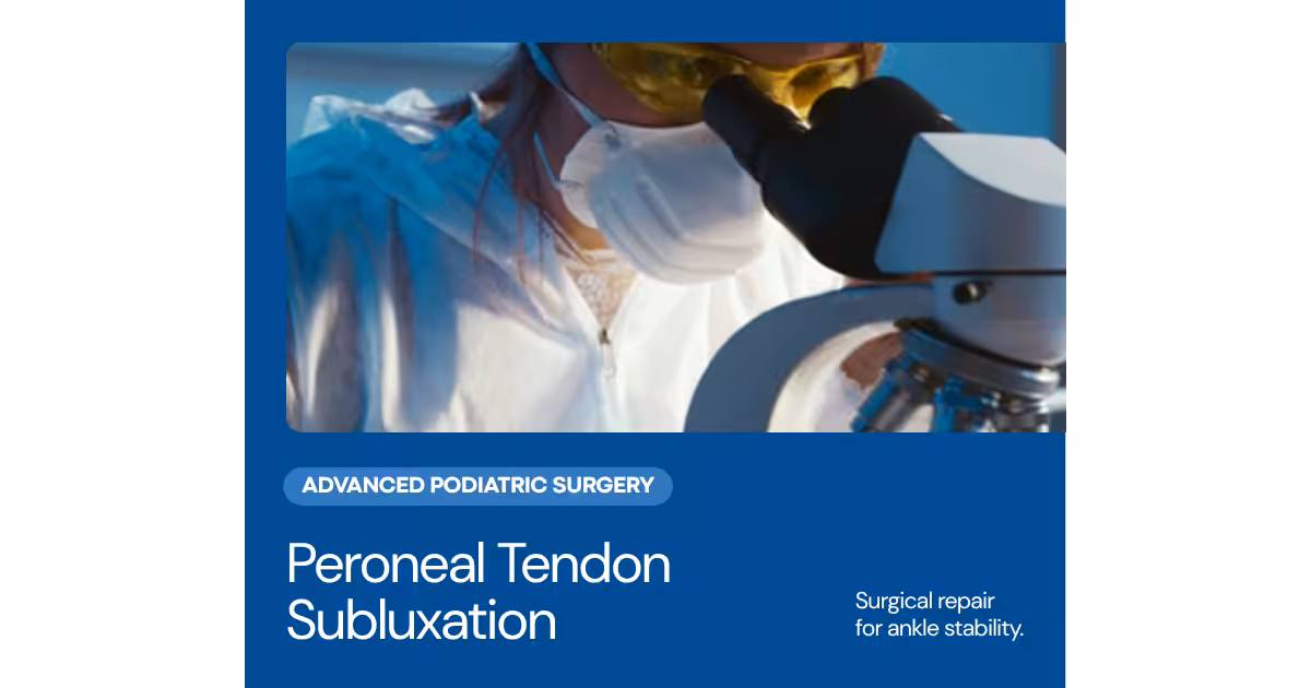
The Peroneal Tendons: Ankle Stabilizers Often Overlooked
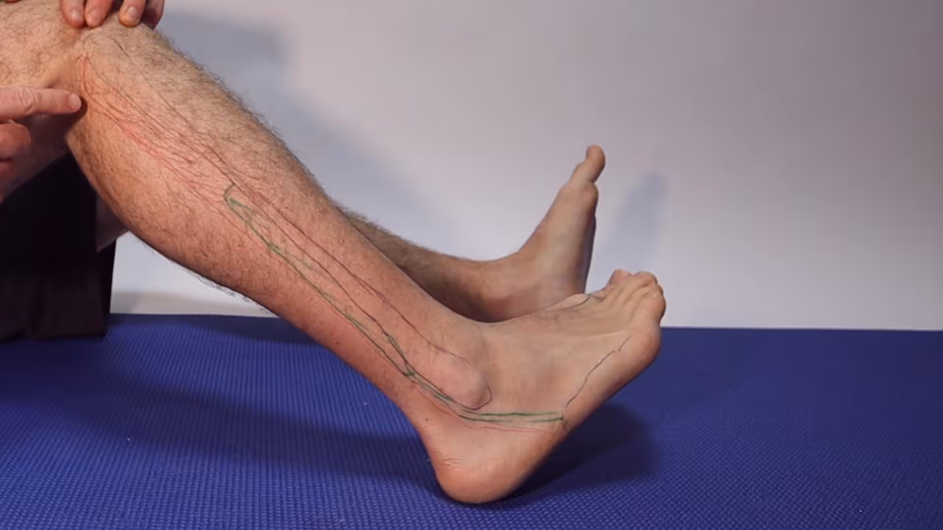
The peroneal tendons — the peroneus longus and peroneus brevis — run along the outer ankle, tucked in a groove behind the lateral malleolus (the bony prominence on the outer ankle). These tendons originate from the peroneal muscles in the outer calf and insert on the base of the fifth metatarsal (brevis) and the plantar surface of the first metatarsal and medial cuneiform (longus). Their primary function is to evert (pronate) the foot and provide dynamic stability against inward rolling — making them the primary muscular defenders against ankle sprains.
Peroneal tendon pathology is substantially underdiagnosed. Most patients who present with persistent outer ankle pain after an ankle sprain are appropriately diagnosed with lateral ligament sprains — but a significant proportion also have concurrent peroneal tendon tears or subluxation that is missed, leading to chronic pain despite apparently appropriate ligament treatment. Studies of patients with chronic lateral ankle instability found peroneal tendon tears in 25-77% depending on the study — remarkably high rates for a condition that’s often not on the radar.
Types of Peroneal Tendon Pathology
Peroneal tendon problems fall into three main categories, often occurring together.
Peroneal tendon tears are either longitudinal splits within the tendon substance (most common) or avulsion injuries at the bony attachment. Peroneus brevis tears are more common than longus tears, typically occurring as longitudinal splits along the tendon as it wraps around the posterior fibula. These splits are caused by the tendon being compressed between the fibula and a sharp edge during ankle inversion (rolling) events. Longitudinal splits may be partial or extend through the full tendon thickness.
Peroneus longus tears most commonly occur at two specific locations: at the fibular tunnel as the tendon wraps around the ankle, and at the lateral border of the cuboid bone (the “cuboid notch”) where the tendon changes direction. A complete peroneus longus rupture at the cuboid notch can cause dramatic forefoot and arch changes because this tendon plantarflexes the first ray — an important biomechanical function for normal foot architecture.
Peroneal tendon subluxation (dislocation) occurs when the superior peroneal retinaculum — the fibrous band that holds the peroneal tendons in their groove behind the fibula — is torn, allowing the tendons to snap in and out of (or completely out of) the groove during foot movement. This produces the characteristic “popping” or “snapping” sensation at the outer ankle that patients often describe after an ankle sprain. The sensation is distinctive — patients can often demonstrate it by dorsiflexing and everting the foot — and should prompt evaluation even though it’s frequently dismissed as “just the tendons snapping.”
Peroneal tendinopathy is chronic degenerative change within the tendon substance from overuse, presenting as diffuse outer ankle and lower leg pain without acute tearing. It’s more common in distance runners, dancers, and people with high-arched feet.
Diagnosis: Getting the Right Imaging
Clinical examination identifies peroneal tendon pathology through tenderness specifically over the tendon course (behind the fibula), pain and weakness with resisted eversion, and reproduction of subluxation with provocative maneuvers. X-rays may show a small fleck of bone behind the fibula — a rim sign indicating peroneal retinaculum avulsion — but are often normal.
MRI provides the most comprehensive assessment of the tendons, identifying split tears, signal changes indicating degeneration, fluid in the tendon sheath, and retinaculum injury. Ultrasound provides dynamic assessment that MRI cannot — specifically, it allows real-time visualization of the tendons subluxing out of the groove with provocative movements, definitively confirming subluxation. Both modalities have roles depending on the specific clinical question.
Conservative Treatment
Early-stage peroneal tendinopathy and small longitudinal tears without subluxation can often be managed conservatively. Rest from aggravating activities, bracing to limit excessive ankle inversion, physical therapy targeting peroneal strengthening and proprioceptive training, and custom orthotics to address underlying biomechanical contributors (high arch, heel varus) provide the foundation of conservative care.
The high-arched (cavus) foot is a major risk factor for peroneal tendon problems because the heel’s natural varus (inward) positioning increases load on the peroneal tendons with every step. Lateral heel posting (wedging) in custom orthotics reduces this varus and the repetitive peroneal loading it causes.
Acute peroneal subluxation — the first episode of the tendons snapping out of place — is sometimes managed non-surgically in a cast for 6 weeks. Results of conservative treatment for acute subluxation are variable; recurrent subluxation after conservative treatment typically warrants surgical stabilization.
Surgical Treatment
Surgery is indicated for peroneal tendon tears with significant symptomatic failure of conservative care and for recurrent peroneal subluxation. The type of surgery depends on the specific pathology.
For longitudinal splits, the torn tendon is repaired by debriding the damaged tissue and tubularizing (closing) the split with sutures. If the brevis tendon is severely damaged (more than 50% of its substance), the remaining viable portion may be tenodesis-fixed to the longus tendon, effectively converting the two-tendon system into a single-tendon system. Results of peroneal tendon repair are generally good, with most patients returning to their pre-injury activity level.
For peroneal subluxation, the retinaculum is repaired and reinforced — often combined with groove deepening (surgically creating a deeper groove behind the fibula to better contain the tendons) for cases with an abnormally shallow groove. Acute retinaculum repair has an excellent success rate when performed within weeks of the initial injury. Chronic subluxation repair is also effective, though the results are slightly less predictable than acute repair.
Recovery from peroneal tendon surgery typically involves 4-6 weeks non-weight-bearing, followed by progressive rehabilitation over 3-6 months. Return to sport typically occurs at 4-6 months for most patients.
Foot or Ankle Pain? We Can Help.
Balance Foot & Ankle — Howell & Bloomfield Township, MI
📅 Book Online
📞 (810) 206-1402
Peroneal Tendon Tear Treatment in Michigan
Peroneal tendon tears and subluxation cause chronic outer ankle pain often misdiagnosed as ankle sprains. At Balance Foot & Ankle, Dr. Tom Biernacki uses ultrasound and MRI to accurately diagnose peroneal injuries — serving Howell and Bloomfield Hills, MI.
Learn About Ankle Injury Treatment → | Book Your Appointment | Call (810) 206-1402
Clinical References
- Dombek MF, Lamm BM, Saltrick K, Mendicino RW, Catanzariti AR. Peroneal tendon tears: a retrospective review. J Foot Ankle Surg. 2003;42(5):250-258.
- Roster B, Michelier P, Giza E. Peroneal tendon disorders. Clin Sports Med. 2015;34(4):625-641.
- Saxena A, Cassidy A. Lateral ankle sprains: can the delay to diagnosis of peroneal tendon injuries be avoided? J Foot Ankle Surg. 2003;42(5):268-274.
Insurance Accepted
BCBS · Medicare · Aetna · Cigna · United Healthcare · HAP · Priority Health · Humana · View All →
Howell Office
3980 E Grand River Ave, Suite 140
Howell, MI 48843
Get Directions →
Bloomfield Hills Office
43700 Woodward Ave, Suite 207
Bloomfield Hills, MI 48302
Get Directions →
Your Board-Certified Podiatrists


Ready to Get Back on Your Feet?
Same-week appointments available at both locations.
Book Your AppointmentMore Podiatrist-Recommended Foot Health Essentials
Hoka Clifton 10

Max-cushion everyday shoe — podiatrist favorite for walking and running.

OOFOS Recovery Slide

Impact-absorbing recovery sandal — wear after long days on your feet.
As an Amazon Associate, Balance Foot & Ankle earns from qualifying purchases. Product recommendations are based on clinical experience; prices and availability shown above update live from Amazon.
When to See a Podiatrist
If foot or ankle pain has been bothering you for more than a few weeks, home care alone may not be enough. Balance Foot & Ankle offers same-week appointments at our Howell and Bloomfield Hills clinics — no referral needed in most cases. Bring your current shoes and a short list of symptoms and we’ll build you a treatment plan in one visit.
Call Balance Foot & Ankle: (810) 206-1402 · Book online · Offices in Howell & Bloomfield Hills
Differential Diagnosis: What Else Could It Be?
Not every case of peroneal tendonitis is straightforward. In our clinic we routinely rule out three look-alike conditions before confirming the diagnosis. If your symptoms don’t match the classic presentation, one of these may explain the pain — which is why physical exam matters more than self-diagnosis.
| Condition | How It Differs |
|---|---|
| Lateral ankle sprain | Acute inversion mechanism, bruising along anterior talofibular ligament, pain with anterior drawer. |
| 5th metatarsal base stress fracture | Point tenderness at 5th metatarsal base, pain with weight-bearing, fracture line on imaging. |
| Sinus tarsi syndrome | Deep ache in the sinus tarsi, pain reproduced with lateral palpation just anterior to the lateral malleolus. |
Red Flags — When to See a Podiatrist Now
Seek same-day evaluation at Balance Foot & Ankle if you notice any of the following:
- Snapping or popping behind the lateral malleolus (subluxation)
- Inability to evert the foot actively
- Persistent lateral ankle swelling >4 weeks
- Sudden pop with inability to continue walking
Call (810) 206-1402 or request an appointment. Our Howell and Bloomfield Hills offices reserve same-day slots for urgent foot and ankle issues.
In Our Clinic: What We See
Clinical perspective from Dr. Tom Biernacki, DPM — Balance Foot & Ankle, Howell & Bloomfield Hills, MI:
In our clinic, peroneal tendonitis patients usually come in after a recent ankle sprain — the pain started as a “sprain that didn’t fully heal.” They report lateral ankle pain that’s worse with turning the foot outward or walking on uneven surfaces. On exam we palpate specifically along the peroneal tendons behind the fibula and resist eversion. If we feel or see snapping behind the lateral malleolus, that’s peroneal subluxation, which usually needs surgical repair. Isolated peroneal tendonitis responds well to ankle bracing, peroneal eccentric strengthening, and temporary activity modification.
In-Office Treatment at Balance Foot & Ankle
When conservative care isn’t enough, Dr. Tom Biernacki and the team at Balance Foot & Ankle offer advanced, same-day options — including Peroneal Tendon Disorders Treatment in Michigan at our Howell and Bloomfield Hills clinics.
Same-day appointments available. Call (810) 206-1402 or book online.
Dr. Tom Biernacki, DPM is a double board-certified podiatrist and foot & ankle surgeon at Balance Foot & Ankle Specialists in Southeast Michigan. With over a decade of clinical experience, he specializes in heel pain, bunions, diabetic foot care, sports injuries, and minimally invasive surgery. Dr. Biernacki is a member of the APMA and ACFAS, and his patient education content on MichiganFootDoctors.com and YouTube has reached over one million views.
- Diagnosis and Treatment of Plantar Fasciitis (PubMed / AAFP)
- Heel Pain (APMA)
- Hallux Valgus (Bunions): Evaluation and Management (PubMed)
- Bunions (Mayo Clinic)
Recommended Products from Dr. Tom