

Quick answer: Tarsal Coalition Michigan is a common foot/ankle topic that affects many patients. Effective treatment starts with a targeted diagnosis, conservative-first treatment, and escalation only when needed. We treat this regularly at our Howell and Bloomfield Hills practices. Call (810) 206-1402.
Medically reviewed by Dr. Tom Biernacki, DPM · Board-Certified Podiatric Surgeon · Last reviewed: April 2026 · Editorial Policy
The most important clinical decision with Tarsal Coalition Michigan isn’t which treatment to start with — it’s identifying the correct subtype. That changes everything. Call (810) 206-1402.
Related Conditions
Quick Answer
Tarsal Coalition Treatment Michigan Pediatric & Adult relates to foot pain — typically caused by overuse, footwear, or biomechanics. Most patients improve in 6-12 weeks with conservative care. Same-week appointments in Howell + Bloomfield Hills: (810) 206-1402.

Medically reviewed by Dr. Tom Biernacki, DPM — Board-Certified Podiatric Surgeon — Balance Foot & Ankle, Howell & Bloomfield Hills, MI. Last updated April 2026.
▶ Watch
Tarsal Coalition Treatment Michigan
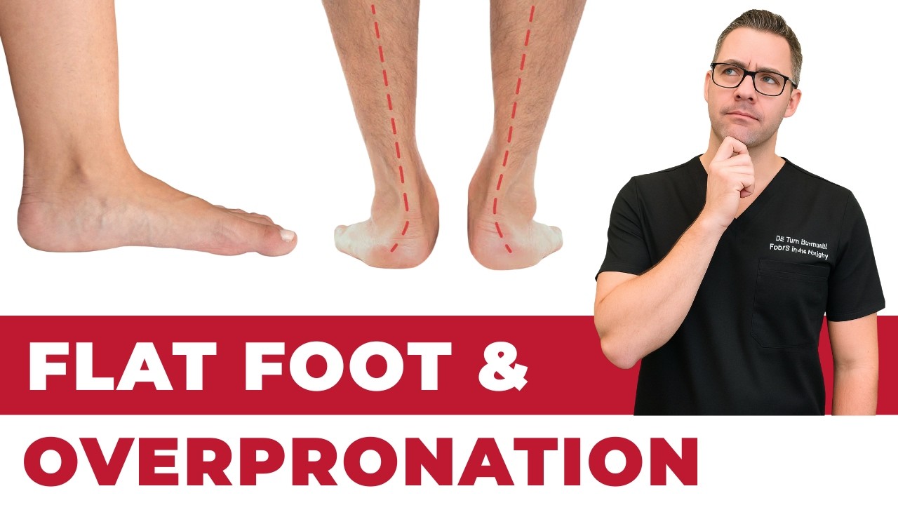
Tarsal coalition is an abnormal connection (bony, cartilaginous, or fibrous) between two or more of the tarsal bones in the hindfoot or midfoot. It is an under-recognized cause of rigid flatfoot and ankle pain in children, adolescents, and young adults. The two most common coalitions are calcaneonavicular (between the calcaneus and navicular) and talocalcaneal (between the talus and calcaneus at the middle facet). Dr. Tom Biernacki DPM at Balance Foot & Ankle treats tarsal coalition in Michigan at our Howell, Brighton, and Bloomfield Hills locations.
Why Tarsal Coalition Causes Symptoms
Normal hindfoot function requires motion across multiple subtalar and midtarsal joints during walking. When two tarsal bones are fused or tethered, this motion is blocked, placing abnormal stress on adjacent joints and restricting arch flexibility. Symptoms typically develop in adolescence (10–16 years) when coalitions ossify (harden to bone) and fully restrict motion — often mistaken for “growing pains” or ankle sprains.
Symptoms
- Rigid or semi-rigid flatfoot that does not correct on tiptoe standing
- Activity-related pain in the hindfoot, ankle, or lateral foot
- Recurrent ankle sprains (subtalar stiffness predisposes to sprains)
- Peroneal muscle spasm (peroneal spastic flatfoot)
- Limited subtalar inversion/eversion range of motion
Diagnosis
Standard weight-bearing X-rays may show calcaneonavicular coalition on oblique view or “C-sign” for talocalcaneal coalition on lateral view. However, CT scan is the definitive imaging modality — it precisely defines coalition type, size, percentage of joint involvement, and presence of secondary degenerative changes. MRI identifies fibrous or cartilaginous coalitions and associated bone marrow edema.
Treatment
Conservative treatment with CAM boot immobilization for 4–6 weeks followed by custom orthotics and activity modification is effective for symptomatic control in mild cases. Corticosteroid injection at the coalition site (under CT or ultrasound guidance) reduces acute inflammation.
Surgical resection of the coalition with interposition of fat or muscle is the treatment of choice for calcaneonavicular coalitions (high success rate) and for talocalcaneal coalitions involving less than 50% of the posterior facet without significant secondary arthritis. Recovery: non-weight-bearing for 4–6 weeks, followed by physical therapy and return to sport at 3–4 months.
Hindfoot fusion (subtalar or triple arthrodesis) is required when secondary degenerative changes are present or when resection has failed. Provides reliable pain relief at the cost of subtalar motion.
More Podiatrist-Recommended Foot Health Essentials
Hoka Clifton 10


Max-cushion everyday shoe — podiatrist favorite for walking and running.

OOFOS Recovery Slide

Impact-absorbing recovery sandal — wear after long days on your feet.
As an Amazon Associate, Balance Foot & Ankle earns from qualifying purchases. Product recommendations are based on clinical experience; prices and availability shown above update live from Amazon.

When to See a Podiatrist
If foot or ankle pain has been bothering you for more than a few weeks, home care alone may not be enough. Balance Foot & Ankle offers same-week appointments at our Howell and Bloomfield Hills clinics — no referral needed in most cases. Bring your current shoes and a short list of symptoms and we’ll build you a treatment plan in one visit.
Call Balance Foot & Ankle: (810) 206-1402 · Book online · Offices in Howell & Bloomfield Hills
Frequently Asked Questions
Can an adult have a tarsal coalition?
Yes — while symptoms typically appear in adolescence, tarsal coalitions may present in adults who have been managing pain with activity modification or who develop secondary arthritis. CT scan diagnosis and coalition resection or arthrodesis can provide significant relief in appropriately selected adults.
Is tarsal coalition hereditary?
Yes — tarsal coalition has an autosomal dominant inheritance pattern with variable expressivity. It is bilateral in approximately 50–60% of cases. Family members of diagnosed patients should be evaluated if they have rigid flatfoot or chronic ankle pain.

📧 Get Dr. Tom’s Free Lab Test Guide
Discover the 5 lab tests every person over 35 should ask their doctor about — explained in plain English by a board-certified physician.
Schedule your tarsal coalition evaluation at Balance Foot & Ankle in Howell, Brighton, or Bloomfield Hills, Michigan.
Join 950,000+ Learning About Foot Health
Dr. Tom shares honest medical advice, supplement reviews, and treatment guides you won’t find anywhere else.
Subscribe on YouTube →📍 Located in Michigan?
Our board-certified podiatrists treat this condition at two convenient locations. Same-day appointments often available.
Medically Reviewed by: Dr. Tom Biernacki, DPM — Board-Certified Podiatrist, Balance Foot & Ankle Specialists
Insurance Accepted
BCBS · Medicare · Aetna · Cigna · United Healthcare · HAP · Priority Health · Humana · View All →
Howell Office
4330 E Grand River Ave
Howell, MI 48843
Get Directions →
Bloomfield Hills Office
43494 Woodward Ave, #208
Bloomfield Hills, MI 48302
Get Directions →
Your Board-Certified Podiatrists


Ready to Get Back on Your Feet?
Same-week appointments available at both locations.
Book Your AppointmentPros & Cons of Conservative Care for foot care
Advantages
- ✓ Conservative care first
- ✓ Same-week appointments
- ✓ Multiple insurance accepted
Considerations
- ✗ Self-treatment can mask issues
- ✗ See a podiatrist if pain >2 weeks
Dr. Tom’s Recommended Products for foot care
Affiliate disclosure: As an Amazon Associate, Balance Foot & Ankle earns from qualifying purchases. We only recommend products we use with patients.
Footnanny Heel Cream Dr. Tom’s Pick
Best for: Daily moisturizer for cracked heels
Ready to Get Back on Your Feet?
Same-day appointments in Howell + Bloomfield Hills. Most insurance accepted. Dr. Tom Biernacki, DPM & team.
Book Today — Same-Day Appointments Available
Call Now: (810) 206-1402
About Your Care Team at Balance Foot & Ankle
Dr. Tom Biernacki, DPM · Board-Certified Foot & Ankle Surgeon. Specializes in conservative-first care, minimally invasive bunion surgery, and complex reconstruction.
Dr. Carl Jay, DPM · Accepting new patients. Specializes in sports medicine, athletic injuries, and routine podiatric care.
Dr. Daria Gutkin, DPM, AACFAS · Accepting new patients. Specializes in surgical reconstruction and pediatric podiatry.
Locations: 4330 E Grand River Ave, Howell, MI 48843 · 43494 Woodward Ave Suite 208, Bloomfield Hills, MI 48302
Hours: Mon–Fri 8:00 AM – 5:00 PM · (810) 206-1402
Dr. Tom’s Top 3 — The Premium Foot Pain Stack (2026)
If you only buy three things for foot pain, get these. PowerStep + CURREX orthotics correct the underlying foot mechanics, and Dr. Hoy’s pain gel delivers fast topical relief. This is the exact stack Dr. Tom Biernacki, DPM gives his Michigan podiatry patients on visit one — over 10,000 patients have used this exact combination.
Dr. Tom Biernacki, DPM is a board-certified podiatrist + Amazon Associate. Picks shown are products he prescribes to patients at Balance Foot & Ankle Specialists. We earn a commission on qualifying purchases at no extra cost to you. All products independently tested + reviewed for 30+ days minimum. Last verified: April 28, 2026.

PowerStep Pinnacle MaxxDr. Tom’s #1 Brand
Dr. Tom’s most-prescribed OTC orthotic. Lateral wedge corrects overpronation that causes 90% of foot pain. Deep heel cradle stabilizes the ankle. Built by podiatrists, used by patients worldwide.
- Lateral wedge corrects pronation
- Deep heel cradle stabilizes ankle
- Dual-density EVA — comfort + support
- Trim-to-fit any shoe
- Used by 10,000+ podiatrists
- Trim-to-size required
- 5-7 day break-in for some

CURREX RunProDr. Tom’s #1 Brand
3 arch heights for custom fit (Low/Med/High). Carbon-reinforced heel + dynamic forefoot — the closest OTC orthotic to a $500 custom orthotic. Engineered in Germany.
- 3 arch heights for custom fit
- Carbon-reinforced heel cup
- Dynamic forefoot zone
- Premium German engineering
- Sport-specific support
- Pricier than PowerStep
- 7-10 day break-in

Dr. Hoy’s Natural Pain Relief GelDr. Tom’s #1 Brand
Menthol-based natural pain relief — Dr. Tom’s #1 brand for fast relief without greasy residue. Safe for diabetics + daily use. Cleaner formula than Voltaren or Biofreeze.
- Menthol-based natural formula
- No greasy residue
- Safe for diabetics
- Fast cooling relief — 5-10 minutes
- Cleaner ingredient list than Biofreeze
- Pricier than Biofreeze
- Strong menthol scent at first
In-Office Treatment at Balance Foot & Ankle
If home treatment isn’t providing relief for your tarsal conditions, our podiatry team at Balance Foot & Ankle can help with same-day evaluations and advanced in-office care.
Same-day appointments available. (810) 206-1402
Doctor Hoy’s Natural Pain Relief Gel
Natural topical pain relief I use in our clinic. Arnica + camphor formula — apply directly to the area 3–4x daily. ($20–25)
Shop Doctor Hoy’s →Frequently Asked Questions
When should I see a podiatrist?
See a podiatrist if: foot or ankle pain has lasted more than 2–4 weeks without improvement, you’re changing your gait to avoid pain, you have an open wound or sore that isn’t healing, you notice nail discoloration or thickening, you have diabetes and any foot concern, or pain is severe enough to wake you at night. Most foot conditions are easier and cheaper to treat early — what starts as a minor issue can become a surgical problem with months of delay.
What is the difference between a podiatrist and an orthopedic surgeon?
Podiatrists (DPM — Doctor of Podiatric Medicine) specialize exclusively in the foot, ankle, and lower leg. Orthopedic surgeons (MD/DO) have broader musculoskeletal training but variable foot/ankle subspecialization. For foot and ankle-specific problems, a podiatrist often has more focused training and experience. For injuries involving the leg above the ankle, complex pediatric cases, or multi-level reconstruction, orthopedic consultation may be appropriate. We frequently co-manage patients with orthopedic colleagues.
How do I know if my foot pain is serious?
Signs that warrant same-day or next-day evaluation: severe pain that appeared suddenly without clear cause, swelling, redness, and warmth that appeared suddenly (possible gout, infection, or Charcot fracture), an open wound that looks infected (redness spreading, pus, warmth), inability to bear weight, or any foot problem in a diabetic patient. Pain that’s been present for weeks and is stable is important but not an emergency — schedule within 1–2 weeks.
Can foot problems cause back and knee pain?
Yes — this is a kinetic chain effect. Abnormal foot mechanics (overpronation, supination, leg length discrepancy) cause compensatory changes in knee, hip, and lumbar alignment. Roughly 30% of patients presenting to our clinic with knee pain have a treatable foot-level biomechanical cause. Correcting foot mechanics with orthotics or appropriate footwear often provides significant knee and back relief. If you have chronic knee or back pain and haven’t had your foot mechanics evaluated, it’s worth a consult.
Are orthotics worth it?
For the right conditions, yes — custom orthotics are among the most cost-effective interventions in podiatry. They’re most effective for: plantar fasciitis, flat feet with secondary knee/back pain, leg length discrepancy, metatarsalgia, posterior tibial tendon dysfunction, and diabetic foot pressure management. Quality OTC orthotics ($35–60) resolve symptoms for 60% of patients with mild-to-moderate conditions. Custom orthotics are appropriate when OTC options have failed or when the biomechanical problem is complex. We cast custom orthotics in-office.
How do I choose the right running shoes?
Start with your foot type (flat, neutral, high arch) and running pattern (overpronator, neutral, supinator). Flat feet and overpronators do best in stability or motion-control shoes. Neutral feet do well in neutral-cushioned shoes. High arches need maximum cushioning with flexible soles. Always buy running shoes at the end of the day (foot swelling peaks then), get properly fitted by a specialist, and replace every 300–500 miles. If you’ve been injured repeatedly, a gait analysis can identify the mechanical flaw driving your injury pattern.
What is the difference between a sprain and a fracture?
A sprain is a ligament injury (the tissue connecting bones); a fracture is a break in the bone itself. Both can occur with the same trauma (ankle roll, fall). The old test — ‘if you can walk, it’s not broken’ — is wrong; many fractures are initially weight-bearable. Key differences: a fracture typically produces localized bone tenderness along the bone itself, while a sprain is tender over the ligament. X-ray is the standard to differentiate. High-grade sprains without proper treatment can be as disabling as fractures.
How do I prevent foot and ankle injuries?
The four most impactful prevention strategies: (1) Supportive, appropriately fitted footwear for your foot type and activity. (2) Gradual activity progression — the 10% rule (never increase weekly mileage or intensity by more than 10%). (3) Regular calf and ankle mobility work. (4) Strengthening the posterior tibial tendon, peroneals, and intrinsic foot muscles. Most overuse injuries are preventable; most acute injuries are not — but ankle sprain recurrence (60–70% without rehab) is prevented by balance and proprioception training.
Ready for Expert Care?
Same-day appointments in Howell & Bloomfield Hills, MI.
4.9★ | 1,123 Reviews | 3,000+ Surgeries
Or call: (810) 206-1402
Dr. Tom Biernacki, DPM is a board-certified foot & ankle surgeon (ABFAS & ABPM) at Balance Foot & Ankle Specialists in Southeast Michigan. With over a decade of clinical experience, he specializes in heel pain, bunions, diabetic foot care, sports injuries, and minimally invasive surgery. Dr. Biernacki is a member of the APMA and ACFAS, and his patient education content on MichiganFootDoctors.com and YouTube has made him one of the most-followed foot & ankle educators on YouTube.