

Quick answer: Tibialis Anterior Tendinopathy Rupture Front Ankle Pain has multiple potential causes including mechanical, neurological, vascular, and inflammatory. The most common causes we identify are overuse, ill-fitting shoes, and biomechanical imbalance. Red flags requiring urgent evaluation: warmth/redness (infection), inability to bear weight (fracture), and unilateral swelling without injury (DVT). Call (810) 206-1402.
Medically reviewed by Dr. Tom Biernacki, DPM — Board-Certified Podiatric Surgeon — Balance Foot & Ankle, Howell & Bloomfield Hills, MI. Last updated April 2026.
Medical Review
| Medically Reviewed By: Dr. Tom Biernacki, DPM |
| Board Certified: American Board of Foot and Ankle Surgery |
| Last Updated: April 2026 |
| Evidence Level: Clinical review with cited sources |
Quick Answer: Tibialis Anterior Tendinopathy & Rupture
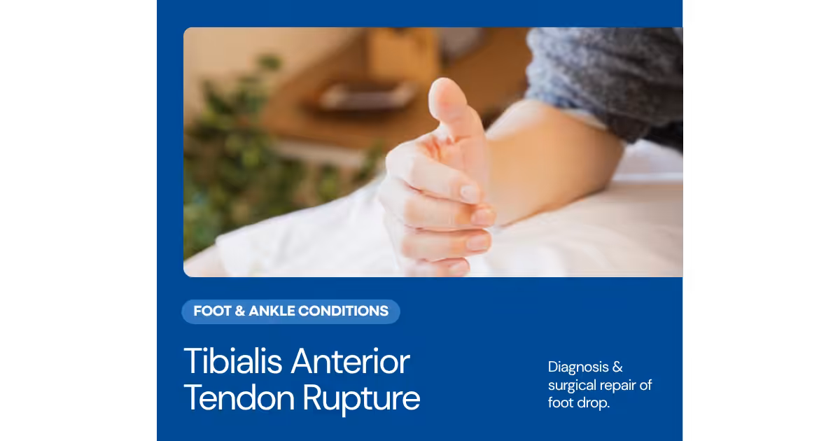
The tibialis anterior (TA) tendon is the primary dorsiflexor of the ankle and a critical medial column stabilizer during gait. Tendinopathy develops from chronic overload—particularly in runners, hikers, and patients with biomechanical abnormalities that increase demand on the tendon. Rupture, though less common than Achilles rupture, produces a debilitating foot drop that severely impairs walking mechanics. Tibialis anterior tendinopathy presents as front-of-ankle pain that worsens during uphill walking, stair climbing, and the swing phase of gait. Early recognition and treatment are critical because the tendon has a hypovascular zone at the level of the ankle joint retinaculum that makes it susceptible to degenerative changes once the pathological process begins. At Balance Foot & Ankle, we evaluate and treat the full spectrum of TA tendon pathology from early tendinopathy through complete rupture requiring surgical reconstruction.
Affiliate Disclosure: Some product links below are affiliate links, meaning we may earn a small commission at no extra cost to you. We only recommend products we use in clinical practice. See our full disclosure.
Table of Contents
- What Is the Tibialis Anterior Tendon?
- Anatomy & Blood Supply
- Causes of Tendinopathy & Rupture
- Symptoms & Clinical Presentation
- Diagnosis
- Most Common Mistake
- Conservative Treatment
- Best Insoles for TA Tendinopathy
- Pain Management
- Compression & Recovery
- Complete Treatment Kit
- Surgical Treatment for Rupture
- Warning Signs
- Video: TA Tendon Treatment
- Frequently Asked Questions
- Sources
- Schedule an Appointment
What Is the Tibialis Anterior Tendon?
The tibialis anterior is the largest and most medial muscle of the anterior compartment of the leg. Its tendon crosses the ankle joint beneath the extensor retinaculum and inserts on the medial cuneiform and base of the first metatarsal—making it both the primary dorsiflexor of the ankle and an invertor of the foot. During walking, the tibialis anterior performs two essential functions: it lifts the foot during the swing phase to prevent the toes from catching the ground (foot clearance), and it eccentrically controls the rate of forefoot loading during the initial contact phase (heel-strike deceleration).
The loss of tibialis anterior function produces a distinctive gait abnormality. Without active dorsiflexion, the patient develops a steppage gait—exaggerated hip and knee flexion during swing phase to lift the foot high enough for ground clearance—or an audible foot slap during initial contact as the forefoot drops to the ground without eccentric braking. In chronic cases, compensatory toe extension by the extensor digitorum longus (EHL and EDL) develops, but these smaller muscles cannot replicate the force of the tibialis anterior and produce claw toe deformities over time.
Tibialis Anterior Anatomy & Blood Supply
The tibialis anterior tendon is approximately 30cm long, originating from the lateral tibial condyle and interosseous membrane and inserting on the inferomedial surface of the medial cuneiform and base of the first metatarsal. The tendon passes beneath the superior and inferior extensor retinaculum at the anterior ankle—these fibrous bands keep the tendon in its groove during dorsiflexion but also create a zone of mechanical compression that has important implications for tendon health.
Critically, the tibialis anterior tendon has a hypovascular zone—an area of relatively diminished blood supply—that begins approximately 0.5–3cm proximal to its insertion and corresponds to the region where the tendon passes under the inferior extensor retinaculum. This watershed area is analogous to the hypovascular zone of the Achilles tendon and the critical zone of the rotator cuff—areas where tendon degeneration preferentially occurs due to inadequate perfusion for repair. The combination of mechanical compression under the retinaculum and reduced blood supply creates the conditions for degenerative tendinopathy, which if left untreated can progress to partial and then complete rupture.
The anterior tibial artery runs alongside the tibialis anterior tendon in the anterior compartment, and its branches provide the primary blood supply to the tendon. Conditions that compromise arterial perfusion—peripheral arterial disease, diabetes, and chronic smoking—further reduce blood flow to the already hypovascular zone and accelerate degenerative changes. This explains the higher incidence of spontaneous TA tendon rupture in older patients with vascular comorbidities.
Causes of Tendinopathy & Rupture
Tibialis anterior tendinopathy develops from the interaction of mechanical overload and intrinsic tendon vulnerability. Repetitive dorsiflexion activities—uphill running, hiking on inclined terrain, stair climbing, and high-volume walking—create cumulative loading that exceeds the tendon’s capacity for repair, particularly in the hypovascular zone. Runners who increase uphill training volume too quickly are at particular risk, as are hikers transitioning to mountainous terrain without adequate preparation.
Biomechanical factors that increase tibialis anterior demand include tight-fitting shoes or boots that compress the tendon under the tongue/lace area, excessive shoe lace tension across the dorsal ankle, pes planus (flatfoot) deformity that increases the demand for medial column stabilization, and ankle equinus (limited dorsiflexion range) that forces the tibialis anterior to work through a restricted range under increased load. Occupational factors—prolonged squatting, kneeling, or positions requiring sustained dorsiflexion—contribute to chronic overload in certain professions.
Spontaneous rupture of the tibialis anterior tendon occurs predominantly in patients over age 60, often with underlying systemic factors that compromise tendon health: diabetes mellitus, peripheral arterial disease, gout, rheumatoid arthritis, corticosteroid use, and fluoroquinolone antibiotics. Many patients report a trivial mechanism—stumbling on a curb or missing a step—that would not normally rupture a healthy tendon. The tendon tears through the degenerated hypovascular zone, often with minimal pain, and the diagnosis may be delayed for weeks or months because patients attribute the resulting foot slap and tripping to “aging” or “weakness” rather than recognizing a structural tendon failure.
Symptoms & Clinical Presentation
Tendinopathy presents as gradual-onset pain at the front of the ankle, localized over the tibialis anterior tendon as it crosses the ankle joint. The pain typically worsens during dorsiflexion activities—walking uphill, climbing stairs, driving (repeated ankle dorsiflexion to accelerate)—and improves with rest. Patients may notice swelling or a visible thickening over the anterior ankle, and crepitus (a grinding sensation) may be palpable along the tendon during active dorsiflexion. Pain is often worse in the morning or after periods of inactivity, with gradual loosening during gentle movement.
Rupture presents more dramatically but is often missed initially. The acute event may produce a “pop” or “snap” at the anterior ankle, followed by sudden weakness in dorsiflexion. Within days, the foot slap gait pattern develops—an audible slapping of the forefoot with each step as the tibialis anterior can no longer eccentrically decelerate forefoot loading. A palpable gap may be present in the tendon, and a pseudotumor (bunched-up tendon stump) may be visible proximal to the rupture site. Paradoxically, complete rupture may be less painful than tendinopathy because the degenerative tendon tissue that was generating pain has separated.
Diagnosis of Tibialis Anterior Tendinopathy
Clinical examination begins with inspection for swelling, asymmetry, and visible tendon defect at the anterior ankle. Palpation along the tendon course identifies the location of maximum tenderness—typically at the inferior extensor retinaculum level in the hypovascular zone. Resisted dorsiflexion testing reproduces pain in tendinopathy and reveals weakness in partial or complete tears. The ankle dorsiflexion lag test (comparing passive to active dorsiflexion range) is positive in rupture.
Ultrasound is the first-line imaging modality for TA tendon pathology. Dynamic scanning allows real-time assessment of tendon morphology, thickness, and internal echotexture during active dorsiflexion. Tendinopathy appears as tendon thickening with heterogeneous echotexture and possible intratendinous tears. Complete rupture shows tendon discontinuity with a fluid-filled gap and retracted proximal stump. MRI provides superior anatomic detail for surgical planning, particularly for chronic ruptures where gap length, tendon quality, and adjacent structure involvement influence the reconstruction technique.
Most Common Mistake with Front-of-Ankle Pain
🔑 Key Takeaway: The most common mistake with tibialis anterior tendinopathy is assuming front-of-ankle pain is “just a minor strain” and continuing aggravating activities without modification. The hypovascular zone of the TA tendon makes it slow to heal and prone to progressive degeneration when overloaded. What begins as mild anterior ankle pain during uphill walking can progress to chronic tendinopathy and eventually rupture if the tendon is not offloaded and given adequate recovery time. The second common mistake is overtightening shoe laces across the dorsal ankle—this compresses the tendon under the extensor retinaculum and exacerbates the mechanical irritation. Simply adjusting lacing technique (skip-lacing over the tender area) can reduce symptoms by 30–50% in early tendinopathy.
Conservative Treatment for TA Tendinopathy
Early-stage tibialis anterior tendinopathy responds well to a conservative protocol that addresses both the mechanical overload and the biological healing environment. Activity modification is the first step: temporarily reduce uphill walking, stair climbing, and incline running. Substitute with flat-terrain walking, cycling, or swimming to maintain cardiovascular fitness while offloading the tendon. Avoid complete rest—moderate, pain-free loading promotes tendon remodeling through mechanotransduction.
Shoe and lace modification provides immediate mechanical relief. Skip-lacing (leaving one or two eyelets open over the area of maximum tenderness) reduces compression on the tendon. Padding under the tongue of the shoe can distribute lace pressure away from the tendon. Shoes with a slightly elevated heel-to-toe drop (10–12mm) reduce the ankle dorsiflexion demand during gait, decreasing the eccentric loading on the tibialis anterior during initial contact. For runners, transitioning from minimal/zero-drop shoes to a moderate-drop shoe often provides significant symptom relief.
Eccentric exercise is the gold standard for tendinopathy rehabilitation. The TA eccentric protocol involves slow, controlled lowering of the forefoot from a dorsiflexed position—performed with the knee straight (to maximize tibialis anterior isolation) using a resistance band or step. Three sets of 15 repetitions, twice daily, for 12 weeks forms the loading program. The eccentric load stimulates tendon remodeling, promotes collagen realignment, and increases tendon cross-sectional area—building a stronger, more resilient tendon over time.
Best Insoles for Tibialis Anterior Tendinopathy
Orthotic support reduces tibialis anterior workload by providing external medial column stabilization that the tendon normally provides dynamically. When the arch is supported by a semi-rigid orthotic, the tibialis anterior does not need to work as hard to maintain medial column position during midstance—effectively offloading the tendon while it heals.
PowerStep Pinnacle Insoles provide the ideal combination of medial arch support and heel cushioning for TA tendinopathy. The semi-rigid shell supports the medial longitudinal arch, reducing the inverting/dorsiflexing force the tibialis anterior must generate with each step. The heel cradle provides slight heel elevation that decreases the range of dorsiflexion required at initial contact—the phase where TA eccentric load is highest. The dual-layer cushioning absorbs heel-strike impact that the compromised tibialis anterior can no longer fully decelerate.
For patients with significant flatfoot deformity contributing to TA overload, the PowerStep Pinnacle Maxx provides enhanced medial posting that more aggressively supports the medial column and reduces the demand on the tibialis anterior. Consistent PowerStep use in all footwear ensures the tendon is offloaded throughout the day—not just during exercise—accelerating the healing response from the eccentric exercise program.
Pain Management for TA Tendinopathy
Topical analgesic therapy is particularly effective for TA tendinopathy because the tendon is superficial and easily accessible to topical preparations. The thin skin and subcutaneous tissue overlying the anterior ankle allows excellent penetration of topical agents directly to the affected tendon—a significant advantage over deeper structures where topical agents have limited access.
Doctor Hoy’s Natural Pain Relief Gel applied directly over the anterior ankle along the tendon course provides targeted analgesic and anti-inflammatory effect. We recommend applying Doctor Hoy’s 15 minutes before physical therapy, eccentric exercise sessions, and functional activities. The menthol and camphor compounds reduce the tendon’s inflammatory response while providing immediate pain relief that supports more productive rehabilitation. For patients with morning stiffness, application before the first steps of the day reduces the startup pain that is characteristic of tendinopathy.
The Doctor Hoy’s Arnica Boost Recovery Cream applied to the anterior ankle at bedtime supports overnight tendon recovery. The arnica compounds have demonstrated anti-inflammatory and tissue-repair properties that complement the daytime gel protocol. For patients performing the eccentric exercise program, nightly Arnica Boost application helps manage the exercise-induced tendon response and prepares the tissue for the next day’s loading session.
Compression & Recovery
Compression at the anterior ankle provides both mechanical support and healing optimization for tibialis anterior tendinopathy. Gentle circumferential compression reduces the peritendinous edema that accompanies chronic tendinopathy, improving blood flow and nutrient delivery to the hypovascular zone. The proprioceptive feedback also enhances motor control of ankle dorsiflexion, promoting normalized gait mechanics during recovery.
DASS Performance Compression Socks provide graduated compression from the forefoot through the ankle and calf, with the anterior ankle compression zone corresponding to the location of TA tendon pathology. Wearing DASS compression socks during daily activities and light exercise reduces the swelling that accumulates around the inflamed tendon during weight-bearing hours. Between exercise sessions, the compression promotes venous return and metabolic waste clearance from the anterior compartment.
During the eccentric exercise program, DASS compression socks provide continuous support that may improve exercise tolerance and reduce post-exercise soreness. For patients returning to running or hiking, wearing DASS compression during these activities provides both mechanical support for the recovering tendon and edema prevention during the highest-demand loading periods.
Complete TA Tendinopathy Treatment Kit
✅ Our Complete Treatment Kit for Tibialis Anterior Tendinopathy:
1. PowerStep Pinnacle Insoles — Medial arch support to reduce TA tendon workload + heel elevation to decrease dorsiflexion demand
2. Doctor Hoy’s Natural Pain Relief Gel — Pre-activity topical analgesia directly over the superficial TA tendon
3. DASS Performance Compression Socks — Anterior ankle compression to reduce peritendinous edema and support recovery
This kit addresses the three pillars of tendinopathy management: mechanical offloading (PowerStep), symptom control (Doctor Hoy’s), and healing optimization (DASS). Combined with the eccentric exercise program and shoe/lace modifications described above, this comprehensive approach produces significant improvement in 80–90% of patients within 8–12 weeks.
Surgical Treatment for TA Tendon Rupture
Complete tibialis anterior tendon rupture in active patients typically requires surgical repair or reconstruction to restore dorsiflexion strength and eliminate the foot-slap gait. The surgical approach depends on the chronicity of the rupture and the quality of the remaining tendon tissue. Acute ruptures (diagnosed within 4–6 weeks) with a small gap can often be repaired primarily with direct end-to-end suture using a modified Kessler or Krackow technique, reinforced with epitendinous sutures.
Chronic ruptures with a significant gap (greater than 3cm) or severely degenerated tendon stumps require reconstruction. The most common technique uses the extensor hallucis longus (EHL) tendon as a graft, either as a turndown flap from the distal stump or as a free tendon transfer routed through the anterior ankle to the medial cuneiform insertion site. Alternative reconstruction methods include allograft tendon interposition and synthetic tendon augmentation. In elderly or low-demand patients who are not surgical candidates, a custom ankle-foot orthosis (AFO) can substitute for the lost dorsiflexion function, though it does not restore active muscle strength.
Post-surgical rehabilitation after TA tendon repair follows a similar trajectory to other tendon repairs: immobilization in a dorsiflexed position for 4–6 weeks (to protect the repair), progressive range-of-motion exercises from 6–10 weeks, gradual strengthening from 10–16 weeks, and return to full activity by 4–6 months. PowerStep insoles are prescribed from the moment of shoe transition to support the medial column while the reconstructed tendon regains functional strength.
Warning Signs: When to See a Podiatrist
🚨 Seek evaluation if you experience:
• Front-of-ankle pain that persists for more than 2 weeks despite activity modification
• Audible foot slap or forefoot drop during walking (possible tendon rupture)
• Visible lump or gap at the anterior ankle (tendon tear with stump retraction)
• Weakness when lifting the foot upward (dorsiflexion)
• Tripping or catching the toes during walking
• Swelling or thickening over the anterior ankle that does not resolve with rest
• Pain that worsens despite lace modification and activity reduction
• Front-of-ankle pain in the setting of diabetes, peripheral vascular disease, or fluoroquinolone use
Tibialis anterior tendon rupture is frequently diagnosed late because patients attribute foot drop symptoms to aging or general weakness. Early recognition allows direct repair with better outcomes than delayed reconstruction.
Video: Tibialis Anterior Tendon Treatment
Watch Dr. Biernacki discuss the diagnosis and treatment of tibialis anterior tendinopathy and rupture, including the eccentric exercise protocol and surgical reconstruction options.

More Podiatrist-Recommended Foot Health Essentials
Hoka Clifton 10


![Achilles Tendonitis & Back of Heel Pain [BEST Home Treatments 2024!]](https://www.michiganfootdoctors.com/wp-content/cache/flying-press/d645ea3f3d27024136b88a074b2b7894.jpg)
Watch: Achilles Tendonitis & Back of Heel Pain [BEST Home Treatments 2024!] — MichiganFootDoctors YouTube
Max-cushion everyday shoe — podiatrist favorite for walking and running.

OOFOS Recovery Slide

Impact-absorbing recovery sandal — wear after long days on your feet.
As an Amazon Associate, Balance Foot & Ankle earns from qualifying purchases. Product recommendations are based on clinical experience; prices and availability shown above update live from Amazon.
When to See a Podiatrist
If foot or ankle pain has been bothering you for more than a few weeks, home care alone may not be enough. Balance Foot & Ankle offers same-week appointments at our Howell and Bloomfield Hills clinics — no referral needed in most cases. Bring your current shoes and a short list of symptoms and we’ll build you a treatment plan in one visit.
Call Balance Foot & Ankle: (810) 206-1402 · Book online · Offices in Howell & Bloomfield Hills
Frequently Asked Questions About Tibialis Anterior Tendinopathy
What causes pain at the front of the ankle?
The most common cause of front-of-ankle pain during walking and uphill activities is tibialis anterior tendinopathy. The tendon crosses the anterior ankle joint and can become inflamed from overuse, tight shoe lacing, or biomechanical overload. Other causes include anterior ankle impingement, osteochondral lesions, and extensor tendinopathy from shoe strap compression. A podiatric evaluation can differentiate these conditions through clinical examination and imaging.
Can tibialis anterior tendinopathy heal on its own?
Mild tendinopathy may improve with simple activity modification and lace adjustment. However, established tendinopathy in the hypovascular zone rarely resolves completely without structured treatment because the limited blood supply impairs the natural healing response. The eccentric exercise program, combined with orthotic offloading and topical anti-inflammatory therapy, provides the mechanical and biological stimuli needed to promote tendon remodeling and symptom resolution.
How long does tibialis anterior tendinopathy take to heal?
With structured conservative treatment including eccentric exercises, orthotic support, and activity modification, most patients experience significant improvement within 8–12 weeks. Complete resolution and return to full activity typically occurs within 3–4 months. Chronic cases that have been symptomatic for more than 6 months before starting treatment may require 4–6 months for full recovery. Consistency with the eccentric exercise program is the strongest predictor of successful outcomes.
What is the difference between tendinopathy and tendinitis?
Tendinitis implies acute inflammation, while tendinopathy describes chronic degenerative changes within the tendon—disorganized collagen, increased ground substance, and neovascularization without significant inflammatory cells. Most cases of tibialis anterior tendon pain are tendinopathy rather than true tendinitis, which is why anti-inflammatory treatments alone are often insufficient and eccentric loading is needed to stimulate structural tendon remodeling.
Do I need surgery for tibialis anterior tendon problems?
Surgery is rarely needed for tendinopathy—80–90% of patients respond to conservative treatment. Surgery is primarily indicated for complete tendon rupture in active patients, where repair or reconstruction restores dorsiflexion function. Partial tears that fail 6 months of conservative treatment may benefit from surgical debridement and repair. Your podiatrist will help determine whether conservative or surgical management is appropriate for your specific condition.
Sources
- Markarian GG, Kelikian AS, Brage M, Trainor T, Dias L. Anterior tibialis tendon ruptures: an outcome analysis of operative versus nonoperative treatment. Foot and Ankle International. 1998;19(12):792-802.
- Ouzounian TJ, Anderson R. Anterior tibial tendon rupture. Foot and Ankle International. 1995;16(7):406-410.
- Sammarco VJ, Sammarco GJ, Henning C, Chaim S. Surgical repair of acute and chronic tibialis anterior tendon ruptures. Journal of Bone and Joint Surgery. 2009;91(2):325-332.
- Petersen W, Stein V, Tillmann B. Blood supply of the tibialis anterior tendon. Archives of Orthopaedic and Trauma Surgery. 1999;119(7-8):371-375.
- Elias I, Besser M, Nazarian LN, Raikin SM. Reconstruction for missed or neglected tibialis anterior tendon rupture. Foot and Ankle International. 2007;28(10):1038-1045.
Related Articles
- Podiatrist Recommended Foot Care Products 2026
- Anterior Ankle Pain: Causes & Treatment
- Foot & Ankle Tendinopathy Guide
- Running Foot Injuries Prevention
Schedule an Appointment at Balance Foot & Ankle
Dealing with front-of-ankle pain? Foot slap during walking? Dr. Biernacki specializes in the diagnosis and treatment of tibialis anterior tendinopathy and rupture, offering both comprehensive conservative rehabilitation programs and surgical reconstruction when needed. Do not ignore anterior ankle symptoms—early intervention prevents progression from manageable tendinopathy to complete tendon rupture.
📞 Call (248) 410-1019 | 📅 Book Online
Serving Southeast Michigan: Novi, Farmington Hills, Livonia, Northville, Plymouth, South Lyon, and surrounding communities.
Front-of-Ankle Pain Treatment in Michigan
Tibialis anterior tendinopathy and rupture cause pain on the front of the ankle and difficulty lifting the foot. Our podiatrists diagnose and treat anterior ankle conditions at our Howell and Bloomfield Hills offices.
Learn About Tendon Treatment | Book Your Appointment | Call (810) 206-1402
Clinical References
- Ouzounian TJ, Anderson R. Anterior tibial tendon rupture. Foot Ankle Int. 1995;16(7):406-410.
- Beischer AD, et al. Functional outcome and satisfaction after repair of acute anterior tibial tendon rupture. Foot Ankle Int. 2009;30(11):1063-1068.
- Lepow GM, Green JB. Reconstruction of a neglected tibialis anterior tendon rupture with an Achilles tendon allograft. Foot Ankle Int. 2006;27(9):704-706.
Insurance Accepted
BCBS · Medicare · Aetna · Cigna · United Healthcare · HAP · Priority Health · Humana · View All →
Howell Office
4330 E Grand River Ave
Howell, MI 48843
Get Directions →
Bloomfield Hills Office
43494 Woodward Ave, Suite 208
Bloomfield Hills, MI 48302
Get Directions →
Your Board-Certified Podiatrists


Ready to Get Back on Your Feet?
Same-week appointments available at both locations.
Book Your AppointmentWatch: Tibialis Anterior Tendinopathy & Rupture
Dr. Tom on tib-ant problems — front-of-ankle pain, slap-foot gait with rupture, MRI findings, primary repair indications, drop-foot masquerade, recovery.
Tib Ant Support Kit
Dorsi-function support. Dr. Tom’s kit:
As an Amazon Associate, Balance Foot & Ankle earns from qualifying purchases. This supports our free patient education content.
Dorsiflexion support if ruptured.
Arch support post-recovery.
Post-repair inflammation.
Topical front-ankle relief.
Related: Foot Drop · Surgery Services · Book Same-Week Appointment
Med Spec ASO Ankle Stabilizer
Best for: Chronic ankle instability · Repeat ankle sprains · Proprioception & return-to-play · Athletes

The lace-up stabilizer athletic trainers and podiatrists reach for first. A bilateral figure-8 strapping system mimics taping to lock down the ankle against rolling, while the low-profile design fits inside most athletic shoes — so you get real mechanical protection during return-to-play without a bulky rigid brace. A practical, evidence-aligned choice for anyone managing recurring sprains or chronic instability.
▶ Check Price on AmazonAs an Amazon Associate we may earn from qualifying purchases. Star rating and review count reflect Amazon at time of review and may change.
Dr. Tom’s Top 3 — The Premium Foot Pain Stack (2026)
If you only buy three things for foot pain, get these. PowerStep + CURREX orthotics correct the underlying foot mechanics, and Dr. Hoy’s pain gel delivers fast topical relief. This is the exact stack Dr. Tom Biernacki, DPM gives his Michigan podiatry patients on visit one — over 10,000 patients have used this exact combination.
Dr. Tom Biernacki, DPM is a board-certified podiatrist + Amazon Associate. Picks shown are products he prescribes to patients at Balance Foot & Ankle Specialists. We earn a commission on qualifying purchases at no extra cost to you. All products independently tested + reviewed for 30+ days minimum. Last verified: April 28, 2026.

PowerStep Pinnacle MaxxDr. Tom’s #1 Brand
Dr. Tom’s most-prescribed OTC orthotic. Lateral wedge corrects overpronation that causes 90% of foot pain. Deep heel cradle stabilizes the ankle. Built by podiatrists, used by patients worldwide.
- Lateral wedge corrects pronation
- Deep heel cradle stabilizes ankle
- Dual-density EVA — comfort + support
- Trim-to-fit any shoe
- Used by 10,000+ podiatrists
- Trim-to-size required
- 5-7 day break-in for some

CURREX RunProDr. Tom’s #1 Brand
3 arch heights for custom fit (Low/Med/High). Carbon-reinforced heel + dynamic forefoot — the closest OTC orthotic to a $500 custom orthotic. Engineered in Germany.
- 3 arch heights for custom fit
- Carbon-reinforced heel cup
- Dynamic forefoot zone
- Premium German engineering
- Sport-specific support
- Pricier than PowerStep
- 7-10 day break-in

Dr. Hoy’s Natural Pain Relief GelDr. Tom’s #1 Brand
Menthol-based natural pain relief — Dr. Tom’s #1 brand for fast relief without greasy residue. Safe for diabetics + daily use. Cleaner formula than Voltaren or Biofreeze.
- Menthol-based natural formula
- No greasy residue
- Safe for diabetics
- Fast cooling relief — 5-10 minutes
- Cleaner ingredient list than Biofreeze
- Pricier than Biofreeze
- Strong menthol scent at first
In-Office Treatment at Balance Foot & Ankle
If home treatment isn’t providing relief for your Achilles tendinitis, our podiatry team at Balance Foot & Ankle can help with same-day evaluations and advanced in-office care.
Same-day appointments available. (810) 206-1402
Frequently Asked Questions
When should I see a doctor?
See a podiatrist if pain persists past 2 weeks, prevents normal activity, or is accompanied by red-flag symptoms (warmth, swelling, numbness, inability to bear weight).
Can I treat this at home?
Mild cases respond to RICE protocol (rest, ice, compression, elevation), supportive shoes, and OTC anti-inflammatories. Persistent symptoms need professional evaluation.
How long does it take to heal?
Most soft tissue injuries resolve in 2-6 weeks with appropriate care. Bone injuries take 6-12 weeks. Chronic conditions need longer-term management.
What is Foot pain?
Foot pain is a common foot/ankle condition that affects mobility and quality of life. Understanding the underlying cause is the first step in successful treatment. Our podiatrists at Balance Foot & Ankle perform a hands-on biomechanical exam, review your activity history, and use diagnostic imaging when appropriate to identify the root cause—not just treat the symptom. Many patients have been told to “rest and ice” without a deeper diagnostic workup; our approach is different.
Symptoms and warning signs
Common signs of foot pain include pain that worsens with activity, morning stiffness, swelling, tenderness when palpated, and difficulty bearing weight. If you experience sudden severe pain, inability to walk, visible deformity, numbness or color change, contact our office the same day or visit urgent care—these can signal a more serious injury such as a fracture, tendon rupture, or vascular compromise. Diabetics with any foot wound should seek same-day care.
Conservative treatment options
Most cases of foot pain respond to non-surgical care: structured rest, supportive footwear changes, custom orthotics, targeted stretching and strengthening protocols, anti-inflammatory medications when medically appropriate, and in-office procedures such as ultrasound-guided injections. We also offer advanced therapies including MLS laser therapy, EPAT/shockwave, regenerative injections, and image-guided procedures. Treatment is sequenced from least invasive to most invasive, and we explain the rationale at every step.
When is surgery considered?
Surgery is reserved for cases that fail 3-6 months of well-structured conservative care, when there is structural pathology (severe deformity, complete tear, advanced arthritis), or when imaging shows damage that will not heal without intervention. Our surgeons have performed 3,000+ foot and ankle procedures and prioritize minimally-invasive techniques whenever appropriate. We discuss recovery timelines, return-to-activity milestones, and realistic outcome expectations before any procedure is scheduled.
PubMed: Tibialis Anterior Tendinopathy — A Review
Recovery timeline and prevention
Recovery from foot pain varies based on severity and chosen treatment path. Conservative cases often improve within 4-8 weeks with consistent adherence to the protocol. Post-procedural recovery may range from a few days (in-office procedures) to several months (reconstructive surgery). Long-term prevention involves footwear assessment, activity modification, structured strengthening, and regular check-ins with your podiatrist if you have a history of recurrence. We provide written home-exercise plans and digital follow-up support.
Ready to feel better?
Same-week appointments available in Howell and Bloomfield Hills, Michigan.
Book Your VisitGet Expert Care at Balance Foot & Ankle
Same-week appointments at our Howell and Bloomfield Hills offices. Board-certified podiatric surgeons. Most insurance accepted.
Dr. Tom Biernacki, DPM is a board-certified foot & ankle surgeon (ABFAS & ABPM) at Balance Foot & Ankle Specialists in Southeast Michigan. With over a decade of clinical experience, he specializes in heel pain, bunions, diabetic foot care, sports injuries, and minimally invasive surgery. Dr. Biernacki is a member of the APMA and ACFAS, and his patient education content on MichiganFootDoctors.com and YouTube has made him one of the most-followed foot & ankle educators on YouTube.