

Quick answer: Venous Stasis Ulcer Foot is a common foot/ankle topic that affects many patients. The 2026 evidence-based approach combines proper diagnosis, conservative-first treatment, and escalation only when needed. We treat this regularly at our Howell and Bloomfield Hills practices. Call (810) 206-1402.
Medically Reviewed | Dr. Tom Biernacki, DPM | Board-Certified Podiatric Surgeon | Balance Foot & Ankle, Michigan

The most important clinical decision with Venous Stasis Ulcer Foot isn’t which treatment to start with — it’s identifying the correct subtype. That changes everything. Call (810) 206-1402.
What Causes Venous Stasis Ulcers?
Venous stasis ulcers account for approximately 70–90% of all chronic lower extremity ulcers and represent the end-stage complication of chronic venous insufficiency — a condition in which the venous valves of the leg fail to adequately return blood from the feet and lower legs to the heart. When venous blood pools in the lower leg (venous hypertension), the elevated pressure forces fluid, proteins, and red blood cells through the capillary walls into the surrounding tissue. Hemosiderin (from degraded red blood cells) deposits in the skin, producing the characteristic brown discoloration of chronic venous insufficiency.
The prolonged tissue congestion — inadequate oxygen and nutrient delivery combined with toxic accumulation of inflammatory mediators — progressively damages the skin, producing the lipodermatosclerosis (skin hardening and brown discoloration) that predisposes to ulceration. A minor trauma — a scratch, insect bite, or bump against furniture — triggers full-thickness skin breakdown in tissue that lacks the normal capacity for wound healing. The result is a painful, weeping ulcer that, without appropriate compression therapy, will not heal because the underlying venous hypertension that created it persists.
Risk factors for venous stasis ulcers include prior deep vein thrombosis (DVT), varicose veins, obesity, prolonged standing or sitting occupations, advanced age, and previous leg injuries. Patients with all four limb chronic venous disease have a lifetime ulcer risk exceeding 25%. Recurrence rates after healing without sustained compression therapy exceed 70% within five years — emphasizing that treatment must address the underlying venous disease, not just the wound surface.
Recognizing Venous Stasis Ulcers: Symptoms and Diagnosis
Venous stasis ulcers have a characteristic appearance that distinguishes them from arterial ulcers and diabetic neuropathic ulcers. They are typically located in the gaiter zone — the area between the ankle and mid-calf, particularly around the medial malleolus (inner ankle). The wound base is usually shallow with irregular borders and yellow fibrinous slough; surrounding skin shows the brown hemosiderin staining, varicosities, and dermatitis of chronic venous disease. Pain is typically positional — worsened by dependency (leg hanging down) and relieved by elevation.
Arterial ulcers, by contrast, occur distally (toes, heel, dorsal foot) and are associated with pale, cool skin, absent pulses, and extreme pain out of proportion to wound size. Diabetic neuropathic ulcers occur at pressure points (metatarsal heads, heel) in insensate skin without the surrounding venous stasis changes. Accurate classification determines treatment — applying compression therapy to arterial insufficiency is dangerous and contraindicated.
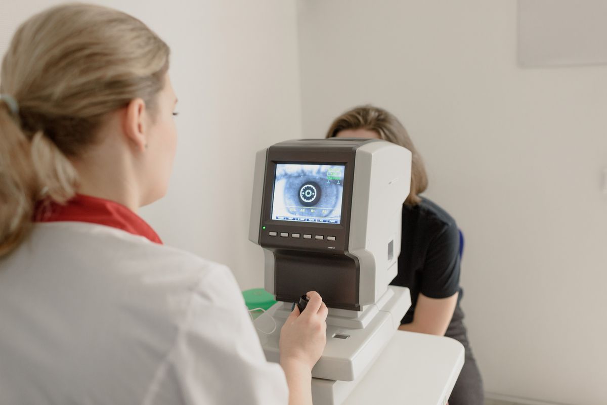
Ankle-brachial index (ABI) measurement using Doppler ultrasound is the essential first diagnostic step in any lower leg ulcer. ABI above 0.8 confirms adequate arterial inflow and safe application of compression; ABI below 0.6 indicates significant arterial disease requiring vascular surgery consultation before any compression is applied. Venous duplex ultrasound maps the extent of valvular incompetence and identifies any residual deep vein thrombosis.
Venous Stasis Ulcer Treatment and Wound Care
Sustained graduated compression therapy is the single most important treatment for venous stasis ulcers — and the treatment most commonly under-applied. High-compression multilayer bandaging (achieving 35–40 mmHg at the ankle) or compression stockings reduce venous hypertension, force fluid back into the vascular system, reduce edema, and create the circulatory conditions that allow wound healing. Studies consistently demonstrate that appropriate compression doubles healing rates compared to standard wound dressings alone.
Local wound care involves debridement of necrotic tissue and slough (removing the physical barrier to wound healing), selection of the appropriate wound dressing to maintain a moist wound healing environment (hydrocolloid, foam, alginate, or antimicrobial silver dressings based on wound status), and surveillance for wound infection (increasing pain, perilesional warmth and erythema, purulent exudate, failure to progress). Biofilm-disrupting strategies — including sharp debridement, cadexomer iodine, or medical-grade honey preparations — are important for stalled wounds.
Advanced wound care modalities for stalled venous ulcers include skin substitutes (bioengineered tissue equivalents that deliver growth factors to the wound bed), split-thickness skin grafting, and negative pressure wound therapy (wound VAC). After healing, sustained compression garment use — ideally for life — is the only effective strategy for preventing recurrence. Patients with significant venous reflux may benefit from endovenous ablation (laser or radiofrequency) to eliminate incompetent saphenous veins and reduce the venous hypertension driving recurrence. Dr. Tom Biernacki provides comprehensive lower extremity wound care at Balance Foot and Ankle, including diabetic wound management, vascular assessment, and evidence-based wound care protocols.
Dr. Tom's Product Recommendations

DASS Medical Grade Compression Socks
⭐ Highly Rated
Graduated medical compression socks that reduce venous pooling and lower leg swelling — the most important tool for both preventing venous stasis ulcers and maintaining compression after healing.
Dr. Tom says: “https://m.media-amazon.com/images/I/71ZrLssb9XL._AC_SL1500_.jpg”
DASS Medical
4.7
Disclosure: We earn a commission at no extra cost to you.

PowerStep Pinnacle Arch Support Insoles
⭐ Highly Rated
Supportive insoles for venous insufficiency patients who need comfortable footwear during wound care and recovery — improving gait mechanics that support venous return.
Dr. Tom says: “https://m.media-amazon.com/images/I/81K+DSvd0VL._AC_SL1500_.jpg”
PowerStep
4.6
Disclosure: We earn a commission at no extra cost to you.
✅ Pros / Benefits
- Graduated compression therapy doubles venous ulcer healing rates compared to dressings alone
- ABI testing reliably identifies patients safe for compression before treatment
- Advanced wound care modalities provide options for chronic non-healing wounds
- Endovenous ablation reduces recurrence by addressing the root venous pathology
❌ Cons / Risks
- Without sustained lifelong compression, recurrence rates exceed 70% within five years
- Patients with mixed arterial-venous disease require modified compression to avoid arterial compromise
- Wound care requires consistent, long-term patient engagement for 3–6 months or more
Dr. Tom Biernacki’s Recommendation
Venous ulcers are some of the most undertreated wounds I see — patients wrap them in gauze and wonder why they don’t heal after months. The wound surface is almost secondary; the driver is the venous hypertension underneath. Until you fix the compression, the ulcer will cycle: partially heal, break down, partially heal. My patients with venous ulcers get Doppler assessment, appropriate compression, proper debridement and dressings, and a plan for lifelong compression maintenance.
— Dr. Tom Biernacki, DPM | Board-Certified Podiatric Surgeon | Balance Foot & Ankle
Frequently Asked Questions
How long does a venous stasis ulcer take to heal?
With appropriate compression and wound care, most venous stasis ulcers heal in 3–6 months. Larger, chronic, or complicated ulcers may take 12+ months. Without adequate compression, healing may not occur regardless of wound dressing.
Are venous ulcers dangerous?
They can become dangerous if infected, but the primary morbidity is chronic pain, disability, and recurrence. Untreated venous disease progresses and ulcers enlarge. Prompt podiatric and vascular evaluation is important.
Can venous ulcers be prevented?
Yes. Wearing graduated compression stockings daily, elevating legs when sitting, and avoiding prolonged standing are the most effective preventive strategies for those with chronic venous insufficiency.
American Academy of Dermatology: Venous Stasis Dermatitis
Get Expert Care at Balance Foot & Ankle
Same-week appointments at our Howell and Bloomfield Hills offices. Board-certified podiatric surgeons. Most insurance accepted.
Dr. Tom Biernacki, DPM is a board-certified foot & ankle surgeon (ABFAS & ABPM) at Balance Foot & Ankle Specialists in Southeast Michigan. With over a decade of clinical experience, he specializes in heel pain, bunions, diabetic foot care, sports injuries, and minimally invasive surgery. Dr. Biernacki is a member of the APMA and ACFAS, and his patient education content on MichiganFootDoctors.com and YouTube has made him one of the most-followed foot & ankle educators on YouTube.