
Quick answer: Treatment for ankle arthroscopy procedure treatment recovery follows a stepwise approach: 1) conservative care first (rest, ice, supportive footwear, OTC anti-inflammatories), 2) physical therapy and targeted exercises, 3) in-office treatments (injections, custom orthotics) if conservative fails at 4-6 weeks, 4) surgery for refractory cases. Most patients resolve at step 1 or 2. Call (810) 206-1402.
Medically reviewed by Dr. Tom Biernacki, DPM — Board-Certified Podiatric Surgeon — Balance Foot & Ankle, Howell & Bloomfield Hills, MI. Last updated April 2026.
▶ Watch
Medically Reviewed by Dr. Tom Biernacki, DPM — Board-Certified Podiatrist, Balance Foot & Ankle Specialists, Michigan. Last updated April 2026.
Ankle Arthroscopy: Seeing Inside the Joint Without Open Surgery
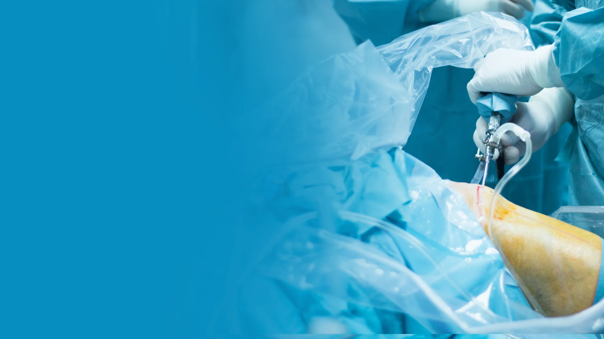
Ankle arthroscopy is a minimally invasive surgical technique that allows a foot and ankle surgeon to directly visualize, diagnose, and treat conditions within the ankle joint through small incisions—typically two portals of 4–5 millimeters. A small camera (arthroscope) is introduced through one portal to provide real-time video visualization of the joint interior on a monitor, while surgical instruments are introduced through the second portal to perform the required procedure. Compared to open ankle surgery, arthroscopy offers smaller incisions, less soft tissue disruption, faster recovery, reduced infection risk, and the ability to address multiple pathologies in a single procedure.
Conditions Treated with Ankle Arthroscopy
Osteochondral Lesions of the Talus (OLTs)
Arthroscopic debridement and microfracture of osteochondral lesions is one of the most common ankle arthroscopy indications. The damaged cartilage is debrided to a stable rim, and the underlying subchondral bone is perforated with an arthroscopic awl to create bleeding that stimulates fibrocartilage formation in the defect. This is effective for lesions under 150mm squared with good surrounding cartilage.
Anterior Ankle Impingement
Arthroscopic excision of anterior osteophytes (bone spurs) and synovial meniscoid tissue that impinge during dorsiflexion reliably resolves anterior impingement pain in athletes and active patients who have failed conservative management.
Posterior Ankle Impingement and Os Trigonum Excision
Two-portal posterior ankle arthroscopy allows excision of the os trigonum, release of FHL tenosynovitis, and posterior ankle spur removal without the risks of open posteromedial or posterolateral approaches. Return to sport is typically 6–10 weeks following posterior arthroscopy—significantly faster than open surgery.
Synovitis and Loose Bodies
Chronic ankle synovitis (from inflammatory arthritis, post-traumatic inflammation, or impingement) responds well to arthroscopic synovectomy—systematic removal of hypertrophied synovial tissue. Loose bodies (fragments of cartilage or bone that float within the joint) are readily identified and removed arthroscopically, eliminating the mechanical locking, catching, and pain they cause.
Ankle Fracture Reduction Assistance
Arthroscopy is increasingly used during ankle fracture fixation to directly assess articular surface reduction quality and identify occult chondral injuries that are not visible on fluoroscopy alone.
The Procedure
Ankle arthroscopy is performed under spinal or general anesthesia with a thigh tourniquet as an outpatient procedure. The surgeon establishes anteromedial and anterolateral portals (or posterior portals for posterior pathology) and distends the joint with sterile saline to create working space. The joint is systematically examined, and the required treatment—debridement, microfracture, spur resection, synovectomy, or loose body removal—is performed under continuous arthroscopic visualization. Total operating time is typically 30–60 minutes depending on the pathology addressed.
Recovery
Recovery after ankle arthroscopy depends on the specific procedure performed. For diagnostic arthroscopy and simple synovectomy, weight-bearing in a boot begins within days and patients typically return to normal activity in 2–4 weeks. Microfracture for OLTs requires 6 weeks of non-weight-bearing. Spur excision for impingement allows weight-bearing within a week, with return to sport at 4–8 weeks. Most patients are surprised by the limited postoperative pain and rapid functional recovery compared to their expectations for “ankle surgery.”
Ready to Relieve Your Foot Pain?
Board-certified podiatrists serving Southeast Michigan. Same-week appointments available.
Expert Foot & Ankle Care in Michigan
From chronic conditions to acute injuries, Dr. Tom Biernacki provides comprehensive podiatric care using the latest evidence-based treatments at Balance Foot & Ankle in Howell and Bloomfield Hills.
View All Treatment Options | Book Your Appointment | Call (810) 206-1402
Clinical References
- Thomas MJ, et al. “The population prevalence of foot and ankle pain in middle and old age: a systematic review.” Pain. 2011;152(12):2870-2880.
- Hill CL, et al. “Prevalence and correlates of foot pain in a population-based study: the North West Adelaide health study.” J Foot Ankle Res. 2008;1(1):2.
- Riskowski JL, et al. “Measures of foot function, foot health, and foot pain.” Arthritis Care Res. 2011;63(S11):S229-S236.
Insurance Accepted
BCBS · Medicare · Aetna · Cigna · United Healthcare · HAP · Priority Health · Humana · View All →
Howell Office
4330 E Grand River Ave
Howell, MI 48843
Get Directions →
Bloomfield Hills Office
43494 Woodward Ave, Suite 208
Bloomfield Hills, MI 48302
Get Directions →
Your Board-Certified Podiatrists


Ready to Get Back on Your Feet?
Same-week appointments available at both locations.
Book Your AppointmentMore Podiatrist-Recommended Foot Health Essentials
Top-Rated Arch Support Insole
Universal podiatrist-recommended insert for pain relief and prevention.
Foot Massage Ball
Daily 3-minute roll reduces most forms of foot and heel pain.
Moisture-Wicking Sock
Prevents fungus, blisters, and odor — the basics matter.
As an Amazon Associate, Balance Foot & Ankle earns from qualifying purchases. Product recommendations are based on clinical experience; prices and availability shown above update live from Amazon.


When to See a Podiatrist
If foot or ankle pain has been bothering you for more than a few weeks, home care alone may not be enough. Balance Foot & Ankle offers same-week appointments at our Howell and Bloomfield Hills clinics — no referral needed in most cases. Bring your current shoes and a short list of symptoms and we’ll build you a treatment plan in one visit.
Call Balance Foot & Ankle: (810) 206-1402 · Book online · Offices in Howell & Bloomfield Hills
In-Office Treatment at Balance Foot & Ankle
When conservative care isn’t enough, Dr. Tom Biernacki and the team at Balance Foot & Ankle offer advanced, same-day options — including Ankle Arthroscopy Michigan at our Howell and Bloomfield Hills clinics.
Same-day appointments available. Call (810) 206-1402 or book online.
What is Foot pain?
Foot pain is a common foot/ankle condition that affects mobility and quality of life. Understanding the underlying cause is the first step in successful treatment. Our podiatrists at Balance Foot & Ankle perform a hands-on biomechanical exam, review your activity history, and use diagnostic imaging when appropriate to identify the root cause—not just treat the symptom. Many patients have been told to “rest and ice” without a deeper diagnostic workup; our approach is different.
Symptoms and warning signs
Common signs of foot pain include pain that worsens with activity, morning stiffness, swelling, tenderness when palpated, and difficulty bearing weight. If you experience sudden severe pain, inability to walk, visible deformity, numbness or color change, contact our office the same day or visit urgent care—these can signal a more serious injury such as a fracture, tendon rupture, or vascular compromise. Diabetics with any foot wound should seek same-day care.
Conservative treatment options
Most cases of foot pain respond to non-surgical care: structured rest, supportive footwear changes, custom orthotics, targeted stretching and strengthening protocols, anti-inflammatory medications when medically appropriate, and in-office procedures such as ultrasound-guided injections. We also offer advanced therapies including MLS laser therapy, EPAT/shockwave, regenerative injections, and image-guided procedures. Treatment is sequenced from least invasive to most invasive, and we explain the rationale at every step.
When is surgery considered?
Surgery is reserved for cases that fail 3-6 months of well-structured conservative care, when there is structural pathology (severe deformity, complete tear, advanced arthritis), or when imaging shows damage that will not heal without intervention. Our surgeons have performed 3,000+ foot and ankle procedures and prioritize minimally-invasive techniques whenever appropriate. We discuss recovery timelines, return-to-activity milestones, and realistic outcome expectations before any procedure is scheduled.
Recovery timeline and prevention
Recovery from foot pain varies based on severity and chosen treatment path. Conservative cases often improve within 4-8 weeks with consistent adherence to the protocol. Post-procedural recovery may range from a few days (in-office procedures) to several months (reconstructive surgery). Long-term prevention involves footwear assessment, activity modification, structured strengthening, and regular check-ins with your podiatrist if you have a history of recurrence. We provide written home-exercise plans and digital follow-up support.
Ready to feel better?
Same-week appointments available in Howell and Bloomfield Hills, Michigan.
Book Your VisitIn-Office Treatment at Balance Foot & Ankle
If home treatment isn’t providing relief for your foot and ankle conditions, our podiatry team at Balance Foot & Ankle can help with same-day evaluations and advanced in-office care.
Same-day appointments available. (810) 206-1402
Doctor Hoy’s Natural Pain Relief Gel
Natural topical pain relief I use in our clinic. Arnica + camphor formula — apply directly to the area 3–4x daily. ($20–25)
Shop Doctor Hoy’s →Dr. Tom Biernacki, DPM is a board-certified foot & ankle surgeon (ABFAS & ABPM) at Balance Foot & Ankle Specialists in Southeast Michigan. With over a decade of clinical experience, he specializes in heel pain, bunions, diabetic foot care, sports injuries, and minimally invasive surgery. Dr. Biernacki is a member of the APMA and ACFAS, and his patient education content on MichiganFootDoctors.com and YouTube has made him one of the most-followed foot & ankle educators on YouTube.