
The most important clinical decision with Fifth Metatarsal Fracture Jones isn’t which treatment to start with — it’s identifying the correct subtype. That changes everything. Call (810) 206-1402.
Dr. Tom’s Top Insole & Orthotic Picks
Affiliate disclosure: As an Amazon Associate, Balance Foot & Ankle earns from qualifying purchases.
Dr. Tom’s Top Pain Relief Picks — Dr. Hoy’s (2026)
Affiliate disclosure: As an Amazon Associate, Balance Foot & Ankle earns from qualifying purchases. I personally use Dr. Hoy’s in my practice for patients who need topical relief.
| Product | Best For | Dr. Tom’s Take | Get It |
|---|---|---|---|
| Dr. Hoy’s Natural Pain Relief Gel 3.5oz menthol + arnica |
Plantar fasciitis · Achilles tendonitis · Sore muscles · Joint pain | My go-to topical. Cooling-then-warming sensation. No greasy residue. Non-NSAID alternative. | Buy Now |
| Dr. Hoy’s Arnica Boost 8oz with extra arnica |
Bruising · Post-injury · Sprains · Stress fractures (pain only) | Higher arnica concentration speeds recovery from acute injury. Use 4x daily for first 7 days. | Buy Now |
| Dr. Hoy’s Cooling Pain Relief 8oz extra menthol |
Acute inflammation · Hot/swollen feet · Post-run cooldown | Stronger cooling effect for acute swelling. Pair with ice for first 48 hours after injury. | Buy Now |
| Dr. Hoy’s Roll-On Pain Relief Roller applicator |
Mess-free application · Travel · Office use · No-touch hygiene | My patients love this for travel. Glides on without hand contact — cleanest application available. | Buy Now |
| Dr. Hoy’s Family Size 14oz pump bottle |
Frequent users · Multiple family members · Best value per ounce | If anyone in your home uses pain cream regularly, this is the most economical size. Same formula. | Buy Now |
Why I recommend Dr. Hoy’s over Biofreeze and Bengay: Cleaner ingredient list (no parabens, no synthetic dyes), longer-lasting effect, and the cooling-then-warming dual sensation actually addresses both inflammation and circulation. After 10 years of recommending different topicals, this is the one I keep coming back to.
Quick Compare: Dr. Tom’s Top Running Shoes
| Shoe | Best For | Watch Out For | Buy | ||||||||||||||||||||||||
|---|---|---|---|---|---|---|---|---|---|---|---|---|---|---|---|---|---|---|---|---|---|---|---|---|---|---|---|
| Hoka Bondi 9 | Plantar fasciitis, max cushion | Heavy, tall stack | Buy | ||||||||||||||||||||||||
| Brooks Ghost 17 | Neutral runners, first running shoe | Not for 200+lb runners | Buy | ||||||||||||||||||||||||
| Brooks Adrenaline GTS 23 | Flat feet, overpronation | Snug toe box | Buy | ||||||||||||||||||||||||
| Altra Torin 8 | Wide feet, bunions, Morton’s toe | Zero-drop transition | Buy | ||||||||||||||||||||||||
| Hoka Clifton 10 | Daily training, lighter Hoka | Less cushion than Bondi | Buy | ||||||||||||||||||||||||
| NB 990v6 | Senior fall prevention, 6E width |
Dr. Tom’s Top Pain Relief Picks — Dr. Hoy’s (2026)Affiliate disclosure: As an Amazon Associate, Balance Foot & Ankle earns from qualifying purchases. I personally use Dr. Hoy’s in my practice for patients who need topical relief.
Why I recommend Dr. Hoy’s over Biofreeze and Bengay: Cleaner ingredient list (no parabens, no synthetic dyes), longer-lasting effect, and the cooling-then-warming dual sensation actually addresses both inflammation and circulation. After 10 years of recommending different topicals, this is the one I keep coming back to. | Buy |
For full detailed reviews with pros/cons/Dr. Tom’s tips, see our complete shoe guide.
Medically reviewed by Dr. Tom Biernacki, DPM · Board-Certified Podiatric Surgeon · Last reviewed: April 2026 · Editorial Policy
Related Conditions
Quick Answer
Fifth Metatarsal Fracture 2026: Jones vs Avulsion DPM relates to foot/ankle injury — typically caused by trauma or twist. Most patients improve in 4-8 weeks with conservative care. Same-week appointments in Howell + Bloomfield Hills: (810) 206-1402.
Medically reviewed by Dr. Tom Biernacki, DPM — Board-certified foot & ankle surgeon, 3,000+ surgeries performed. Updated April 2026 with current clinical evidence. This article reflects real practice experience from Balance Foot & Ankle Specialists in Howell and Bloomfield Hills, Michigan.
Quick Answer
Most foot and ankle problems respond to conservative care — proper footwear, supportive inserts, activity modification, and targeted stretching — within 4-8 weeks. Persistent pain beyond that window, or any symptom that prevents walking, warrants a podiatric evaluation to rule out fracture, tendon tear, or systemic cause.
Watch: Dr. Tom Biernacki, DPM
✅ Medically reviewed by Dr. Tom Biernacki, DPM — Board-certified podiatrist specializing in foot & ankle surgery. View credentials.
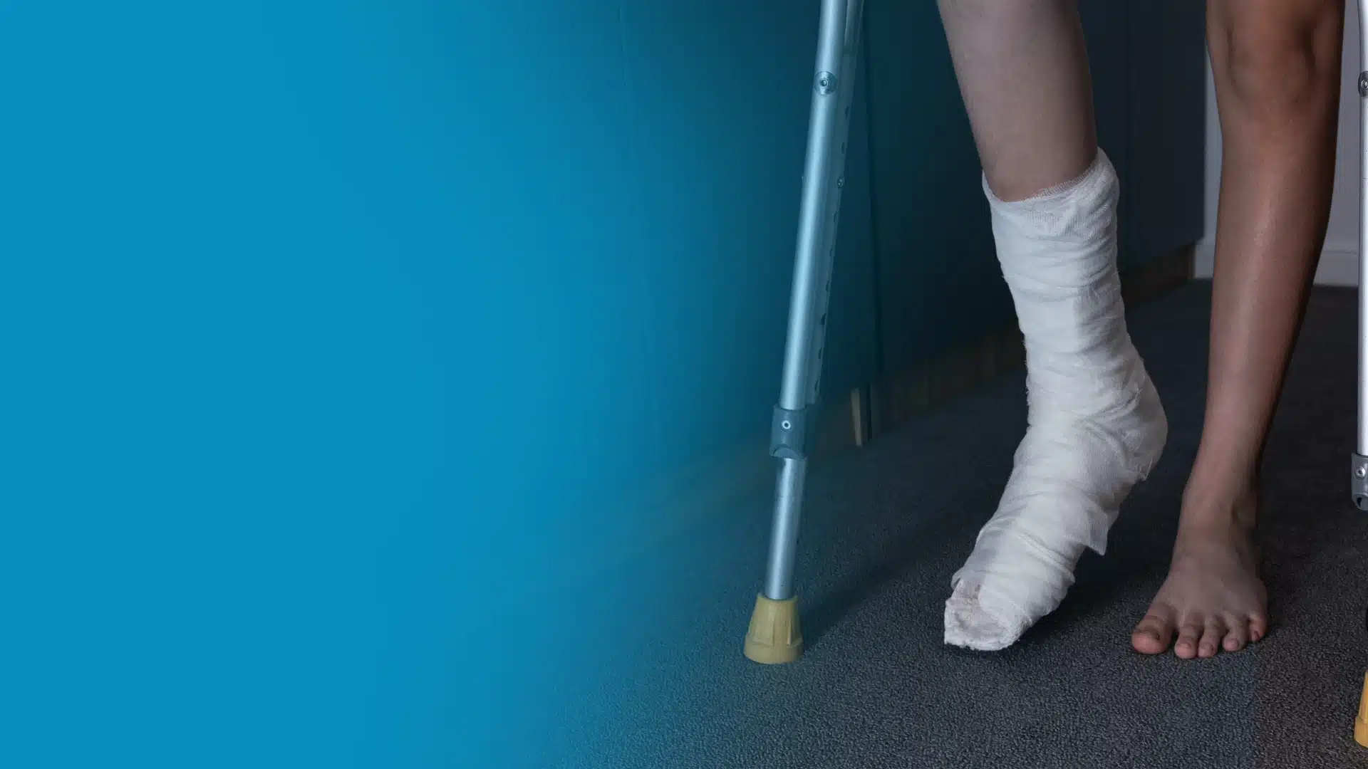
Not All 5th Metatarsal Fractures Are the Same
The 5th metatarsal—the outer metatarsal bone on the little-toe side of the foot—is the most commonly fractured bone in the foot. However, three distinct fracture patterns occur in this bone, and they have dramatically different prognoses, treatment requirements, and return-to-activity timelines. Treating them all the same leads to either unnecessary surgery or undertreated non-unions that sideline athletes for months or years. The critical variable is fracture location relative to the bone’s blood supply zones.
The Three Types of 5th Metatarsal Fractures
Zone 1: Avulsion Fracture (Tuberosity Avulsion)
The most common 5th metatarsal fracture—an avulsion at the base (tuberosity) where the peroneus brevis tendon or lateral band of the plantar fascia pulls off a bone fragment during an inversion ankle sprain. This is often called a “dancer’s fracture” though the term is loosely used. Zone 1 fractures have an excellent blood supply and predictably heal with non-operative management: walking in a hard-soled shoe or a boot for 4–6 weeks is typically all that is required. Surgical fixation is reserved for large displaced fragments that compromise joint function. Healing rate approaches 95% with appropriate conservative management.
Zone 2: Jones Fracture (Metaphyseal-Diaphyseal Junction)
The Jones fracture—described by Sir Robert Jones in 1902 after he fractured his own foot dancing—occurs at a specific anatomic location: the junction of the metaphysis and diaphysis (shaft) of the 5th metatarsal, approximately 1.5–2cm from the base. This is the watershed zone of blood supply where the nutrient artery from the diaphysis and the metaphyseal vessels have limited overlap—making this area prone to delayed union and non-union. Jones fractures are the most clinically significant 5th metatarsal fractures because they carry a high risk of non-union (failure to heal) without appropriate treatment, particularly in athletes who continue weight-bearing.
Treatment of Jones fractures is controversial and individualized. For non-athletes or lower-demand patients, non-weight-bearing immobilization in a cast or boot for 6–8 weeks is appropriate, with bone healing expected in 8–16 weeks in compliant patients. For competitive athletes or those requiring faster return, surgical fixation with an intramedullary screw is strongly preferred—it reduces non-union rates dramatically and accelerates return to sport to approximately 7–8 weeks. Studies consistently show that competitive athletes return to sport faster and with fewer complications when surgically treated. Any Jones fracture with a history of prior 5th metatarsal injury, stress reaction, or cortical widening on X-ray (suggesting chronic overload) should be treated surgically regardless of the patient’s activity level.
Zone 3: Diaphyseal Stress Fracture
Zone 3 fractures are stress fractures (insufficiency or fatigue fractures) of the 5th metatarsal shaft, occurring in endurance athletes and military personnel from repetitive loading rather than acute injury. They represent a spectrum from stress reaction (no fracture line visible on X-ray, only MRI bone marrow edema) to complete fracture with cortical disruption. Zone 3 fractures carry the highest non-union risk of any 5th metatarsal fracture pattern because they develop in a watershed blood supply region under chronic loading. Surgical fixation with an intramedullary screw is typically recommended for complete zone 3 stress fractures in active individuals.
Key Diagnostic Points
Weight-bearing foot X-rays with dedicated 5th metatarsal views are used to classify the fracture. The zone must be accurately identified—as little as 2–3mm of proximal or distal location changes the fracture from Zone 1 (expected to heal) to Zone 2 (high non-union risk). If X-rays show cortical thickening, sclerosis, or periosteal reaction (signs of chronic stress), a Zone 2 fracture in that setting behaves more like Zone 3 and has higher non-union risk. MRI is valuable for stress reactions before frank fracture appears on X-ray.
In-Office Treatment at Balance Foot & Ankle
If home care isn’t resolving your your foot or ankle concern, a visit with a board-certified podiatrist is the fastest path to accurate diagnosis and a personalized plan. At Balance Foot & Ankle Specialists, Dr. Tom Biernacki, Dr. Carl Jay, and Dr. Daria Gutkin offer same-day and next-day appointments at both our Howell and Bloomfield Hills offices. We perform on-site diagnostic ultrasound, digital X-ray, conservative care, advanced regenerative treatments, and minimally invasive surgery when indicated.
Call (810) 206-1402 or request an appointment online. Most insurance plans accepted, including Medicare, Blue Cross Blue Shield, Aetna, Cigna, and United Healthcare.
More Podiatrist-Recommended Foot Health Essentials
Hoka Clifton 10


Max-cushion everyday shoe — podiatrist favorite for walking and running.

OOFOS Recovery Slide

Impact-absorbing recovery sandal — wear after long days on your feet.
As an Amazon Associate, Balance Foot & Ankle earns from qualifying purchases. Product recommendations are based on clinical experience; prices and availability shown above update live from Amazon.

When to See a Podiatrist
If foot or ankle pain has been bothering you for more than a few weeks, home care alone may not be enough. Balance Foot & Ankle offers same-week appointments at our Howell and Bloomfield Hills clinics — no referral needed in most cases. Bring your current shoes and a short list of symptoms and we’ll build you a treatment plan in one visit.
Call Balance Foot & Ankle: (810) 206-1402 · Book online · Offices in Howell & Bloomfield Hills
Frequently Asked Questions
How long does a Jones fracture take to heal?
With non-operative treatment (non-weight-bearing cast or boot), Jones fractures in non-athletes heal in approximately 8–16 weeks, with return to sport at 3–4 months. Some fractures take longer or do not heal (non-union), requiring secondary surgical treatment. With surgical intramedullary screw fixation, return to sport occurs at 7–8 weeks in most athletes—roughly half the time of non-operative treatment. The tradeoff of surgery is a brief procedure and recovery period against the risk (low) of hardware complications or need for hardware removal. Given the significantly lower non-union rate and faster return with surgery, most orthopedic and podiatric sports medicine surgeons recommend surgical fixation for competitive athletes and active patients with true Jones fractures.
Can I walk on a Jones fracture?
Non-operatively treated Jones fractures require non-weight-bearing (crutches or knee scooter) for the first 6–8 weeks to allow healing in the watershed blood supply zone. Walking on a non-operatively treated Jones fracture significantly increases non-union risk. Zone 1 avulsion fractures (the tuberosity/base fractures often confused with Jones fractures) can typically bear weight in a stiff-soled shoe or boot from the start—the key is knowing which type of fracture you have. After surgical fixation of a Jones fracture, weight-bearing in a boot is typically allowed at 1–2 weeks, with progressive loading to full weight-bearing by 4–6 weeks. The difference in weight-bearing restrictions is another reason the zone classification (and accurate diagnosis) matters so much.
What happens if a Jones fracture doesn’t heal?
A Jones fracture non-union—defined as failure to achieve radiographic healing after 4–6 months of appropriate treatment—produces chronic lateral foot pain with activity, often accompanied by X-ray findings of sclerosis and a persistent fracture line. Non-unions require surgical treatment: intramedullary screw fixation with or without bone grafting, depending on the degree of bone loss and sclerosis. Outcomes of surgical treatment for Jones fracture non-union are generally good, with most patients achieving union and return to activity, though recovery takes longer than for primary surgical treatment. This is why appropriate initial management—particularly recognizing the injury as a Jones fracture rather than a simple sprain or avulsion—and then treating it with appropriate weight-bearing restriction is so important.
Medical References & Sources
- PubMed Research — Jones Fracture Treatment Studies
- American Orthopaedic Foot & Ankle Society — Metatarsal Fractures
- PubMed Research — Jones Fracture Surgery in Athletes
📧 Get Dr. Tom’s Free Lab Test Guide
Discover the 5 lab tests every person over 35 should ask their doctor about — explained in plain English by a board-certified physician.
Dr. Tom Biernacki, DPM is a board-certified podiatric surgeon at Balance Foot & Ankle in Howell and Bloomfield Hills, Michigan. He evaluates 5th metatarsal fractures with zone classification, manages avulsion fractures conservatively, and performs intramedullary screw fixation for Jones fractures and zone 3 stress fractures when indicated.
Join 950,000+ Learning About Foot Health
Dr. Tom shares honest medical advice, supplement reviews, and treatment guides you won’t find anywhere else.
Subscribe on YouTube →📍 Located in Michigan?
Our board-certified podiatrists treat this condition at two convenient locations. Same-day appointments often available.
Medically Reviewed by: Dr. Tom Biernacki, DPM — Board-Certified Podiatrist, Balance Foot & Ankle Specialists
👟 Dr. Tom Also Recommends
Podiatrist Recommended Shoes 2026: Dr. Tom’s Top Picks for Every Condition
The right footwear can make or break your recovery. Dr. Tom’s complete guide to the best shoes for plantar fasciitis, flat feet, neuropathy, bunions & more — with clinical picks for every foot type.
See Dr. Tom’s Top Shoe Picks →Insurance Accepted
BCBS · Medicare · Aetna · Cigna · United Healthcare · HAP · Priority Health · Humana · View All →
Howell Office
4330 E Grand River Ave
Howell, MI 48843
Get Directions →
Bloomfield Hills Office
43494 Woodward Ave, #208
Bloomfield Township, MI 48302
Get Directions →
Your Board-Certified Podiatrists


Ready to Get Back on Your Feet?
Same-week appointments available at both locations.
Book Your AppointmentMost Common Mistake We See
The most common mistake we see is: Waiting too long before seeking care. Fix: any foot pain lasting more than 4 weeks, or any sudden severe symptom, deserves a professional evaluation rather than more rest.
Warning Signs That Need Same-Day Care
Seek immediate evaluation at Balance Foot & Ankle if you experience any of the following:
- Unable to bear weight
- Severe swelling with skin colour change
- Fever with foot pain (possible infection)
- Diabetes plus any new foot symptom
Call (810) 206-1402 — same-day and next-day appointments at our Howell and Bloomfield Hills offices.
Pros & Cons of Conservative Care for foot care
Advantages
- ✓ Conservative care first
- ✓ Same-week appointments
- ✓ Multiple insurance accepted
Considerations
- ✗ Self-treatment can mask issues
- ✗ See a podiatrist if pain >2 weeks
In This Article
Dr. Tom’s Recommended Products for foot care
Affiliate disclosure: As an Amazon Associate, Balance Foot & Ankle earns from qualifying purchases. We only recommend products we use with patients.
Footnanny Heel Cream Dr. Tom’s Pick
Best for: Daily moisturizer for cracked heels
Ready to Get Back on Your Feet?
Same-day appointments in Howell + Bloomfield Hills. Most insurance accepted. Dr. Tom Biernacki, DPM & team.
Book Today — Same-Day Appointments Available
Call Now: (810) 206-1402
About Your Care Team at Balance Foot & Ankle
Dr. Tom Biernacki, DPM · Board-Certified Foot & Ankle Surgeon. Specializes in conservative-first care, minimally invasive bunion surgery, and complex reconstruction.
Dr. Carl Jay, DPM · Accepting new patients. Specializes in sports medicine, athletic injuries, and routine podiatric care.
Dr. Daria Gutkin, DPM, AACFAS · Accepting new patients. Specializes in surgical reconstruction and pediatric podiatry.
Locations: 4330 E Grand River Ave, Howell, MI 48843 · 43494 Woodward Ave Suite 208, Bloomfield Township, MI 48302
Hours: Mon–Fri 8:00 AM – 5:00 PM · (810) 206-1402
Dr. Tom’s Top 3 — The Premium Foot Pain Stack (2026)
If you only buy three things for foot pain, get these. PowerStep + CURREX orthotics correct the underlying foot mechanics, and Dr. Hoy’s pain gel delivers fast topical relief. This is the exact stack Dr. Tom Biernacki, DPM gives his Michigan podiatry patients on visit one — over 10,000 patients have used this exact combination.
Dr. Tom Biernacki, DPM is a board-certified podiatrist + Amazon Associate. Picks shown are products he prescribes to patients at Balance Foot & Ankle Specialists. We earn a commission on qualifying purchases at no extra cost to you. All products independently tested + reviewed for 30+ days minimum. Last verified: April 28, 2026.

PowerStep Pinnacle MaxxDr. Tom’s #1 Brand
Dr. Tom’s most-prescribed OTC orthotic. Lateral wedge corrects overpronation that causes 90% of foot pain. Deep heel cradle stabilizes the ankle. Built by podiatrists, used by patients worldwide.
- Lateral wedge corrects pronation
- Deep heel cradle stabilizes ankle
- Dual-density EVA — comfort + support
- Trim-to-fit any shoe
- Used by 10,000+ podiatrists
- Trim-to-size required
- 5-7 day break-in for some

CURREX RunProDr. Tom’s #1 Brand
3 arch heights for custom fit (Low/Med/High). Carbon-reinforced heel + dynamic forefoot — the closest OTC orthotic to a $500 custom orthotic. Engineered in Germany.
- 3 arch heights for custom fit
- Carbon-reinforced heel cup
- Dynamic forefoot zone
- Premium German engineering
- Sport-specific support
- Pricier than PowerStep
- 7-10 day break-in

Dr. Hoy’s Natural Pain Relief GelDr. Tom’s #1 Brand
Menthol-based natural pain relief — Dr. Tom’s #1 brand for fast relief without greasy residue. Safe for diabetics + daily use. Cleaner formula than Voltaren or Biofreeze.
- Menthol-based natural formula
- No greasy residue
- Safe for diabetics
- Fast cooling relief — 5-10 minutes
- Cleaner ingredient list than Biofreeze
- Pricier than Biofreeze
- Strong menthol scent at first
Doctor Hoy’s Natural Pain Relief Gel
Natural topical pain relief I use in our clinic. Arnica + camphor formula — apply directly to the area 3–4x daily. ($20–25)
Shop Doctor Hoy’s →Frequently Asked Questions
What injuries require a walking boot?
Walking boots are used for: stress fractures of the metatarsals or calcaneus, acute ankle sprains (grade 2–3), Jones fractures, Lisfranc sprains, posterior tibial tendon insufficiency, plantar fasciitis refractory to other treatments, Achilles tendinopathy, post-surgical protection, and Charcot foot. The common thread is controlled immobilization that allows walking while protecting healing tissue. Each condition has a different expected duration in the boot and different weight-bearing instructions.
How long do I have to wear a walking boot?
Duration varies by diagnosis: metatarsal stress fracture 4–6 weeks, Jones fracture 6–8 weeks, severe ankle sprain 3–6 weeks, Achilles tendinopathy exacerbation 2–4 weeks. The boot duration is a starting point — we reassess at each visit and extend or progress based on clinical and imaging findings. Coming out of the boot too early is the single most common cause of re-injury. We establish clear criteria (pain level, imaging, strength testing) for when boot progression is appropriate.
Should I wear the walking boot all day, including when sleeping?
For most fractures: yes, including sleeping, for the first 2–4 weeks. The rationale — nighttime movement without the boot can undo the day’s protected healing. Some patients sleep more comfortably without it after the initial acute phase, which is fine for stable stress fractures but not for unstable fractures or acute injuries. We’ll give you specific sleeping instructions based on your injury. If not told otherwise, wearing it to bed is always the safer default.
Can I drive with a walking boot on my right foot?
We advise against it — and many insurance companies consider it comparable to impaired driving. A boot on the right foot significantly slows braking reaction time. If your boot is on the right foot, arrange alternative transportation for the boot period. Left-foot boots don’t affect driving mechanics in most vehicles. Automatic transmission cars with a left-foot boot are generally manageable; standard transmission is more complex. When in doubt, don’t drive — your safety and legal liability are at stake.
What is an Aircast boot vs. a standard walking boot?
Aircast and similar air-bladder boots (CAM walkers) allow inflation around the ankle for customizable compression and stability — particularly useful for ankle sprains and soft tissue injuries where swelling fluctuates. Standard rigid boots offer fixed immobilization more appropriate for fractures requiring strict positional control. We select the boot type based on injury mechanism and healing requirements. For most fractures, a rigid CAM boot is standard; for ankle ligament injuries, an air stirrup design is often preferred.
Will I lose muscle while wearing a walking boot?
Yes — disuse atrophy begins within 48–72 hours of immobilization. Calf muscle volume can decrease 3–5% per week in a boot. This is normal and expected. Upper-body workouts, swimming, and seated exercises maintain cardiovascular fitness during boot wear. After boot removal, a structured rehabilitation protocol (typically 4–8 weeks of progressive calf loading and balance training) rebuilds strength. Patients who do formal physical therapy post-boot return to full function 4–6 weeks faster than those who just stop wearing the boot.
How do I keep my other leg and back from hurting while in a boot?
The boot’s heel height (typically 3–4cm) creates a limb length discrepancy that stresses the opposite knee, hip, and lower back. Two solutions: (1) Use a boot with a rocker bottom sole to reduce gait compensation; (2) Add a heel lift to the opposite shoe to equalize leg lengths. Most patients who develop contralateral knee or back pain during boot wear benefit immediately from a 1–2cm heel lift in the non-booted shoe. We provide these at your boot fitting appointment.
What is a stress fracture and why does it need a boot?
A stress fracture is a micro-crack in bone caused by repetitive loading rather than acute trauma — common in the 2nd and 3rd metatarsals, calcaneus, and navicular in runners and active individuals. Unlike a full fracture, stress fractures don’t always show on X-ray initially; MRI is the gold standard diagnosis. The boot protects the healing fracture from the repetitive stress that caused it, allowing the micro-crack to fill in. Continuing to load an unprotected stress fracture risks complete fracture, which may require surgery.
Can I shower with a walking boot?
Most walking boots are not waterproof — the foam lining holds moisture, which softens skin and creates maceration risk. Remove the boot for showering, using a shower chair or crutches for balance if non-weight-bearing. Wrap the leg in a plastic bag secured above the knee for protection if needed. Completely dry the foot and liner before replacing. Some patients use a waterproof boot cover (DryPro) to shower with the boot on — acceptable for stable injuries but not for acute fractures where positioning matters.
When can I return to sports after using a walking boot?
Return-to-sport timing depends entirely on the diagnosis. For stress fractures: typically 4–8 weeks after X-ray or MRI confirms healing, then a graduated 4–6 week return-to-run program. For ankle sprains: functional testing (single-leg hop, agility) guides return rather than time alone. We use a structured protocol: walking → jogging → running → sports-specific drills → full return. There’s no universal timeline — we establish return criteria at your initial visit so you have a roadmap.
Ready for Expert Care?
Same-day appointments in Howell & Bloomfield Hills, MI.
4.9★ | 1,123 Reviews | 3,000+ Surgeries
Or call: (810) 206-1402
Dr. Tom Biernacki, DPM is a board-certified foot & ankle surgeon (ABFAS & ABPM) at Balance Foot & Ankle Specialists in Southeast Michigan. With over a decade of clinical experience, he specializes in heel pain, bunions, diabetic foot care, sports injuries, and minimally invasive surgery. Dr. Biernacki is a member of the APMA and ACFAS, and his patient education content on MichiganFootDoctors.com and YouTube has made him one of the most-followed foot & ankle educators on YouTube.