

Quick answer: Treatment for high ankle sprain syndesmosis injury diagnosis treatment follows a stepwise approach: 1) conservative care first (rest, ice, supportive footwear, OTC anti-inflammatories), 2) physical therapy and targeted exercises, 3) in-office treatments (injections, custom orthotics) if conservative fails at 4-6 weeks, 4) surgery for refractory cases. Most patients resolve at step 1 or 2. Call (810) 206-1402.
Medically reviewed by Dr. Tom Biernacki, DPM — Board-Certified Podiatric Surgeon — Balance Foot & Ankle, Howell & Bloomfield Hills, MI. Last updated April 2026.
Medically Reviewed by Dr. Tom Biernacki, DPM — Board-Certified Podiatrist, Balance Foot & Ankle Specialists, Michigan. Last updated April 2026.
High Ankle Sprain: Syndesmosis Injury Diagnosis and Treatment
Quick Answer: A high ankle sprain damages the syndesmosis, the ligament complex that binds the tibia and fibula together just above the ankle joint. Unlike common lateral ankle sprains, high ankle sprains take significantly longer to heal, often 6 to 12 weeks or more, and are frequently misdiagnosed as simple sprains. Accurate diagnosis is critical because untreated syndesmosis injuries can lead to chronic ankle instability, arthritis, and persistent pain that limits athletic and daily activities.
Medically Reviewed by: Dr. Tom Biernacki, DPM — Board-Certified Podiatrist at Balance Foot & Ankle Specialists, with over a decade of clinical experience treating ankle injuries including syndesmosis injuries in athletes and active patients.
Affiliate Disclosure: This page contains affiliate links. We may earn a small commission at no extra cost to you if you purchase through these links. We only recommend products we use in our clinical practice.
Table of Contents
- What Is a High Ankle Sprain?
- Anatomy of the Syndesmosis
- How High Ankle Sprains Happen
- Symptoms: High vs. Low Ankle Sprain
- How High Ankle Sprains Are Diagnosed
- Grading System for Syndesmosis Injuries
- Conservative Treatment Protocol
- When Surgery Is Needed
- Recovery Timeline
- Rehabilitation and Physical Therapy
- Pain and Swelling Management
- Return to Sports After High Ankle Sprain
- Chronic Syndesmosis Instability
- Complete Recovery Kit
- Most Common Mistake
- Warning Signs
- Prevention Strategies
- High Ankle Sprains in Specific Sports
- Long-Term Outlook
- Video Guide
- Frequently Asked Questions
- Sources
- Book Your Evaluation
If your ankle injury is taking much longer to heal than you expected, or if the pain is located higher up than a typical ankle sprain, you may have a high ankle sprain. This injury is one of the most commonly missed diagnoses in sports medicine, and the consequences of missed or delayed treatment are significant. High ankle sprains account for approximately 10 to 20% of all ankle sprains but receive far less attention than the more common lateral ankle sprain. Understanding how this injury differs from a regular sprain, why it requires a different treatment approach, and what recovery realistically looks like helps you get the right care and set appropriate expectations.
What Is a High Ankle Sprain? Understanding Syndesmosis Injury
A high ankle sprain injures the syndesmosis, the fibrous joint where the lower ends of the tibia and fibula meet just above the ankle joint. Unlike a lateral ankle sprain that damages the ligaments on the outside of the ankle, a high ankle sprain damages the ligaments that hold these two leg bones together. The syndesmosis acts as a critical stabilizer that prevents the tibia and fibula from spreading apart during weight-bearing. When this structure is damaged, the ankle mortise, the bony socket that holds the talus, can widen, allowing abnormal movement that causes pain and progressive joint damage.
The term high ankle sprain refers to the location of the injury, which is above the ankle joint itself, as opposed to the more common low ankle sprain where the lateral ligaments at the ankle joint level are damaged. This anatomical distinction is important because the syndesmosis has a limited blood supply and heals more slowly than the lateral ankle ligaments. This is why high ankle sprains consistently require two to three times longer to recover than lateral sprains of comparable severity.
Anatomy of the Syndesmosis: Why It Matters
The syndesmosis complex consists of four structures that work together to maintain the tibia-fibula relationship. The anterior inferior tibiofibular ligament, commonly abbreviated AITFL, is the most frequently injured component and runs diagonally across the front of the ankle just above the joint. The posterior inferior tibiofibular ligament, or PITFL, provides posterior stability and is the strongest component. The interosseous membrane extends between the tibia and fibula along most of their length, and the transverse tibiofibular ligament provides additional posterior restraint.
These structures collectively maintain the ankle mortise width within one to two millimeters of precision. Even small increases in mortise width from syndesmosis injury alter the contact mechanics of the ankle joint, reducing the surface area over which forces are distributed and increasing contact pressure on the remaining cartilage. Research shows that just one millimeter of lateral talar shift reduces tibiotalar contact area by approximately 42%, dramatically increasing the pressure on the remaining contact surface. This is why even seemingly minor syndesmosis injuries can lead to significant long-term consequences if not properly treated.
How High Ankle Sprains Happen: Injury Mechanisms
High ankle sprains typically result from external rotation forces applied to the foot while the ankle is in dorsiflexion. The classic mechanism involves the foot being planted and twisted outward while the body rotates over the ankle. In football, this commonly occurs when a player is tackled from behind while the foot is planted, rotating the leg over the fixed foot. In skiing, it happens during forward falls with the foot locked in the ski boot. In soccer, it occurs during sliding tackles that catch the foot and rotate it externally.
The dorsiflexion component is key to understanding why this injury affects the syndesmosis rather than the lateral ligaments. When the ankle is dorsiflexed, the wider anterior portion of the talus is wedged into the ankle mortise, creating a natural splint that protects the lateral ligaments. The rotational force must then be absorbed by the syndesmosis instead. This is why high ankle sprains are more common in sports that involve cutting, pivoting, and contact where the foot is loaded in dorsiflexion, and less common in simple inversion injuries that occur with the ankle in a neutral or plantarflexed position.
Symptoms: How to Tell High vs. Low Ankle Sprain
Distinguishing a high ankle sprain from a common lateral ankle sprain is critical because the treatment and timeline differ substantially. High ankle sprain pain is located above the ankle joint, along the front or inner aspect of the lower leg between the tibia and fibula. The pain typically worsens with external rotation of the foot, dorsiflexion under load, and pushing off during walking. Swelling may be less dramatic than a lateral sprain initially, which contributes to the injury being underestimated.
In contrast, a lateral ankle sprain causes pain over the outer ankle bone area, typically below and in front of the fibula. The swelling and bruising are concentrated around the lateral malleolus. The squeeze test is a key clinical differentiator: compressing the tibia and fibula together at mid-calf level reproduces pain at the ankle in high ankle sprains but not in lateral sprains. If your ankle sprain was caused by a rotational mechanism, the pain is located higher than the typical lateral ankle, and the injury is taking longer than expected to heal, a high ankle sprain should be suspected.
How High Ankle Sprains Are Diagnosed
Accurate diagnosis requires clinical examination combined with appropriate imaging. The physical examination includes several provocative tests specific to the syndesmosis. The squeeze test compresses the tibia and fibula at mid-calf, which transmits force to the syndesmosis and reproduces pain if injured. The external rotation stress test applies rotational force to the dorsiflexed foot and reproduces syndesmotic pain. The crossed-leg test, where the patient crosses the injured leg over the other knee, applies a gravity-driven external rotation force that causes syndesmotic pain.
Imaging begins with weight-bearing ankle radiographs. The tibiofibular clear space, measured on the anteroposterior view, should be less than 6 millimeters. Widening beyond this threshold suggests syndesmosis disruption. The tibiofibular overlap should exceed 6 millimeters on the AP view. MRI is the most sensitive imaging study for diagnosing syndesmosis injuries, revealing ligament tears, bone bruising patterns, and the extent of interosseous membrane involvement. Stress radiographs, taken while applying external rotation force, can demonstrate dynamic instability that is not apparent on standard static views.
Grading System for Syndesmosis Injuries
High ankle sprains are classified into three grades based on severity. Grade I injuries involve a sprain of the AITFL without significant fiber disruption. The syndesmosis is stable on examination and stress testing. These injuries cause pain and mild functional limitation but respond to conservative treatment with a relatively faster recovery of four to six weeks. There is no widening on radiographs.
Grade II injuries involve a partial tear of the syndesmotic ligaments with some degree of laxity but maintained structural integrity. The ankle mortise may show subtle widening on stress views. Recovery takes six to twelve weeks with conservative management, though some Grade II injuries require prolonged immobilization or even surgical stabilization. Grade III injuries represent complete disruption of the syndesmosis with clear instability and mortise widening. These injuries almost always require surgical fixation to restore ankle stability and prevent long-term joint damage.
Conservative Treatment Protocol for High Ankle Sprains
Grade I and stable Grade II syndesmosis injuries are managed conservatively with a structured protocol that respects the slower healing capacity of the syndesmotic ligaments. The initial phase focuses on protecting the healing ligaments while controlling pain and swelling. A walking boot or cast immobilizes the ankle for two to four weeks, preventing the external rotation forces that stress the syndesmosis. DASS graduated compression socks worn inside the boot help manage swelling and support circulation during the immobilization period.
Pain management during the acute phase combines ice therapy, elevation, and topical treatment. Doctor Hoy’s Natural Pain Relief Gel applied to the anterior ankle and lower leg provides soothing topical relief for the deep aching pain characteristic of syndesmosis injuries. As pain improves, gentle range-of-motion exercises begin within pain-free limits. The transition from boot to supportive shoe occurs when weight-bearing is comfortable, typically at three to six weeks. PowerStep Pinnacle insoles in the transition shoe provide stability and cushioning as you resume normal walking.
When Surgery Is Needed for High Ankle Sprains
Surgical intervention is indicated for Grade III syndesmosis injuries with clear instability and mortise widening, and for Grade II injuries that fail to stabilize with conservative treatment. The standard surgical technique involves reducing the tibia and fibula to their anatomic position and holding them together with one or two syndesmotic screws placed across the joint. These screws prevent the bones from spreading apart while the ligaments heal, typically over eight to twelve weeks.
Newer techniques use suture-button constructs such as the TightRope system, which provide flexible fixation that allows normal physiological motion at the syndesmosis while maintaining reduction. The advantage of suture-button fixation is that it does not typically require removal, unlike syndesmotic screws that may need to be removed at three to four months if they limit ankle motion or break. Your surgeon recommends the fixation method best suited to your injury pattern and activity level. Regardless of the technique used, surgical stabilization followed by structured rehabilitation produces reliable outcomes for unstable syndesmosis injuries.
Recovery Timeline for High Ankle Sprains
The recovery timeline for high ankle sprains is significantly longer than for lateral ankle sprains, which is one of the most important points for patients to understand. Grade I injuries typically allow return to normal activities at four to six weeks and return to sports at six to eight weeks. Grade II injuries require six to twelve weeks for daily activities and eight to sixteen weeks for sports return. Grade III injuries treated surgically may require twelve to twenty weeks for full recovery and sports return.
The slow healing reflects the limited blood supply to the syndesmotic ligaments compared to the lateral ankle ligaments. During recovery, DASS compression socks help manage the persistent swelling that characterizes syndesmosis healing. As you transition to regular shoes, PowerStep Pinnacle insoles provide stability and cushioning that support the healing ankle. The most common error is returning to activity too soon because pain has improved, even though the ligaments have not fully healed.
Rehabilitation and Physical Therapy Protocol
Rehabilitation after a high ankle sprain progresses through phases that respect the healing timeline. The early phase, during boot immobilization, includes toe curls, towel scrunches, and isometric ankle exercises that maintain muscle activation without stressing the syndesmosis. Aquatic therapy can begin once wounds are healed for surgical patients, providing gentle resistance in a reduced-gravity environment.
The intermediate phase begins when the boot is removed and includes progressive range-of-motion exercises, proprioception training on balance boards, and resistance band strengthening in all planes of motion. Doctor Hoy’s Natural Pain Relief Gel applied before and after therapy sessions helps manage the soreness that accompanies progressive rehabilitation. The advanced phase incorporates sport-specific agility drills, plyometric training, and progressive return-to-play testing. Full clearance requires demonstrating normal strength, range of motion, balance, and performance on functional testing without pain or compensatory movement patterns.
Pain and Swelling Management
Pain from syndesmosis injuries has a characteristic deep, aching quality that differs from the sharp pain of lateral ankle sprains. The pain is often most noticeable with the first steps in the morning, when climbing stairs, and when pushing off the affected foot during walking. A multimodal approach combining compression, ice, elevation, and topical pain relief provides the most effective management throughout the recovery process.
Doctor Hoy’s Natural Pain Relief Gel applied along the anterior lower leg between the tibia and fibula targets the specific area of syndesmotic pain. The natural ingredients provide soothing relief without the gastrointestinal concerns of prolonged oral anti-inflammatory use. DASS compression socks address the persistent swelling that often accompanies syndesmosis healing, with graduated compression helping push fluid out of the ankle and lower leg to reduce that heavy, congested feeling that slows rehabilitation progress.
Return to Sports After High Ankle Sprain
Return to sports after a high ankle sprain should be criteria-based rather than time-based. The athlete must demonstrate full pain-free range of motion, at least 90% strength compared to the uninjured side on isokinetic testing, normal proprioception on balance testing, ability to perform sport-specific movements at full speed without pain or apprehension, and symmetrical performance on functional hop tests. Returning before meeting these criteria significantly increases the risk of re-injury and chronic instability.
For the initial return-to-sport period, wear PowerStep Pinnacle insoles in your sport shoes for optimal ankle support and cushioning. Consider ankle bracing or taping for the first several weeks back. Apply Doctor Hoy’s Natural Pain Relief Gel before activity and ice after. Progress from practice to full competition gradually. Many professional athletes report that high ankle sprains affect their performance for several months beyond the initial return date, with full confidence and peak performance returning three to six months after initial return to play.
Chronic Syndesmosis Instability: When Healing Goes Wrong
Chronic syndesmosis instability develops when the initial injury heals with persistent laxity, allowing the ankle mortise to widen subtly with weight-bearing activities. This condition causes ongoing anterior ankle pain, difficulty with push-off activities, reduced athletic performance, and progressive tibiotalar arthritis from the altered joint mechanics. Chronic instability is more common when the initial injury was misdiagnosed as a simple lateral sprain and treated with a shorter, less protective recovery protocol.
Treatment of chronic syndesmosis instability often requires surgical reconstruction of the syndesmotic ligaments, as the stretched or scarred ligaments cannot tighten on their own. Various reconstruction techniques use autograft or allograft tissue to recreate the stability of the original syndesmosis. Outcomes are generally good but inferior to those achieved with proper treatment of the acute injury, which underscores why we accurate initial diagnosis and appropriate treatment from the start.
Complete High Ankle Sprain Recovery Kit
Our Complete High Ankle Sprain Recovery Kit
These three products support each phase of syndesmosis injury recovery from acute treatment through return to full activity:
- PowerStep Pinnacle Orthotic Insoles — Essential when transitioning from boot to shoe. The structured support and cushioning help stabilize the healing ankle while reducing impact forces. Continue using during return to sports for ongoing ankle support and injury prevention.
- Doctor Hoy’s Natural Pain Relief Gel — Target the specific syndesmotic pain area along the anterior lower leg. Apply before and after physical therapy sessions and before athletic activity during return to play. Natural ingredients provide sustained topical relief.
- DASS Graduated Compression Socks — Address the persistent swelling that characterizes syndesmosis healing. Graduated compression promotes fluid drainage and supports circulation throughout the extended recovery period. Wear daily during rehabilitation.
This combination provides ankle stability (PowerStep), targeted pain relief (Doctor Hoy’s), and swelling management (DASS) for comprehensive recovery from one of the most underestimated ankle injuries.
Most Common Mistake
🔑 Most Common Mistake: Treating a high ankle sprain like a regular lateral ankle sprain and returning to activity too soon. High ankle sprains take two to three times longer to heal than lateral sprains. The syndesmosis has limited blood supply and heals slowly. Athletes who return to play at the three to four week mark, which would be appropriate for a moderate lateral sprain, frequently re-injure or develop chronic instability from a syndesmosis injury. Respect the longer timeline, follow your rehabilitation protocol completely, and meet objective return-to-play criteria before resuming sports.
Warning Signs: When to Seek Urgent Evaluation
⚠️ Seek evaluation if you experience:
- Ankle sprain pain located above the ankle joint rather than below
- Pain that worsens when twisting or rotating the foot outward
- Ankle sprain not improving after three weeks of standard treatment
- Pain when squeezing the lower leg bones together at mid-calf
- Difficulty pushing off the affected foot during walking
- A sense of ankle weakness or instability with pivoting movements
- Persistent swelling above the ankle joint
- Pain when climbing stairs that is located higher than the ankle bones
These patterns suggest syndesmosis involvement rather than a simple lateral sprain. Accurate diagnosis changes the treatment approach and prevents chronic problems from inadequate management.
Prevention Strategies for High Ankle Sprains
While high ankle sprains cannot be completely prevented, several strategies reduce risk. Ankle strengthening with resistance band exercises in all planes of motion builds the muscular support that protects the syndesmosis. Proprioception training on balance boards and unstable surfaces improves the neuromuscular control that helps your ankle react to potentially injurious forces before damage occurs. Flexibility training for the calf and ankle improves the joint’s ability to absorb forces safely.
Sport-specific strategies include wearing appropriate footwear with adequate ankle support and PowerStep insoles for optimal foot positioning. Prophylactic ankle taping or bracing may reduce syndesmosis injury risk in high-risk sports, particularly for athletes with a history of ankle injury. Proper warm-up before activity prepares the muscles and ligaments for the demands ahead. Understanding the injury mechanism helps athletes recognize and avoid the positions of vulnerability, particularly dorsiflexion combined with external rotation forces.
High Ankle Sprains in Specific Sports
High ankle sprains are most common in football, where they account for up to 25% of ankle injuries due to the rotational tackles and planted foot mechanisms. Soccer sees frequent syndesmosis injuries from slide tackles and pivoting movements. Ice hockey’s skating mechanics and board contact create high-risk scenarios. Skiing’s fixed boot position makes dorsiflexion-rotation injuries common during falls. Basketball and lacrosse have moderate syndesmosis injury rates from cutting and contact mechanisms.
In professional sports, high ankle sprains are among the most frustrating injuries for athletes and teams because of the extended recovery timeline. NFL players with high ankle sprains miss an average of four to six weeks, compared to one to two weeks for lateral sprains. The injury often lingers beyond the return-to-play date, with athletes reporting reduced cutting ability, speed, and power for weeks to months after initial return. This pattern underscores why comprehensive rehabilitation and patience are essential for complete recovery.
Long-Term Outlook After High Ankle Sprain
The long-term prognosis for properly treated high ankle sprains is generally favorable. Grade I and II injuries treated with appropriate immobilization and rehabilitation typically achieve full recovery without long-term consequences. Grade III injuries treated with surgical stabilization also have good outcomes when rehabilitation is completed. The key variable is the accuracy and timeliness of diagnosis and treatment, as delayed or inadequate treatment significantly worsens long-term outcomes.
Long-term preventive care includes maintaining ankle strength and flexibility through ongoing exercise, wearing PowerStep orthotic insoles for daily activities and sports, and being vigilant for signs of recurrence. Some patients experience mild intermittent symptoms in cold weather or with high-demand activities for up to a year after injury, which is considered normal healing progression. If pain persists beyond twelve months or worsens over time, evaluation for chronic instability or early arthritis is warranted.
Video: Ankle Sprain Recovery Guide
Watch Dr. Biernacki discuss ankle injury evaluation and rehabilitation principles:

More Podiatrist-Recommended Ankle Sprain Essentials
Stability Walking/Running Shoe
Brooks Adrenaline GTS 25 — lateral support during recovery walking.
KT Tape for Ankle Support
KT Tape — proprioceptive support for athletic return-to-play.
Supportive Insole


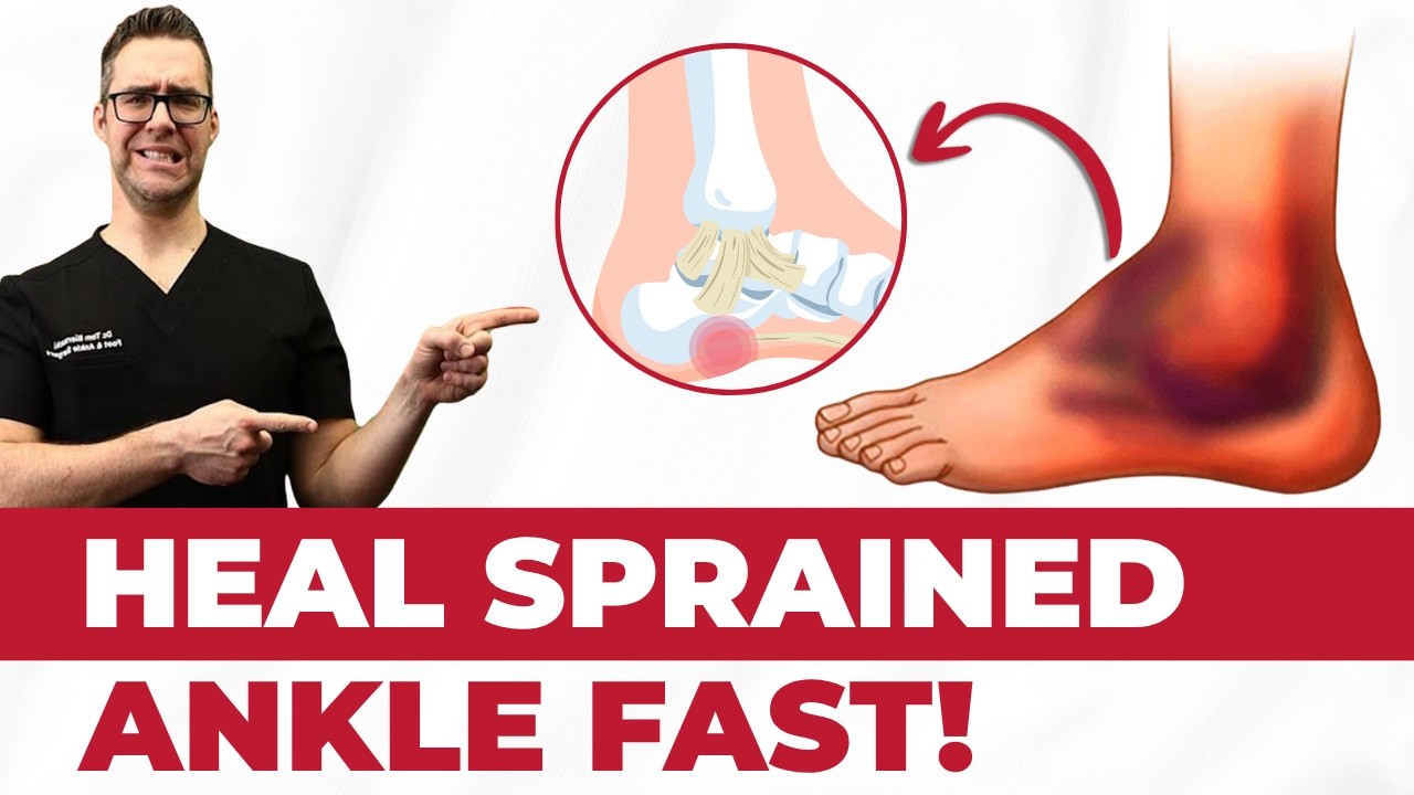
Watch: Fix TWISTED Ankle, ROLLED Ankle or SPRAINED Ankle Ligaments FASTER! — MichiganFootDoctors YouTube
PowerStep Pinnacle — arch support reduces re-injury risk during recovery.
As an Amazon Associate, Balance Foot & Ankle earns from qualifying purchases. Product recommendations are based on clinical experience; prices and availability shown above update live from Amazon.
When to See a Podiatrist
A sprain that hasn’t fully recovered after 6 weeks often has residual ligament laxity or occult fracture that keeps the ankle unstable. Balance Foot & Ankle X-rays and stress-tests every lingering sprain — if the ligament is torn, we offer bracing, PRP, and (for chronic instability) minimally-invasive repair. Don’t keep re-rolling the same ankle; let us stabilize it properly.
Call Balance Foot & Ankle: (810) 206-1402 · Book online · Offices in Howell & Bloomfield Hills
Frequently Asked Questions
How long does a high ankle sprain take to heal?
High ankle sprains take 2-3 times longer than lateral sprains. Grade I: 4-6 weeks. Grade II: 6-12 weeks. Grade III (surgical): 12-20 weeks. The syndesmosis has limited blood supply and heals slowly. Many patients report continued improvement for several months beyond initial return to activity.
How can I tell if I have a high ankle sprain or regular sprain?
High ankle sprain pain is located above the ankle joint between the tibia and fibula, worsens with foot rotation, and is reproduced by squeezing the calf bones together. Regular lateral sprain pain is below and around the outer ankle bone. If your sprain involved a twisting mechanism and the pain is higher than typical, suspect a high ankle sprain and seek evaluation.
Do high ankle sprains require surgery?
Not always. Grade I and stable Grade II injuries respond to conservative treatment with immobilization and rehabilitation. Grade III injuries with clear syndesmosis instability and mortise widening typically require surgical fixation with screws or suture-button devices. Your podiatrist determines the appropriate treatment based on examination and imaging findings.
Can I walk on a high ankle sprain?
Many patients can walk with a high ankle sprain, though with significant pain and a limp. The ability to walk does not indicate the injury is minor. Some athletes initially continue playing on high ankle sprains, which can worsen the injury. Walking should be limited to a boot or supportive brace during the initial healing phase to protect the syndesmosis.
Why is my ankle sprain taking so long to heal?
If your ankle sprain is taking longer than 3-4 weeks to heal, you may have a high ankle sprain that was initially diagnosed as a lateral sprain. High ankle sprains consistently take 2-3 times longer due to the syndesmosis ligaments’ limited blood supply. Seek re-evaluation with syndesmosis-specific examination and potentially MRI to confirm the diagnosis and adjust your treatment plan.
Differential Diagnosis: What Else Could It Be?
Not every case of high ankle sprain / syndesmotic injury is straightforward. In our clinic we routinely rule out three look-alike conditions before confirming the diagnosis. If your symptoms don’t match the classic presentation, one of these may explain the pain — which is why physical exam matters more than self-diagnosis.
| Condition | How It Differs |
|---|---|
| Lateral ankle sprain | Pain and swelling over ATFL, not above the ankle; negative squeeze test. |
| Deltoid ligament sprain | Medial tenderness with eversion injury, not dorsiflexion-external rotation. |
| Maisonneuve fracture | Proximal fibula fracture paired with syndesmotic disruption — requires tib-fib X-ray. |
Red Flags — When to See a Podiatrist Now
Seek same-day evaluation at Balance Foot & Ankle if you notice any of the following:
- Inability to bear weight after ankle injury
- Positive squeeze test above the ankle
- Pain with external rotation of the foot
- Suspected Maisonneuve fracture (proximal fibula pain)
Call (810) 206-1402 or request an appointment. Our Howell and Bloomfield Hills offices reserve same-day slots for urgent foot and ankle issues.
In Our Clinic: What We See
Clinical perspective from Dr. Tom Biernacki, DPM — Balance Foot & Ankle, Howell & Bloomfield Hills, MI:
High ankle sprains present differently than lateral sprains. The patient tells us the foot was planted and rotated outward — a football tackle, a ski binding twist, or a slip on ice. Pain is felt above the ankle, not at the ATFL. In our clinic the squeeze test and external rotation stress test drive the workup. Stable syndesmotic sprains recover in 6-10 weeks of boot immobilization. Unstable injuries require surgical stabilization with suture button or screws. Dr. Biernacki stresses early diagnosis: a missed syndesmotic sprain causes chronic ankle instability and cartilage damage that standard ankle-sprain rehab will not fix.
Sources
- Williams GN, et al. “Syndesmosis Sprains of the Ankle: A Systematic Review.” Sports Medicine, 2007;37(3):263-283.
- Hunt KJ, et al. “High Ankle Sprains and Syndesmosis Injuries in Athletes.” Journal of the American Academy of Orthopaedic Surgeons, 2015;23(11):661-673.
- Nussbaum ED, et al. “Syndesmosis Sprains in the National Football League.” American Journal of Sports Medicine, 2020;48(1):71-77.
- Ramsey PL, Hamilton W. “Changes in Tibiotalar Area of Contact Caused by Lateral Talar Shift.” Journal of Bone and Joint Surgery, 1976;58(3):356-357.
- van den Bekerom MPJ, et al. “Diagnosing Syndesmotic Instability in Ankle Fractures.” BMC Musculoskeletal Disorders, 2015;16:345.
Book Your Ankle Injury Evaluation
Ankle Sprain Not Getting Better?
Dr. Biernacki and the team at Balance Foot & Ankle Specialists provide expert evaluation of ankle injuries including commonly missed high ankle sprains. We use specialized examination techniques and advanced imaging to ensure accurate diagnosis and appropriate treatment for every ankle injury.
Schedule Your Ankle Evaluation →
Same-week appointments available · Most insurance accepted · MRI referral available
Related Resources
- Lateral Ankle Sprain Treatment Guide
- Chronic Ankle Instability
- Ankle Fracture Surgery
- Podiatrist-Recommended Foot Care Products
- Sports Foot and Ankle Injuries
When to See a Podiatrist for a High Ankle Sprain
High ankle sprains are more severe than typical ankle sprains and require specialized diagnosis. If you’re experiencing pain above the ankle joint, especially with rotation or weight-bearing, seek evaluation. At Balance Foot & Ankle, we diagnose and treat syndesmosis injuries at our Howell and Bloomfield Hills offices.
Learn About Our Ankle Sprain Treatment | Book Your Appointment | Call (810) 206-1402
Clinical References
- Williams GN, Jones MH, Amendola A. “Syndesmotic ankle sprains in athletes.” American Journal of Sports Medicine. 2007;35(7):1197-1207.
- Nussbaum ED, Hosea TM, Sieler SD, Incremona BR, Kessler DE. “Prospective evaluation of syndesmotic ankle sprains without diastasis.” American Journal of Sports Medicine. 2001;29(1):31-35.
- Hopkinson WJ, St Pierre P, Ryan JB, Wheeler JH. “Syndesmosis sprains of the ankle.” Foot & Ankle. 1990;10(6):325-330.
Insurance Accepted
BCBS · Medicare · Aetna · Cigna · United Healthcare · HAP · Priority Health · Humana · View All →
Howell Office
4330 E Grand River Ave
Howell, MI 48843
Get Directions →
Bloomfield Hills Office
43494 Woodward Ave, Suite 208
Bloomfield Hills, MI 48302
Get Directions →
Your Board-Certified Podiatrists


Ready to Get Back on Your Feet?
Same-week appointments available at both locations.
Book Your AppointmentWatch: High Ankle Sprain (Syndesmosis Injury)
Dr. Tom on syndesmosis sprains — squeeze test, external rotation test, when surgery is needed, tightrope vs screw fixation, return-to-sport.
Syndesmosis Recovery Kit
Slower than lateral sprain. Dr. Tom’s kit:
As an Amazon Associate, Balance Foot & Ankle earns from qualifying purchases. This supports our free patient education content.
Protected weight-bearing.
Return-to-shoe stability.
Acute inflammation.
Topical ankle relief.
Related: Sprain Grades · Ankle Sprain Care · Book Same-Week Appointment
In-Office Treatment at Balance Foot & Ankle
When conservative care isn’t enough, Dr. Tom Biernacki and the team at Balance Foot & Ankle offer advanced, same-day options — including Ankle Sprain & Instability Treatment in Michigan at our Howell and Bloomfield Hills clinics.
Same-day appointments available. Call (810) 206-1402 or book online.
In-Office Treatment at Balance Foot & Ankle
If home treatment isn’t providing relief for your ankle sprains, our podiatry team at Balance Foot & Ankle can help with same-day evaluations and advanced in-office care.
Same-day appointments available. (810) 206-1402
Frequently Asked Questions
How long does treatment take to work?
Most patients see improvement in 4-8 weeks with consistent conservative care. Persistent symptoms after 8 weeks need imaging and escalation.
When is surgery needed?
Surgery is reserved for cases that fail 3-6 months of conservative care, structural deformities, or fractures requiring stabilization.
OrthoInfo – AAOS: Sprained Ankle
Is this covered by insurance?
Most diagnostic visits and conservative treatments are covered by Medicare and major insurers. Custom orthotics often require diabetic or post-surgical justification.
Dr. Tom Biernacki, DPM is a board-certified foot & ankle surgeon (ABFAS & ABPM) at Balance Foot & Ankle Specialists in Southeast Michigan. With over a decade of clinical experience, he specializes in heel pain, bunions, diabetic foot care, sports injuries, and minimally invasive surgery. Dr. Biernacki is a member of the APMA and ACFAS, and his patient education content on MichiganFootDoctors.com and YouTube has made him one of the most-followed foot & ankle educators on YouTube.