
Medically reviewed by Dr. Tom Biernacki, DPM
Board-certified podiatric surgeon | Balance Foot & Ankle, Howell & Bloomfield Hills, MI
Last reviewed: May 2026

| Fracture Type | Location | Surgery Needed? | Recovery Time | Fixation Method |
|---|---|---|---|---|
| Shaft Fracture (non-displaced) | Mid-shaft MT 1–5 | Usually no | 4–6 weeks boot | Cam boot or hard-sole shoe |
| Shaft Fracture (displaced >3–4mm) | Mid-shaft MT 1–5 | Yes | 8–10 weeks | Intramedullary screw or plate |
| Jones Fracture (Zone II–III) | 5th MT metaphyseal-diaphyseal | Often yes (athletes) | 8–12 weeks | Intramedullary screw (6.5mm) |
| Avulsion Fracture (Zone I) | 5th MT tuberosity | Rarely | 4–6 weeks | Cam boot; rarely ORIF |
| First Metatarsal Fracture | 1st MT (weight-bearing critical) | Often yes (>2mm displacement) | 10–12 weeks | Plate and screws (ORIF) |
| Stress Fracture | 2nd–3rd MT most common | Rarely | 6–8 weeks | Activity restriction, bone stimulator if delayed |
| Surgical Approach | Incision | Hardware | Post-Op WB | Advantages |
|---|---|---|---|---|
| Intramedullary Screw | Single small incision | Solid/cannulated screw | Protected WB in boot | Minimal soft tissue disruption, fast recovery |
| Open Reduction Internal Fixation (ORIF) | Dorsal incision over fracture | Plate + screws | NWB 6–8 weeks | Direct visualization, rigid fixation |
| Percutaneous Pinning (K-wire) | Percutaneous stab incision | K-wire (temporary) | NWB 4–6 weeks | Minimally invasive, used in children |
| Lisfranc ORIF | Dorsal 1–2 incisions | Plate, screws, suture button | NWB 8–12 weeks | Restores arch stability in Lisfranc injuries |
Quick answer: Metatarsal Fracture Surgery Michigan Podiatrist is a common foot/ankle topic that affects many patients. The 2026 evidence-based approach combines proper diagnosis, conservative-first treatment, and escalation only when needed. We treat this regularly at our Howell and Bloomfield Hills practices. Call (810) 206-1402.
Foot pain isn't resolving?
Same-week appointments at Howell & Bloomfield Hills
Medically Reviewed | Dr. Tom Biernacki, DPM | Board-Certified Podiatric Surgeon | Balance Foot & Ankle, Michigan


![Metatarsalgia Treatment [BEST Ball of Foot Pain RELIEF 2024]](https://www.michiganfootdoctors.com/wp-content/cache/flying-press/d7857672675f2bf191a8e896b2f7208b.jpg)
Watch: Metatarsalgia Treatment [BEST Ball of Foot Pain RELIEF 2024] — MichiganFootDoctors YouTube
Metatarsal Fracture Surgery in Michigan
The metatarsals — the five long bones forming the midfoot and forefoot — are among the most commonly fractured bones in the foot. Whether you’ve sustained a Jones fracture pushing off on the basketball court, a 5th metatarsal avulsion fracture rolling your ankle, a stress fracture from high-mileage running, or an acute fracture from direct trauma, understanding your fracture type and treatment options is critical to achieving a complete, reliable recovery. Dr. Tom Biernacki at Balance Foot & Ankle PLLC provides expert metatarsal fracture management for patients across Michigan, with surgical intervention when necessary to achieve the bone healing and functional outcomes that conservative treatment cannot reliably deliver.
Metatarsal Anatomy and Fracture Patterns
The five metatarsals articulate proximally with the tarsal bones and distally with the proximal phalanges at the metatarsophalangeal joints. Each metatarsal has a head, neck, shaft (diaphysis), and base. Fractures occur at different anatomic zones with markedly different healing biology and treatment implications. The first metatarsal bears the greatest weight loads and requires anatomic reduction for any displaced fracture. The central metatarsals (2nd, 3rd, 4th) are relatively protected and most fractures heal conservatively. The 5th metatarsal is by far the most surgically significant, with three distinct fracture zones at its base carrying vastly different prognoses.
5th Metatarsal Fractures: Zone Classification
The 5th metatarsal base is the site of three distinct fracture patterns that are frequently confused but have dramatically different treatment requirements:
Zone I: Avulsion Fracture (Pseudo-Jones)
Zone I avulsion fractures occur at the very tip of the 5th metatarsal tuberosity where the peroneus brevis tendon and lateral band of the plantar fascia insert. These fractures occur with ankle inversion injuries, pulling off a small fragment. Despite their dramatic appearance on X-ray, Zone I fractures have an excellent blood supply and heal reliably with conservative management — hard-soled shoe or boot for 3–6 weeks. Surgery is rarely required unless a large fragment is displaced more than 2 mm.
Zone II: Jones Fracture
The true Jones fracture occurs at the metaphyseal-diaphyseal junction of the 5th metatarsal — approximately 1.5–2 cm from the tip of the tuberosity. This zone is a watershed area between the metaphyseal blood supply proximally and the diaphyseal nutrient artery distally, creating a zone of relative avascularity. Jones fractures have a notorious propensity for delayed union, non-union, and re-fracture with conservative treatment alone. In active patients and athletes, primary intramedullary screw fixation is strongly recommended over conservative management to achieve reliable union and early return to activity. Re-fracture rates after non-surgical Jones fracture healing can reach 25–50%.
Zone III: Diaphyseal Stress Fracture
Zone III fractures occur in the proximal 5th metatarsal diaphysis and represent stress fractures from repetitive loading rather than acute injury. They present with progressive forefoot pain in runners, dancers, and military recruits. Acute diaphyseal fractures with cortical disruption require surgical fixation; chronic stress reactions without complete fracture may respond to rest and offloading, but high-demand athletes typically benefit from early surgical stabilization to accelerate return to activity and prevent progression to complete fracture.
Jones Fracture Surgery: Intramedullary Screw Fixation
Surgical treatment of Jones fractures involves intramedullary screw fixation — a technically elegant procedure performed through a small stab incision at the tip of the 5th metatarsal tuberosity. Under fluoroscopic guidance, a guidewire is placed down the medullary canal of the 5th metatarsal, followed by reaming and insertion of a partially threaded intramedullary screw (typically 4.5 mm or 5.5 mm cannulated solid cortical screw). The screw provides compression at the fracture site and maintains fracture alignment while bone healing occurs, allowing earlier return to weight-bearing than conservative treatment. Careful screw selection — size, thread length, and position within the medullary canal — is critical to optimal fixation and long-term durability. Dr. Biernacki uses fluoroscopy to confirm perfect intramedullary screw position and appropriate compression before closing.
Acute Metatarsal Shaft Fractures (Metatarsals 1–4)
Acute fractures of the metatarsal shafts from direct trauma or crush injuries are evaluated for displacement, angulation, and shortening. Isolated closed fractures with less than 3–4 mm of displacement and under 10 degrees of angulation are managed conservatively with a below-knee walking cast or boot for 4–6 weeks. Displaced fractures — particularly those involving the first metatarsal or multiple metatarsals simultaneously — require ORIF with plates and screws to restore the weight-bearing architecture of the forefoot. Multiple metatarsal fractures create a flail forefoot; even seemingly small residual deformities at the metatarsal heads create pressure abnormalities causing chronic callus and transfer metatarsalgia.
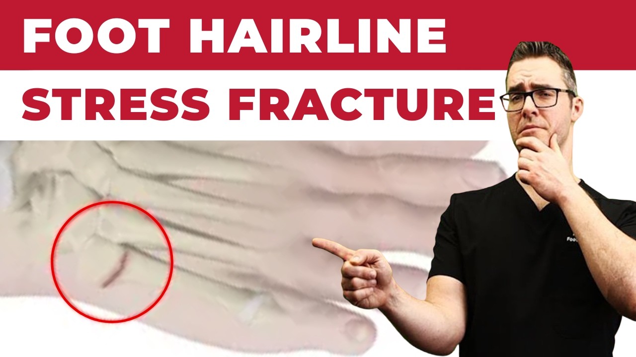
Metatarsal Stress Fractures
Stress fractures of the metatarsals — most commonly the 2nd and 3rd metatarsal shafts — result from repetitive cyclic loading exceeding the bone’s remodeling capacity. They present as progressive forefoot pain without a discrete injury event in runners, military recruits, dancers, and patients who have recently increased their activity level. Most respond to 4–6 weeks of offloading in a walking boot with activity modification. MRI is the imaging gold standard for early detection before X-ray findings develop. Bisphosphonate therapy or vitamin D/calcium supplementation may be indicated for patients with evidence of metabolic bone disease contributing to stress fracture susceptibility.
Recovery After Metatarsal Fracture Surgery
Recovery from Jones fracture intramedullary screw fixation follows a structured protocol. Weight-bearing in a boot typically begins at 1–2 weeks postoperatively. Transition to a regular shoe at 6–8 weeks when radiographic healing is confirmed. Running and sport-specific activity at 10–14 weeks. Full return to competitive sport at 3–4 months. Serial X-rays confirm progressive fracture healing and screw position throughout recovery. Bone stimulator therapy may be recommended for patients with risk factors for delayed healing (smokers, nutritional deficiencies, Zone III lesions).
Non-Union: When Bones Don’t Heal
Jones fracture non-union — failure to achieve bony healing after 3–4 months — occurs in a meaningful percentage of conservatively managed cases and occasionally after surgical fixation with inadequate screw size or position. Non-union presents as persistent pain, tenderness to palpation, and failure of X-ray progression. Treatment requires revision surgery with larger intramedullary screw fixation, curettage of the non-union site, and often autogenous bone grafting from the calcaneus or distal tibia to stimulate healing biology. Non-union of the Jones fracture is one of the most compelling reasons to pursue primary surgical fixation in active patients.
Dr. Tom's Product Recommendations
Aircast AirSelect Walker Boot
⭐ Highly Rated
Pneumatic walker boot with pre-inflated air cells for customized compression and support. Standard immobilization device for metatarsal fractures during the conservative management and post-surgical protected weightbearing phases.
Dr. Tom says: “”Wore this for my Jones fracture surgery recovery. The air bladders kept swelling down and I could actually walk comfortably to the car.””
Post-surgical metatarsal fracture immobilization and conservative metatarsal fracture management
Confirm with Dr. Biernacki whether weight-bearing is permitted in the boot for your specific fracture pattern
Disclosure: We earn a commission at no extra cost to you.
Calcium + Vitamin D3 Supplement by Nature Made
⭐ Highly Rated
Calcium citrate plus vitamin D3 supporting bone mineralization and fracture healing. Adequate calcium (1200mg/day) and vitamin D (1000–2000 IU/day) are the foundational nutritional requirements for optimal bone fracture repair and stress fracture prevention.
Dr. Tom says: “”My podiatrist told me I was vitamin D deficient and that contributed to my stress fractures. Supplementing made a measurable difference on follow-up labs.””
Bone fracture healing support and stress fracture prevention in patients with nutritional deficiencies
Check vitamin D levels with your primary care provider before supplementing — dose depends on baseline level
Disclosure: We earn a commission at no extra cost to you.
✅ Pros / Benefits
- Intramedullary screw fixation of Jones fractures provides reliable union with earlier return to activity vs. conservative treatment
- ORIF of displaced first metatarsal fractures restores forefoot weight-bearing architecture and prevents chronic metatarsalgia
- Surgical stabilization of diaphyseal stress fractures allows athletes to return to training significantly faster than conservative management
- Primary fixation prevents the significantly higher failure rates of Jones fracture non-union revision surgery
❌ Cons / Risks
- Zone I (avulsion) fractures virtually never require surgery and heal reliably conservatively — avoid over-treatment
- Return to sport after Jones fracture fixation typically takes 3–4 months even with optimal surgical management
- Screw breakage (rare) or stress risers at screw tip can cause re-fracture with premature return to high-impact activity
- Smokers and patients with vitamin D deficiency have significantly slower fracture healing regardless of treatment approach
Dr. Tom Biernacki’s Recommendation
I see Jones fractures mis-managed constantly — patients sent home in a boot for months that eventually end up needing revision surgery for non-union. In active patients, primary screw fixation is almost always the right answer. It’s a small, elegant procedure with a clear pathway back to sport. Don’t waste months hoping a Jones fracture heals on its own if you’re an active person.
— Dr. Tom Biernacki, DPM | Board-Certified Podiatric Surgeon | Balance Foot & Ankle
Frequently Asked Questions
How do I know if I have a Jones fracture vs. a regular ankle sprain?
Both involve outer foot pain from an inversion injury. A Jones fracture causes specific point tenderness at the base of the 5th metatarsal, not the lateral ankle ligaments. X-ray confirms the diagnosis. If you can feel the bone hurting under your pinky toe, get an X-ray.
Do all Jones fractures need surgery?
In truly sedentary patients with low functional demands, conservative management with a boot and strict non-weight-bearing is an option. However, for most active adults, athletes, and workers who need to return to full activity reliably, primary surgical fixation dramatically reduces non-union risk and recovery time.
How long is recovery after Jones fracture screw fixation?
Weight-bearing in a boot begins at 1–2 weeks. Transition to regular shoes at 6–8 weeks. Jogging at 10–12 weeks. Full return to sport at 3–4 months. This is significantly faster than the 3–4 months of conservative treatment that still carries meaningful non-union risk.
Can I walk on a metatarsal fracture?
It depends on the fracture. Many isolated, undisplaced metatarsal shaft fractures can bear weight in a stiff-soled boot. Jones fractures are typically kept non-weight-bearing for 1–2 weeks post-surgery. Dr. Biernacki specifies your exact weight-bearing restrictions at your postoperative visits.
Will I need the screw removed after Jones fracture surgery?
Screw removal is not routinely required. However, some patients develop prominence or hardware irritation from the screw head at the 5th metatarsal tuberosity that requires removal under local anesthesia as an office procedure. Dr. Biernacki discusses hardware removal on a case-by-case basis.
Michigan Foot Pain? See Dr. Biernacki In Person
4.9★ rated | 1,123 Reviews | 3,000+ Surgeries
Same-week appointments · Howell & Bloomfield Hills
📞 (810) 206-1402 Book Online →Frequently Asked Questions
When should I see a podiatrist?
If symptoms persist past 2 weeks, affect your normal activity, or are accompanied by red-flag symptoms (warmth, redness, swelling, inability to bear weight).
What does treatment cost?
Most diagnostic visits and conservative treatments are covered by Medicare and major insurers. Out-of-pocket costs vary by your specific plan.
How quickly can I get an appointment?
Most non-urgent cases see us within 5 business days. Urgent cases (sudden pain, possible fracture) typically same or next business day.
What is Stress fracture?
Stress fracture is a common foot/ankle condition that affects mobility and quality of life. Understanding the underlying cause is the first step in successful treatment. Our podiatrists at Balance Foot & Ankle perform a hands-on biomechanical exam, review your activity history, and use diagnostic imaging when appropriate to identify the root cause—not just treat the symptom. Many patients have been told to “rest and ice” without a deeper diagnostic workup; our approach is different.
Symptoms and warning signs
Common signs of stress fracture include pain that worsens with activity, morning stiffness, swelling, tenderness when palpated, and difficulty bearing weight. If you experience sudden severe pain, inability to walk, visible deformity, numbness or color change, contact our office the same day or visit urgent care—these can signal a more serious injury such as a fracture, tendon rupture, or vascular compromise. Diabetics with any foot wound should seek same-day care.
Conservative treatment options
Most cases of stress fracture respond to non-surgical care: structured rest, supportive footwear changes, custom orthotics, targeted stretching and strengthening protocols, anti-inflammatory medications when medically appropriate, and in-office procedures such as ultrasound-guided injections. We also offer advanced therapies including MLS laser therapy, EPAT/shockwave, regenerative injections, and image-guided procedures. Treatment is sequenced from least invasive to most invasive, and we explain the rationale at every step.
When is surgery considered?
Surgery is reserved for cases that fail 3-6 months of well-structured conservative care, when there is structural pathology (severe deformity, complete tear, advanced arthritis), or when imaging shows damage that will not heal without intervention. Our surgeons have performed 3,000+ foot and ankle procedures and prioritize minimally-invasive techniques whenever appropriate. We discuss recovery timelines, return-to-activity milestones, and realistic outcome expectations before any procedure is scheduled.
Recovery timeline and prevention
Recovery from stress fracture varies based on severity and chosen treatment path. Conservative cases often improve within 4-8 weeks with consistent adherence to the protocol. Post-procedural recovery may range from a few days (in-office procedures) to several months (reconstructive surgery). Long-term prevention involves footwear assessment, activity modification, structured strengthening, and regular check-ins with your podiatrist if you have a history of recurrence. We provide written home-exercise plans and digital follow-up support.
Ready to feel better?
Same-week appointments available in Howell and Bloomfield Hills, Michigan.
Book Your VisitOrthoInfo – AAOS: Metatarsal Fractures
Ready to Get Relief?
Same-day appointments available in Howell & Bloomfield Hills, MI
4.9★ | 1,123 Reviews | 3,000+ Surgeries
Or call: (810) 206-1402
Dr. Tom Biernacki, DPM is a board-certified foot & ankle surgeon (ABFAS & ABPM) at Balance Foot & Ankle Specialists in Southeast Michigan. With over a decade of clinical experience, he specializes in heel pain, bunions, diabetic foot care, sports injuries, and minimally invasive surgery. Dr. Biernacki is a member of the APMA and ACFAS, and his patient education content on MichiganFootDoctors.com and YouTube has made him one of the most-followed foot & ankle educators on YouTube.