
Quick answer: Dark Streak Toenail Subungual Melanoma Michigan is a common nail condition with multiple causes including trauma, fungal infection, biomechanical pressure, and underlying medical conditions. Treatment depends on the cause: trauma resolves as the nail grows out (6-12 months), fungus needs antifungal therapy, and biomechanical issues need shoe and orthotic correction. Call (810) 206-1402.
Medically reviewed by Dr. Tom Biernacki, DPM · Board-Certified Podiatric Surgeon · Last reviewed: April 2026 · Editorial Policy
The most important clinical decision with Dark Streak Toenail Subungual Melanoma Michigan isn’t which treatment to start with — it’s identifying the correct subtype. That changes everything. Call (810) 206-1402.
Quick Answer
Dark Streak in Toenail: Subungual Melanoma vs Hematoma Michi relates to toenail conditions — typically caused by fungal infection or trauma. Most patients improve in 6-12 months for nail regrowth with conservative care. Same-week appointments in Howell + Bloomfield Hills: (810) 206-1402.
Medically reviewed by Dr. Tom Biernacki, DPM — Board-Certified Podiatric Surgeon — Balance Foot & Ankle, Howell & Bloomfield Hills, MI. Last updated April 2026.
▶ Watch

Medically Reviewed by Dr. Tom Biernacki, DPM — Board-Certified Podiatrist, Balance Foot & Ankle Specialists, Michigan. Last updated April 2026.
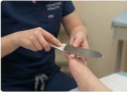
A dark brown or black streak running lengthwise under a toenail must always be evaluated by a podiatrist or dermatologist — it can represent a benign blood collection (subungual hematoma) or one of the most dangerous skin cancers, subungual melanoma. Dr. Tom Biernacki, DPM, at Balance Foot & Ankle in Howell and Bloomfield Hills, Michigan, evaluates all nail pigmentation changes and performs nail biopsy when indicated.
Quick Answer: When Is a Dark Toenail Streak Dangerous?
A dark streak (melanonychia) under a toenail is concerning for subungual melanoma when: it appears without any recalled injury; it is getting wider over weeks or months; it has irregular borders or multiple colors; it extends from the nail onto the surrounding skin (Hutchinson’s sign); or it occurs in a patient over age 50. A dark area that appeared after a clear injury (dropped object, stubbed toe, running descent) and is moving distally with nail growth over 3–4 months is almost always a subungual hematoma (benign). Any ambiguous case requires same-day podiatric or dermatologic evaluation — subungual melanoma delays in diagnosis average 2 years nationally, and stage at diagnosis determines survival.
What Is Melanonychia (Dark Nail Streaks)?
Melanonychia refers to brown or black pigmentation of the nail plate, arising from melanin deposits in the nail matrix or nail bed. It is classified as melanonychia striata (a longitudinal band, the concerning type) or diffuse melanonychia (the entire nail, more commonly benign). Longitudinal melanonychia has many causes: melanocytic activation (common and benign, especially in darker-skinned individuals), melanocytic nevus (a mole in the nail matrix), subungual melanoma (the diagnosis not to miss), nail trauma with hematoma, medications (hydroxychloroquine, AZT, some chemotherapy agents), and systemic conditions (Addison’s disease, HIV). In Fitzpatrick skin types V–VI (darker skin tones), longitudinal melanonychia is often benign melanocyte activation — but it still requires evaluation to exclude melanoma.
Subungual Hematoma: The Common Benign Cause
Subungual hematoma — blood pooling under the nail from trauma — is by far the most common cause of a dark toenail. It is uniformly dark (blue-black), appears within hours to days of a specific traumatic event, and moves distally (toward the nail tip) at the rate of nail growth — approximately 1–2mm per month for toenails. A hematoma that first appeared 3 months ago should be 3–6mm from its original position and well on its way to the nail tip. Painful subungual hematomas occupying more than 50% of the nail plate benefit from trephination (small hole drilled through the nail to decompress the blood) — this provides immediate relief and prevents nail loss from pressure necrosis. Painless hematomas are watched until they grow out.
Subungual Melanoma: The Diagnosis Not to Miss
Subungual melanoma is a rare but deadly form of melanoma arising from melanocytes in the nail matrix or nail bed. It accounts for approximately 1–3% of all melanomas in the general US population but represents a disproportionate percentage of melanomas in darker-skinned individuals (up to 40% in Black patients) — where it is particularly likely to be misdiagnosed as a benign finding. The big toe and thumb are the most commonly affected digits. Warning features (ABCD + E criteria for nail melanoma): Age (peak 5th–7th decade); Brown/black color band wider than 3mm; Change in size, shape, or color over time; Digit — the great toe, thumb, or index finger; and Extension of pigment to the surrounding skin (Hutchinson’s sign) — any periungual pigmentation is a red flag requiring urgent biopsy. 5-year survival for early-stage (in situ) subungual melanoma is >95%. 5-year survival for late-stage (with distant metastasis) is approximately 15%.
The ABCDEF Rule for Nail Pigmentation
The ABCDEF mnemonic assists clinical triage of nail pigmentation: Age and race — peak incidence age 50–70; higher risk in darker skin tones. Band — brown/black band of width ≥3mm, with irregular or blurry borders. Change — rapid increase in width or change in pigmentation pattern. Digit — big toe or thumb most commonly; single digit involvement. Extension — Hutchinson’s sign (pigment spreading to periungual skin). Family or personal history — prior melanoma or family history of melanoma. Three or more of these criteria warrant urgent nail matrix biopsy.
Nail Biopsy: The Definitive Diagnostic Procedure
When clinical evaluation cannot definitively exclude melanoma, nail matrix biopsy is indicated. The procedure is performed under digital block anesthesia — the nail plate is partially or fully avulsed to expose the nail matrix, and a 3mm punch biopsy is taken from the pigmented matrix area. The specimen is sent for dermatopathologic analysis with melanocytic immunohistochemistry (S100, Melan-A, HMB-45). Results typically return in 7–10 business days. Biopsy-related nail changes (ridge, thinning) are usually transient but permanent changes can occur in 5–10% of cases. The risk of permanent nail change from biopsy is substantially outweighed by the risk of missing a melanoma. In our clinic, we refer all suspected melanonychia cases requiring biopsy to dermatology for joint management and ensure there is no delay in the referral process.
Differential Diagnosis: Other Causes of Nail Discoloration
Other nail color changes that are frequently concerning to patients but are generally benign: White nails (leukonychia) — usually nail plate trauma (true leukonychia, in the nail plate itself) or onychomycosis (white surface changes); Yellow-brown thickened nails — onychomycosis (toenail fungus) confirmed by culture; Green discoloration — pseudomonas bacterial infection, typically under a lifted nail plate; Yellow dystrophic nails — yellow nail syndrome (lymphedema, pleural effusions — systemic evaluation needed); Splinter hemorrhages (tiny dark lines, not a band) — most commonly trauma, also seen in bacterial endocarditis; Red streak under a nail — glomus tumor (rare, highly pressure-tender point, benign but requires excision). Any nail change that has been present more than 4–6 weeks and is not clearly explained by an identified traumatic event should be evaluated by a podiatrist or dermatologist.
Hutchinson’s Sign: The Critical Red Flag
Hutchinson’s sign is the extension of brown or black pigmentation from under the nail onto the periungual skin (the cuticle, the skin folds beside the nail, or the fingertip/toe tip skin). It is pathognomonic (essentially diagnostic) for subungual melanoma when present — the melanoma has invaded beyond the nail matrix into the surrounding dermis. Pseudo-Hutchinson’s sign can occur with benign conditions (ethnic nail pigmentation, benign nevus) where the pigment appears to extend but is actually visible through the translucent cuticle without true skin invasion. True Hutchinson’s sign requires urgent biopsy. Any patient with periungual pigmentation surrounding a dark nail streak should be seen same-day or within 1 week at maximum. Call (810) 206-1402.
Book a Nail Pigmentation Evaluation in Michigan
📧 Get Dr. Tom’s Free Lab Test Guide
Discover the 5 lab tests every person over 35 should ask their doctor about — explained in plain English by a board-certified physician.
Dr. Tom Biernacki, DPM, evaluates all nail pigmentation changes and coordinates nail biopsy and dermatology referral when indicated at Balance Foot & Ankle in Howell (4330 E Grand River Ave, Howell MI 48843) and Bloomfield Hills (43494 Woodward Ave #208, Bloomfield Hills MI 48302). Urgent appointments available for Hutchinson’s sign or rapidly changing nail bands. Call (810) 206-1402 or book online →.
Medically reviewed by Dr. Tom Biernacki, DPM — podiatric physician and surgeon, Howell and Bloomfield Hills, Michigan.
Dr. Tom’s Recommended Products for Toenail Problems
📍 Located in Michigan?
Our board-certified podiatrists treat this condition at two convenient locations. Same-day appointments often available.
These are products I personally use and recommend to my patients at Balance Foot & Ankle.
- Nail Tek Intensive Therapy II — Restores brittle, discolored, and damaged nails — base coat that strengthens nail plate after trauma or fungal damage
- Kerasal Fungal Nail Renewal — Visibly improves nail appearance for both fungal and non-fungal discoloration within 8 weeks
- Professional Toenail Clipper — Harperton Nail Nipper — Stainless steel curved jaw cuts thick or ingrown toenails cleanly — prevents nail trauma and subungual hematoma
Affiliate disclosure: As an Amazon Associate, Balance Foot & Ankle earns from qualifying purchases. We only recommend products we trust for our own patients.
Join 950,000+ Learning About Foot Health
Dr. Tom shares honest medical advice, supplement reviews, and treatment guides you won’t find anywhere else.
Subscribe on YouTube →🔗 Related Care & Resources
Treated by Dr. Tom Biernacki DPM — Board-certified podiatric surgeon at Balance Foot & Ankle in Howell & Bloomfield Hills, MI.
Schedule an Appointment → or call (810) 206-1402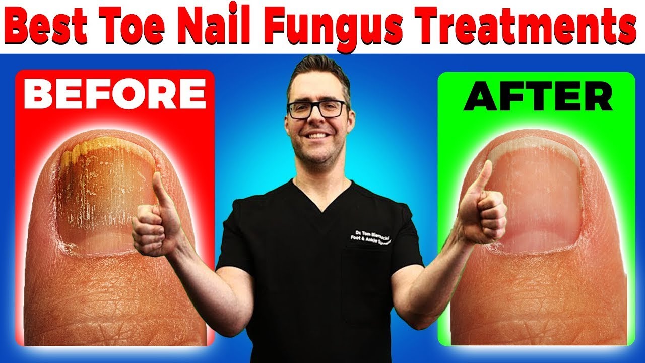
Insurance Accepted
BCBS · Medicare · Aetna · Cigna · United Healthcare · HAP · Priority Health · Humana · View All →
Howell Office
4330 E Grand River Ave
Howell, MI 48843
Get Directions →
Bloomfield Hills Office
43494 Woodward Ave, #208
Bloomfield Hills, MI 48302
Get Directions →
Your Board-Certified Podiatrists


Ready to Get Back on Your Feet?
Same-week appointments available at both locations.
Book Your AppointmentWatch: Dr. Tom explains

Podiatrist-recommended products
As an Amazon Associate, Dr. Tom earns from qualifying purchases.
Reduce nail trauma that mimics pigmented lesions.
View on Amazon →Topical for tender toenail bed after evaluation.
View on Amazon →Ice the toe after biopsy or procedure.
View on Amazon →Reduce forward slide in shoes that drives nail trauma.
View on Amazon →Related resources
Ready to solve this? Book today.
Same-week appointments · Howell & Bloomfield Hills · 4.9★ (1,123+ reviews)
☎ (810) 206-1402Book Online →More Podiatrist-Recommended Foot Health Essentials
Hoka Clifton 10


![Tea Tree Oil Toenail Fungus Home Treatment [Doctor Cure!]](https://www.michiganfootdoctors.com/wp-content/cache/flying-press/a434e04a532dc4826563b11aa0541dd4.jpg)
Watch: Tea Tree Oil Toenail Fungus Home Treatment [Doctor Cure!] — MichiganFootDoctors YouTube
Max-cushion everyday shoe — podiatrist favorite for walking and running.

OOFOS Recovery Slide

Impact-absorbing recovery sandal — wear after long days on your feet.
As an Amazon Associate, Balance Foot & Ankle earns from qualifying purchases. Product recommendations are based on clinical experience; prices and availability shown above update live from Amazon.
When to See a Podiatrist
If foot or ankle pain has been bothering you for more than a few weeks, home care alone may not be enough. Balance Foot & Ankle offers same-week appointments at our Howell and Bloomfield Hills clinics — no referral needed in most cases. Bring your current shoes and a short list of symptoms and we’ll build you a treatment plan in one visit.
Call Balance Foot & Ankle: (810) 206-1402 · Book online · Offices in Howell & Bloomfield Hills
Pros & Cons of Conservative Care for toenail conditions
Advantages
- ✓ Most cases resolve at home
- ✓ Same-week appointments available
- ✓ Permanent fix exists
Considerations
- ✗ Recurrence common without prevention
- ✗ Diabetics need professional care
Dr. Tom’s Recommended Products for toenail conditions
Affiliate disclosure: As an Amazon Associate, Balance Foot & Ankle earns from qualifying purchases. We only recommend products we use with patients.
Tolcylen Antifungal Solution Dr. Tom’s Pick
Best for: Most effective topical for fungus
Ready to Get Back on Your Feet?
Same-day appointments in Howell + Bloomfield Hills. Most insurance accepted. Dr. Tom Biernacki, DPM & team.
Book Today — Same-Day Appointments Available
Call Now: (810) 206-1402
About Your Care Team at Balance Foot & Ankle
Dr. Tom Biernacki, DPM · Board-Certified Foot & Ankle Surgeon. Specializes in conservative-first care, minimally invasive bunion surgery, and complex reconstruction.
Dr. Carl Jay, DPM · Accepting new patients. Specializes in sports medicine, athletic injuries, and routine podiatric care.
Dr. Daria Gutkin, DPM, AACFAS · Accepting new patients. Specializes in surgical reconstruction and pediatric podiatry.
Locations: 4330 E Grand River Ave, Howell, MI 48843 · 43494 Woodward Ave Suite 208, Bloomfield Hills, MI 48302
Hours: Mon–Fri 8:00 AM – 5:00 PM · (810) 206-1402
In-Office Treatment at Balance Foot & Ankle
If home treatment isn’t providing relief for your toenail issues, our podiatry team at Balance Foot & Ankle can help with same-day evaluations and advanced in-office care.
Same-day appointments available. (810) 206-1402
Frequently Asked Questions
How long does it take a toenail to grow back?
6-12 months for a full big toenail. Smaller toenails 4-6 months. Speed varies with age, circulation, and nutrition.
Will this affect other nails?
Trauma affects only the injured nail. Fungal infection can spread without treatment. Systemic causes affect multiple nails simultaneously.
Should I cover the nail or leave it open?
Cover with a breathable bandage during work or activity. Leave open at night for healing. Keep dry and clean.
Our podiatrists treat the underlying cause, not just the symptom. Same-week appointments at our Howell and Bloomfield Hills, Michigan offices.
Ready for Expert Care?
Same-day appointments in Howell & Bloomfield Hills, MI.
4.9★ | 1,123 Reviews | 3,000+ Surgeries
Or call: (810) 206-1402
Dr. Tom Biernacki, DPM is a board-certified foot & ankle surgeon (ABFAS & ABPM) at Balance Foot & Ankle Specialists in Southeast Michigan. With over a decade of clinical experience, he specializes in heel pain, bunions, diabetic foot care, sports injuries, and minimally invasive surgery. Dr. Biernacki is a member of the APMA and ACFAS, and his patient education content on MichiganFootDoctors.com and YouTube has made him one of the most-followed foot & ankle educators on YouTube.