

The most important step with peroneus brevis tendon pain is telling simple tendinitis apart from a partial split tear of the tendon or chronic ankle instability driving the irritation — because a tear or an unstable ankle needs a different plan. That’s why we check the tendon and ankle stability before settling on treatment. We see this at our Howell and Bloomfield Hills offices. Call (810) 206-1402.
Foot or ankle pain that isn’t getting better? A podiatrist can diagnose it in one visit — pick a real appointment time online, 24/7: Howell or Bloomfield Township. Takes about a minute, 24/7.
Peroneus brevis tendonitis hurts on the outside of the ankle near the fifth metatarsal base — and it often gets misdiagnosed as a chronic ankle sprain. The fix targets the tendon directly.
You’re in the right place. Dr. Tom Biernacki, DPM, FACFAS — board-certified foot & ankle surgeon with 3,000+ surgeries — explains exactly what peroneus brevis tendonitis means and what works. Call (810) 206-1402 for same-day appointment at Howell or Bloomfield Township.
Medically reviewed by Tom Biernacki, DPM · Board-Certified Foot & Ankle Surgeon · Balance Foot & Ankle PLLC, Howell & Bloomfield Township, MI · Last updated May 6, 2026 · This article reflects current clinical guidelines and 15+ years of evaluating peroneus brevis tendinopathy and split tears in runners, dancers, and chronic ankle-instability patients.
In This Article
- Quick Answer
- What Is Peroneus Brevis Tendonitis?
- Tendon Anatomy
- Symptoms
- Causes & Risk Factors
- Peroneus Brevis Split Tear
- Diagnosis
- Differential Diagnosis
- Home Treatment
- In-Office Treatment
- Surgical Options
- Footwear & Activity
- When to See a Podiatrist
- Most Common Mistake
- Frequently Asked Questions
- The Bottom Line
- See a Michigan Peroneal Tendon Specialist
- Sources
- Frequently Asked Questions
- What is Tendonitis?
- Symptoms and warning signs
- Conservative treatment options
- When is surgery considered?
- Recovery timeline and prevention
- Tendonitis — Frequently Asked Questions
- Related Conditions
- Frequently Asked Questions
Quick Answer
📋 Medically Reviewed by Dr. Tom Biernacki, DPM
Board-Certified Podiatrist · Balance Foot & Ankle · Last updated: 2026
Peroneus brevis tendonitis is inflammation, degeneration, or longitudinal split tearing of the peroneus brevis tendon as it runs behind the lateral ankle bone (fibula) and attaches to the base of the fifth metatarsal. It causes pain on the outside of the ankle and along the lateral foot, especially with eversion against resistance or after lateral ankle sprains. Treatment is conservative for 6–12 weeks (rest, bracing, custom orthotics with lateral posting, eccentric strengthening), with surgical debridement or tubularization for split tears that fail conservative care.
If you’ve had a string of “ankle sprains” that never quite settle down — and now you have persistent pain right behind your outer ankle bone that gets worse on uneven ground or when you push off to the outside of your foot — there’s an excellent chance you’re dealing with peroneus brevis tendonitis or a split tear of the brevis tendon. Patients tell us, “It feels like my ankle is going to give out,” or, “I can press right behind the ankle bone and feel a thick, painful lump.” In our clinic, peroneus brevis pathology is the most commonly missed cause of chronic lateral ankle pain, and patients are often told for years they have “ankle arthritis” before the tendon is properly imaged.
What Is Peroneus Brevis Tendonitis?
Peroneus brevis tendonitis is inflammation or degenerative tendinosis of the peroneus brevis tendon as it courses behind the lateral malleolus (outer ankle bone), under the peroneal retinaculum, and along the lateral border of the foot to its insertion on the base of the fifth metatarsal. The condition exists on a continuum: acute tenosynovitis (inflammation of the sheath), chronic tendinosis (degeneration of the tendon itself), and frank longitudinal split tearing — by far the most common structural injury of the brevis. Roughly 70–80% of peroneal tendon split tears involve the brevis specifically, which makes this pathology far more common than peroneus longus tendinopathy.
In our clinic, the typical patient is a 40–60-year-old runner, hiker, or basketball player with a history of multiple lateral ankle sprains, a varus hindfoot (slight inward heel posture), or chronic ankle instability. The pain pattern is distinctive: tender directly behind the lateral malleolus and along the peroneal groove, worse with eversion against resistance, worse on uneven terrain, and often accompanied by a palpable thickening or “fullness” along the tendon. Without correct diagnosis, these patients cycle through ankle braces, NSAIDs, and physical therapy for years without addressing the actual problem.
Tendon Anatomy
The peroneus brevis is one of two peroneal tendons that share the retromalleolar groove behind the fibula. The brevis runs deep and anterior, hugging the back of the fibula, while the longus runs superficial and posterior. The brevis muscle originates from the lower two-thirds of the lateral fibula, becomes a tendon at the level of the ankle, and inserts on the lateral tubercle of the fifth metatarsal base. Its primary action is foot eversion (turning the sole outward), and it is a key dynamic stabilizer of the lateral ankle along with the lateral collateral ligaments.
The brevis lies in a tight space against the fibula. Anatomic features that predispose to tearing include a shallow retromalleolar groove, a sharp peroneal tubercle on the calcaneus, a low-lying peroneus brevis muscle belly, the presence of an accessory peroneus quartus, and varus hindfoot alignment. Any of these increases shear and friction at the tendon — the brevis is squeezed against the fibula by the longus on top, and over years this produces the characteristic longitudinal split tear seen on MRI.
Most people search for this condition simply as peroneus brevis pain — an ache or sharp pain along the outer edge of the foot and just behind the outer ankle bone. That peroneus brevis tendon pain usually worsens with walking, running, or rolling the ankle outward, and is frequently mistaken for an ordinary ankle sprain. Here is exactly what that pain feels like and what it means.
Symptoms
Peroneus brevis tendonitis presents with pain on the lateral ankle and outer foot, especially behind the fibula. Pain is sharp with activity (running, cutting, hiking on uneven ground) and dull or aching at rest. Most patients also describe a feeling of lateral ankle instability — the sense that the ankle is going to “roll” again — even when ligamentous instability tests are negative, because a weakened brevis cannot dynamically stabilize the ankle.
- Pain behind the lateral malleolus: sharp local tenderness directly behind the outer ankle bone.
- Pain along the lateral foot: follows the tendon path toward the base of the 5th metatarsal.
- Pain with resisted eversion: turning the sole outward against resistance reproduces the pain.
- Lateral ankle instability: a feeling the ankle will “give out” on uneven terrain, even with intact ligaments.
- Swelling and fullness: visible swelling behind the fibula, sometimes a palpable “string” of the thickened tendon.
- Stiffness: first-step morning pain along the lateral ankle.
- Crepitus: grinding or clicking with ankle motion in advanced tendinosis.
Causes & Risk Factors
Peroneus brevis tendinopathy is an overuse injury driven by repetitive shear and load against the fibula. Most cases develop in patients with predisposing anatomy or biomechanics — anyone with chronic lateral ankle instability, a high-arch (cavus) foot with varus hindfoot, or a sharp peroneal tubercle is at substantially elevated risk. Acute tears do happen, usually with a forced inversion injury (a severe ankle sprain), but most cases we treat are chronic degenerative tears that built up over months to years.
- Chronic lateral ankle instability — recurrent sprains place repeated stress on the brevis.
- Cavovarus (high-arch + varus heel) foot — directs more load to the lateral column.
- Sharp peroneal tubercle on the calcaneus — saws into the brevis with motion.
- Shallow retromalleolar groove — predisposes to peroneal subluxation and tear.
- Low-lying brevis muscle belly — fills the retromalleolar groove and increases pressure.
- Repetitive loading sports: running, hiking, basketball, dance, soccer, tennis.
- Inadequate footwear — minimal lateral support, worn lateral heel.
- Inflammatory arthritis — RA, psoriatic arthritis can drive tenosynovitis.
Key takeaway: Chronic lateral ankle pain after multiple sprains is almost never “just arthritis.” Roughly 70% of peroneal split tears are in the brevis, and the diagnosis is missed for years in patients who never get an MRI of the lateral ankle.
Peroneus Brevis Split Tear
A peroneus brevis split tear is a longitudinal cleavage in the substance of the tendon — typically 1–4 cm long — caused by repeated shear between the brevis and the longus against the fibula. On MRI cross-section the tendon takes on a “C-shape” or “boomerang” configuration as the longus drops into the split. Split tears are classified by Sobel: Grade I (faint horizontal line within the tendon), Grade II (partial-thickness split < 1 cm), Grade III (full-thickness split > 1 cm), Grade IV (multiple splits and complete disruption).
- Grade I: faint splaying of fibers, no measurable defect.
- Grade II: partial-thickness longitudinal split < 1 cm.
- Grade III: full-thickness split 1–2 cm or longer; tendon shape distorted.
- Grade IV: multiple splits and severe degeneration; surgical reconstruction usually required.
Diagnosis
Diagnosis is clinical with confirmation by imaging. We palpate directly behind the fibula and along the tendon to the 5th metatarsal base, looking for sharp local tenderness and a palpable thickened tendon. Resisted eversion against the examiner’s hand — done with the foot in plantarflexion — reproduces pain in essentially every case of peroneus brevis tendinopathy. We also evaluate the entire kinetic chain because the underlying biomechanics (varus hindfoot, ankle instability, peroneal tubercle prominence) drive recurrence and need to be addressed in treatment.
- History: recurrent ankle sprains, chronic lateral ankle pain, “instability” sensation.
- Inspection: swelling behind the fibula, varus hindfoot, callus pattern under the lateral foot.
- Palpation: sharp tenderness directly behind the lateral malleolus and along the tendon.
- Resisted eversion test: pain with the patient pushing the foot outward against resistance — most sensitive bedside finding.
- Single-leg heel raise: assesses tendon function and pain reproduction.
- Anterior drawer + talar tilt: rule out coexisting lateral ligament instability.
- Weight-bearing X-rays: evaluate hindfoot alignment, peroneal tubercle morphology, and rule out ankle arthritis or 5th metatarsal avulsion fracture.
- MRI: gold standard. Shows split tear morphology, tendinosis, and tenosynovitis.
- Diagnostic ultrasound: dynamic assessment of subluxation and real-time visualization of split tears.
Differential Diagnosis
Several conditions mimic peroneus brevis tendonitis. The most common diagnostic confusion is with a chronic lateral ankle sprain, an osteochondral lesion of the talus, or a base-of-fifth-metatarsal stress fracture (Jones fracture). Imaging — particularly MRI — is what distinguishes them, because the physical exam findings overlap substantially.
- Chronic lateral ankle ligament tear (ATFL/CFL): positive anterior drawer or talar tilt; coexists with brevis pathology in >30% of cases.
- Peroneus longus tendinopathy: pain extends further distally under the cuboid; less common than brevis.
- Peroneal subluxation/dislocation: palpable snapping of the tendon over the fibula.
- Osteochondral lesion of the talus (OLT): deep ankle pain, mechanical catching, MRI shows cartilage defect.
- 5th metatarsal fracture (Jones / avulsion): point tenderness on the bone, X-ray confirms.
- Lateral ankle impingement: pain with end-range plantarflexion-inversion; arthroscopic finding.
- Sinus tarsi syndrome: pain in the soft spot between the talus and calcaneus.
- Sural nerve entrapment: burning or shooting pain along the lateral foot.
Home Treatment
Acute and mild peroneus brevis tendonitis respond well to disciplined home care over 4–8 weeks. The goals are to reduce inflammation, offload the lateral column, and address any underlying ankle instability. Two interventions matter most: a structured eccentric eversion program, and a quality OTC orthotic with lateral posting to reduce shear on the tendon.
- Relative rest 7–14 days: stop aggravating sports; substitute swimming, cycling, or pool running.
- Ice the lateral ankle 15–20 minutes 3–4× daily for the first 2 weeks.
- NSAIDs as tolerated — ibuprofen 600 mg TID with food for 7–14 days, or topical diclofenac 1%.
- Compression sleeve for daily wear during activity.
- Lace-up ankle brace when returning to sport — Aircast A60 or ASO are excellent.
- Apply Doctor Hoy’s Natural Pain Relief Gel 2–3× daily along the lateral ankle for menthol and arnica anti-inflammatory effect. (Affiliate)
- Replace your insole with PowerStep Pinnacle Maxx for arch support and a small lateral wedge to offload the brevis. (Affiliate)
- Eccentric eversion exercises with a resistance band — 3 sets of 15, 2× daily — start at week 2.
Affiliate disclosure: As an Amazon Associate, Balance Foot & Ankle PLLC earns from qualifying purchases. We only recommend products we use in clinic.
In-Office Treatment
When home care plateaus or imaging confirms a Grade II+ split tear, we escalate to in-office interventions targeting both the tendon and the underlying mechanics. The cornerstone is custom orthotics with a lateral hindfoot post — these mechanically reduce shear on the brevis and address the cavovarus alignment that drives most cases. We do not use cortisone injection inside the brevis tendon sheath because of accelerated rupture risk.
- Custom prescription orthotics with lateral hindfoot post and 5th metatarsal cutout.
- CAM walker boot for 4–6 weeks with severe Grade II–III tears or significant tenosynovitis.
- Physical therapy: eccentric peroneal strengthening, proprioception, ankle stabilization protocols.
- PRP injection (platelet-rich plasma) — emerging evidence for tendinosis without complete disruption.
- Extracorporeal shock wave therapy (ESWT): useful for chronic recalcitrant tendinosis.
- Avoid intratendinous corticosteroid: case series document accelerated rupture after intratendinous injection. Peritendinous injection only with imaging guidance, if used at all.
Surgical Options
Surgical repair is indicated for Grade III–IV split tears, persistent Grade II tears that fail 3–6 months of conservative care, or any tear with concurrent peroneal subluxation. The procedure addresses both the tendon and the underlying mechanics — a brevis repair without correcting peroneal tubercle morphology, varus heel, or ankle instability has a high recurrence rate. We typically combine debridement and tubularization with peroneal tubercle planing and lateral ligament reconstruction when appropriate.
- Tendon debridement and tubularization: the workhorse procedure. Damaged fibers are excised and the tendon is reformed with running suture into a tubular shape.
- Peroneal tubercle planing: shaves the prominent calcaneal tubercle to reduce ongoing shear.
- Retromalleolar groove deepening: deepens the fibular groove to prevent subluxation and reduce shear.
- Lateral ligament reconstruction (Brostrom): performed concurrently when ATFL/CFL is also incompetent.
- Tendon transfer (FDL or longus-to-brevis): reserved for irreparable Grade IV tears.
- Calcaneal osteotomy: for cavovarus alignment that is the underlying driver — addresses root cause and reduces recurrence.
Footwear & Activity
Footwear is treatment for peroneal tendon pathology. Look for a shoe with a firm heel counter, motion-control or stability midsole, and lateral flare in the heel to prevent late-stage inversion. Worn lateral heels on your existing shoes are a red flag — replace them. Avoid minimalist shoes, ballet flats, and any shoe that tilts the foot toward inversion.
- Stability or motion-control running shoes with a firm heel counter (Brooks Adrenaline GTS, Asics Kayano, Saucony Tempus).
- Hiking boots with ankle support (mid- or high-cut) for trail use.
- Avoid: minimalist shoes, narrow-base sneakers, worn-out running shoes, and high-stack racing shoes.
- Activity: swap running and hiking for swimming, cycling, or elliptical for 6–8 weeks during acute flares.
When to See a Podiatrist
See a podiatrist if you have:
- Lateral ankle pain lasting more than 4 weeks despite rest and OTC bracing
- A history of multiple ankle sprains and persistent feeling of instability
- Pain with eversion against resistance
- A palpable thick or tender “rope” behind the lateral malleolus
- A snapping or popping sensation behind the ankle bone (peroneal subluxation)
- High-arch foot with chronic lateral ankle pain
Same-day evaluation if you have:
- Sudden severe pain with a popping sensation after an inversion injury (acute rupture or fracture)
- Inability to walk or bear weight after an ankle injury
- Visible deformity or open wound
- Numbness or tingling along the lateral foot (sural nerve injury)
Most Common Mistake
The most common mistake we see is treating peroneus brevis tendinopathy as a simple “ankle sprain that won’t heal” for months or years without imaging. These patients cycle through ankle braces, NSAIDs, and physical therapy without ever getting an MRI of the lateral ankle, and the underlying split tear progresses from Grade II to Grade IV over time. If lateral ankle pain persists more than 6–8 weeks despite reasonable conservative care — or if you have recurrent ankle sprains — push for an MRI specifically targeting the peroneal tendons. Catching the tear at Grade II is the single best determinant of conservative-care success; a Grade IV tear almost always needs surgery.
Frequently Asked Questions
Can a peroneus brevis tear heal on its own?
Grade I tendinosis and small Grade II split tears can resolve symptomatically with disciplined conservative care over 8–12 weeks (relative rest, custom orthotics with lateral posting, eccentric strengthening, and bracing). However, the tear itself rarely “heals” structurally — the brevis has limited blood supply at the retromalleolar groove. The goal of conservative care is to control symptoms and stop progression. Grade III–IV tears almost always require surgical repair.
How long does peroneal tendon surgery recovery take?
After tendon debridement and tubularization, most patients are non-weight-bearing in a CAM boot for 2–4 weeks, transition to a walking boot at 4–6 weeks, return to athletic shoes at 8–10 weeks, and resume running at 4–6 months. Adding peroneal tubercle planing or Brostrom does not significantly extend recovery; calcaneal osteotomy adds 4–6 weeks to the timeline.
What is the difference between peroneus brevis and peroneus longus tendonitis?
The brevis runs deep and inserts on the base of the 5th metatarsal — pain stays around the lateral ankle and outer foot. The longus runs superficial, wraps under the cuboid, and inserts on the base of the 1st metatarsal — pain extends further along the bottom of the foot. Both can develop tenosynovitis, tendinosis, and split tears, but the brevis is involved in 70–80% of structural tears.
Can a cortisone shot rupture my peroneus brevis tendon?
Yes — intratendinous corticosteroid injection has been associated with accelerated tendon rupture in case series. We avoid intratendinous injection of the brevis. Peritendinous (around the tendon, inside the sheath) injection under ultrasound guidance is sometimes used carefully for tenosynovitis, but PRP and physical therapy are usually preferred over steroid for tendinopathy.
What shoes are best for peroneus brevis tendonitis?
Choose stability or motion-control running shoes with a firm heel counter and a wide lateral base — Brooks Adrenaline GTS, Asics Kayano, Saucony Tempus, or Hoka Arahi. For hiking, mid- or high-cut boots provide additional ankle support. Avoid minimalist shoes, narrow-base sneakers, and any shoe with worn lateral heels.
Will my ankle ever feel stable again?
Yes, in most cases. Restoring brevis function with eccentric strengthening, addressing cavovarus alignment with custom orthotics, and reconstructing incompetent lateral ligaments when needed reliably restores stability. Patients with Grade IV tears or untreated cavovarus deformities may have residual instability, but the majority of patients we treat return to full activity without bracing within 4–6 months.
The Bottom Line
Peroneus brevis tendonitis and longitudinal split tears are the most common cause of chronic lateral ankle pain after recurrent ankle sprains, and they are routinely missed for years without targeted MRI. Conservative care (relative rest, custom orthotics with lateral hindfoot posting, eccentric eversion strengthening, ankle bracing) resolves most Grade I–II cases over 8–12 weeks. Grade III–IV split tears require surgical debridement and tubularization, often combined with peroneal tubercle planing, retromalleolar groove deepening, lateral ligament reconstruction, and calcaneal osteotomy when cavovarus alignment is the driver. If you have lateral ankle pain that hasn’t quit after multiple sprains, push for an MRI of the lateral ankle.
See a Michigan Peroneal Tendon Specialist
Tom Biernacki, DPM & the Balance Foot & Ankle team have 15+ years of experience evaluating and repairing peroneal tendon pathology and chronic lateral ankle instability across Howell and Bloomfield Township, MI. Same-week appointments available.
📞 Call: (810) 206-1402
Sources
- Sobel M, Geppert MJ, Olson EJ, Bohne WH, Arnoczky SP. The dynamics of peroneus brevis tendon splits: a proposed mechanism, technique of diagnosis, and classification of injury. Foot Ankle. 1992;13(7):413-422.
- Heckman DS, Reddy S, Pedowitz D, Wapner KL, Parekh SG. Operative treatment for peroneal tendon disorders. J Bone Joint Surg Am. 2008;90(2):404-418.
- Roster B, Michelier P, Giza E. Peroneal tendon disorders. Clin Sports Med. 2015;34(4):625-641.
- Selmani E, Gjata V, Gjika E. Current concepts review: peroneal tendon disorders. Foot Ankle Int. 2006;27(3):221-228.
- Vega J, Golanó P, Batista JP, Malagelada F, Pellegrino A. Peroneal tendon tears: surgical treatment and clinical outcome. Knee Surg Sports Traumatol Arthrosc. 2017;25(6):1909-1914.
Frequently Asked Questions
How long does peroneus brevis tendonitis take to heal?
Mild peroneus brevis tendonitis typically resolves in 4–6 weeks with rest, modified footwear, and targeted exercises. A partial split tear takes 8–12 weeks of conservative care. A full longitudinal split requiring surgery has a 4–6 month return-to-activity timeline.
What is the difference between peroneus brevis tendonitis and a peroneus brevis split tear?
Tendonitis is inflammation of the tendon without structural damage — it responds well to conservative care. A split tear is a longitudinal crack in the tendon substance, confirmed on MRI, that often requires surgical débridement if it fails 3+ months of conservative treatment.
Can I run with peroneus brevis tendonitis?
Not during the acute phase. Running on an inflamed peroneal tendon risks progression to a split tear. Once pain-free with walking, a 6-week return-to-run protocol starting with walk/run intervals is appropriate. Lateral stability training should be a prerequisite before return to sport.
Do I need an MRI for peroneus brevis tendonitis?
Not always. If the diagnosis is straightforward (acute after inversion sprain, lateral ankle point tenderness, positive eversion resistance test), initial treatment can begin without imaging. MRI is indicated when pain doesn’t improve after 4–6 weeks of conservative care, or when a split tear is clinically suspected.
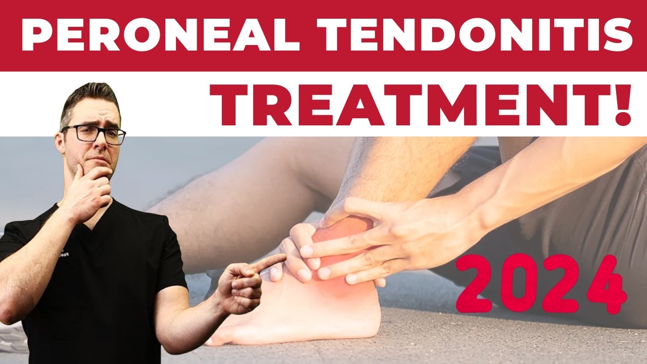
Watch: Peroneal Tendonitis Treatment — Podiatrist Explains

![Peroneal Tendonitis Treatment 2024 [Peroneus Brevis & Longus Pain!]](https://www.michiganfootdoctors.com/wp-content/cache/flying-press/63da3fd3de394bd1f208c4ae18220dcd.jpg)
Dr. Tom covers the full peroneal tendon treatment spectrum — acute splinting, physical therapy progression, bracing, and surgical options for split tears. The clinical decision points that separate conservative candidates from surgical ones are clearly explained.
⚠ The Most Common Mistake We See
Patients with peroneal tendon pain are told it’s “just a sprain” and rest for weeks — then return with the same pain because the underlying cause (ankle instability, supinated foot type, tight peroneals) was never addressed. Peroneal tendonitis does not resolve with rest alone. The tendon heals under load; the instability pattern that caused the injury must be corrected with functional rehab or a splint-only approach fails. If you’ve “rested” for 6+ weeks and it comes back every time you return to activity, you need a structural evaluation — not more rest.
Outer Ankle Pain? Let’s Get an Answer.
Peroneus brevis injuries are frequently misdiagnosed as ankle sprains. Get an accurate diagnosis — including ultrasound imaging if needed — at Balance Foot & Ankle.
4.9★ | 1,123 Reviews | 3,000+ Surgeries
Or call: (810) 206-1402
Tendonitis typically responds best to early podiatrist evaluation, conservative treatments such as supportive footwear and targeted physical therapy, and—when needed—custom orthotics or in-office procedures. Most patients see meaningful improvement within 4-6 weeks of starting a structured treatment plan. Schedule an evaluation at our Howell or Bloomfield Township office for a clinical assessment.
DR. TOM’S RECOMMENDED PRODUCTS
Products I Recommend for This Condition
Before coming in, these are the products I recommend. Affiliate disclosure: I earn a commission at no extra cost to you.
⭐ PowerStep Pinnacle — Best OTC Orthotic
The OTC orthotic I recommend most in clinic. Semi-rigid shell controls rearfoot pronation while dual-layer foam cushions the heel.
Best for: Flat feet, plantar fasciitis, heel pain | Not ideal for: Very narrow shoes
💊 Doctor Hoy’s Natural Pain Relief Gel
Natural topical I use in clinic. Arnica + camphor reduces inflammation at the tissue level — apply 3–4x daily.
Best for: Foot and ankle pain, inflammation | Not ideal for: Open wounds
Persistent pain after 4–6 weeks with conservative care needs evaluation. Same-day appointments →
Ready to feel better?
Same-week appointments available in Howell and Bloomfield Township, Michigan.
Dr. Tom’s Peroneus Brevis Tendonitis Recovery Protocol:
- Doctor Hoy’s Natural Pain Relief Gel — Apply along the lateral ankle and 5th metatarsal insertion — the two sites where peroneus brevis tendinitis hurts most. 3–4× daily. Arnica + camphor reduces tendon sheath inflammation faster than ice alone in most patients.
- PowerStep Pinnacle Insoles — Peroneus brevis tendinitis is driven by chronic inversion stress. Medial arch support controls rear-foot position and reduces the inversion load that strains the peroneal tendon with every step.
- ASO Lace-Up Ankle Brace (Amazon) — Limits the inversion motion that aggravates peroneal tendons during activity. Wear during sport or prolonged walking until the tendon sheath is no longer tender.
Peroneus brevis tears (not just tendinitis) are common and require MRI to differentiate. Persistent lateral ankle pain needs imaging. Learn about our peroneal tendon treatment → or book an evaluation · (810) 206-1402

Tendonitis — Frequently Asked Questions
When should I see a podiatrist for tendonitis?
If symptoms persist beyond 2 weeks of self-care, interfere with daily activity, or worsen suddenly, schedule a podiatrist evaluation. Early intervention typically shortens recovery and prevents chronic compensation patterns that can lead to secondary injuries.
Will I need imaging or surgery?
Most tendonitis cases resolve with conservative care—custom orthotics, supportive shoe changes, anti-inflammatory protocols, and targeted physical therapy. Imaging (X-ray, ultrasound, MRI) is reserved for cases that fail conservative treatment or when structural pathology is suspected. Surgery is rarely the first option.
Does insurance cover tendonitis treatment in Michigan?
Most major Michigan insurance plans (BCBSM, BCN, Priority Health, HAP, Medicare, Medicaid HMOs, United, Aetna, Cigna) cover medically necessary podiatric care. Custom orthotics may have separate DME coverage rules. Our team verifies your specific benefits before your visit.
Related Conditions
Frequently Asked Questions
Related Care at Balance Foot & Ankle
Clinical tendonitis services at our Howell and Bloomfield Township offices.
Get Expert Care at Balance Foot & Ankle
Same-week appointments at our Howell and Bloomfield Township offices. Board-certified podiatric surgeons. Most insurance accepted.
Same-Week Appointments in Howell & Bloomfield Township
Three board-certified podiatric surgeons. 1,123+ five-star reviews. Most insurance accepted.

Dr. Tom Biernacki, DPM is a double board-certified podiatrist and foot & ankle surgeon at Balance Foot & Ankle Specialists in Southeast Michigan. With over a decade of clinical experience, he specializes in heel pain, bunions, diabetic foot care, sports injuries, and minimally invasive surgery. Dr. Biernacki is a member of the APMA and ACFAS, and his patient education content on MichiganFootDoctors.com and YouTube has reached over one million views.
Dr. Tom Biernacki, DPM is a board-certified foot & ankle surgeon (ABFAS & ABPM) at Balance Foot & Ankle Specialists in Southeast Michigan. With over a decade of clinical experience, he specializes in heel pain, bunions, diabetic foot care, sports injuries, and minimally invasive surgery. Dr. Biernacki is a member of the APMA and ACFAS, and his patient education content on MichiganFootDoctors.com and YouTube has made him one of the most-followed foot & ankle educators on YouTube.