
Medically reviewed by Dr. Tom Biernacki, DPM
Board-certified podiatric surgeon | Balance Foot & Ankle, Howell & Bloomfield Hills, MI
Last reviewed: May 2026

| Condition | Structure Impinged | Provocative Activity | Key Finding | Treatment |
|---|---|---|---|---|
| Os Trigonum Syndrome | Os trigonum (accessory bone) compressed between tibia and calcaneus | Plantarflexion (ballet, downhill running) | Lateral posterior ankle pain; os trigonum on X-ray | Rest, injection; endoscopic excision |
| FHL Tendinopathy (stenosing) | Flexor hallucis longus in fibro-osseous tunnel | Repeated push-off, ballet demi-pointe | Pain/triggering behind medial malleolus; positive Strunk test | PT, injection; endoscopic release |
| Soft Tissue Impingement | Posterior capsule / synovium pinched | Plantarflexion | No bony anomaly on imaging; MRI shows synovitis | Injection, arthroscopic debridement |
| Posterior Tibiotalar Impingement | Posterior talofibular ligament excess | Running, jumping | MRI posterior capsular thickening | Arthroscopic debridement |
| Treatment | Evidence | Success Rate | Recovery | Notes |
|---|---|---|---|---|
| Activity Modification + Rest | Strong | 60–70% temporary relief | 4–8 weeks | Reduce plantarflexion-loaded activities |
| Corticosteroid Injection (ultrasound-guided) | Strong | 60–75% | Days to weeks | Diagnostic + therapeutic; avoid FHL tendon |
| Physical Therapy (posterior chain) | Moderate | 50–65% | 6–10 weeks | Addresses tightness perpetuating impingement |
| Endoscopic Os Trigonum Excision | Strong | 85–92% | 4–8 weeks | Gold standard for os trigonum syndrome; two-portal technique |
| Open Os Trigonum Excision | Strong | 80–90% | 8–12 weeks | More exposure; higher soft tissue disruption |
| Endoscopic FHL Release | Moderate-strong | 80–88% | 4–6 weeks | Combined with os trigonum excision when FHL involved |
Foot pain isn’t resolving?
Same-week appointments at Howell & Bloomfield Hills
Medically Reviewed | Dr. Tom Biernacki, DPM | Board-Certified Podiatric Surgeon | Balance Foot & Ankle, Michigan

What Is Posterior Ankle Impingement?
Posterior ankle impingement syndrome (PAIS) describes pain at the back of the ankle that is provoked by plantarflexion — pointing the foot down. The pain arises when structures at the posterior ankle are mechanically compressed or pinched with each plantarflexion movement. This syndrome is particularly common in ballet dancers, gymnasts, soccer players, and any athlete who repetitively loads the ankle in a plantarflexed position.
The posterior ankle is a complex region where the talus, tibia, and calcaneus converge, surrounded by the FHL tendon sheath, posterior capsule, posterior tibiofibular and intermalleolar ligaments, and the retrocalcaneal bursa. Pathology in any of these structures can generate posterior ankle pain that is clinically indistinguishable without careful examination and imaging.
Os Trigonum: The Most Common Cause
During fetal development, a secondary ossification center forms at the posterior process of the talus. In most people, this fuses with the main talar body by age 12. In approximately 10–15% of individuals, it fails to fuse, remaining as a separate accessory bone called the os trigonum. Most os trigona are asymptomatic throughout life.
Impingement occurs when the os trigonum is compressed between the posterior tibia and calcaneus with repeated plantarflexion — creating local bone bruising, synovitis, and fibrocartilage fragmentation. The acute version (os trigonum fracture) occurs with a single traumatic plantarflexion event; the chronic version develops insidiously from repetitive loading.
Clinically, os trigonum syndrome presents as deep posterior ankle pain with forced plantarflexion, tenderness just anterior to the Achilles tendon on both sides of the joint, and characteristic findings on MRI (bone marrow edema at the os trigonum, surrounding soft tissue signal change).
Stieda Process: A Variant Without the Accessory Bone
In patients whose secondary ossification center did fuse, an enlarged lateral talar tubercle (Stieda process or large posterior talar process) can produce identical impingement symptoms. The bone is not accessory — it is part of the talus — but its size makes it vulnerable to impingement. Treatment follows the same algorithm as os trigonum syndrome.
Flexor Hallucis Longus (FHL) Tendinopathy
The FHL tendon runs through a fibro-osseous tunnel at the posterior ankle, directly adjacent to the os trigonum location. FHL tendinopathy — inflammation, partial tear, or triggering (stenosing tenosynovitis) of the FHL — is frequently coexistent with os trigonum syndrome and is sometimes the primary pathology in patients without a demonstrable osseous abnormality.
FHL tendinopathy classically presents with hallux triggering — the big toe catches or locks in mid-range, then releases with an audible or palpable snap. Ballet dancers call this “hallux saltans” or dancer’s tendinopathy. Runners may present with posterior ankle pain that worsens with toe-off. MRI and ultrasound distinguish FHL pathology from bony impingement.
Other Causes of Posterior Ankle Pain
The differential diagnosis for posterior ankle pain is broad and includes conditions Dr. Biernacki systematically evaluates:
- Posterior ankle capsulitis — inflammation of the posterior joint capsule without structural osseous impingement.
- Posterior ankle loose bodies — chondral or osteochondral fragments free in the posterior joint causing catching, locking, and intermittent pain.
- Haglund deformity / retrocalcaneal bursitis — posterior heel pain from bony prominence at the superior calcaneus, distinct from posterior ankle impingement but occasionally coexistent.
- Achilles tendinopathy — insertional or mid-substance Achilles disease generating posterior ankle-area pain that must be distinguished from intra-articular pathology.
- Peroneal tendon pathology — posterior lateral ankle pain from peroneal tendon tears or subluxation.
Diagnosis: Imaging and Clinical Testing
Dr. Biernacki uses a systematic approach to posterior ankle pain diagnosis:
- Clinical examination — the “posterior ankle impingement test” (passive forced plantarflexion reproducing concordant pain) has high sensitivity for PAIS when positive. Palpation locates point tenderness at the lateral vs. medial posterior ankle.
- Weight-bearing radiographs — lateral view typically demonstrates the os trigonum or enlarged lateral talar process. Comparison views help identify acute fracture vs. chronic non-union.
- MRI — the definitive study for soft tissue evaluation. Bone marrow edema at the os trigonum, FHL tendon signal, joint effusion, and capsular pathology all contribute to surgical planning.
- Diagnostic injection — a fluoroscopy- or ultrasound-guided corticosteroid injection targeted to the posterior joint or FHL sheath that produces temporary complete relief confirms the diagnosis and also provides immediate therapeutic benefit.
Non-Surgical Treatment
Initial management of posterior ankle impingement includes:
- Activity modification — reducing or temporarily eliminating plantarflexion-loading activities (ballet, soccer, gymnastics).
- Immobilization — boot walker or cast for 4–6 weeks to reduce acute synovitis.
- Corticosteroid injection — ultrasound- or fluoroscopy-guided injection to the posterior joint reduces inflammation and provides diagnostic confirmation when fully effective.
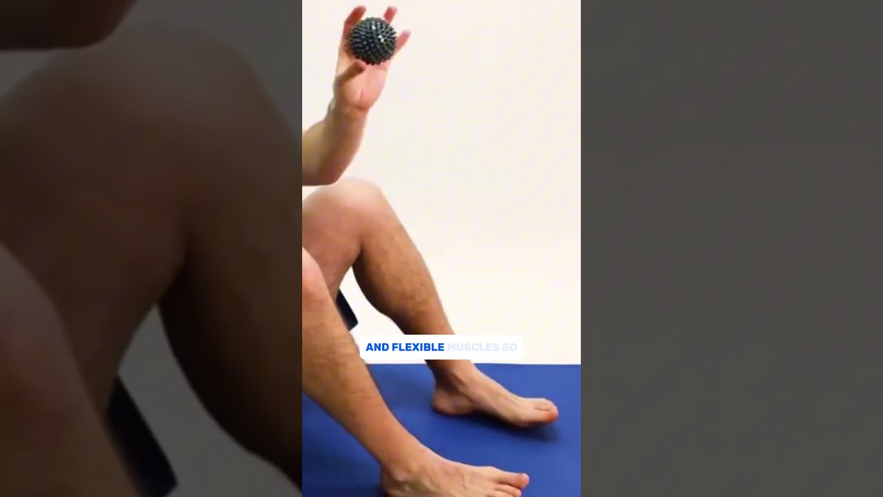
- Physical therapy — posterior chain flexibility, calf stretching, FHL stretching, and proprioceptive training.
Conservative care succeeds in approximately 60% of recreational athletes. Elite and professional athletes — particularly ballet dancers — have lower non-surgical success rates due to the required return to extreme plantarflexion demands.
Surgical Treatment: Os Trigonum Excision
When conservative care fails, os trigonum excision and posterior ankle decompression provides excellent results. Two surgical approaches are used:
Arthroscopic Posterior Ankle Surgery
Performed through two small posterior portals with the patient prone. The arthroscope allows direct visualization of the posterior joint, os trigonum, FHL tendon sheath, and posterior capsule. The os trigonum is excised, FHL tenosynovitis is released if present, and any loose bodies or capsular hypertrophy are addressed. Recovery is faster than open surgery, with most patients weightbearing within 1–2 weeks and return to sport at 2–4 months. Dr. Biernacki prefers the arthroscopic approach when anatomy is suitable.
Open Posterior Ankle Approach
Through a posteromedial or posterolateral incision, the os trigonum is directly accessed and excised. Open surgery provides better visualization in complex cases — large bony fragments, significant FHL pathology requiring formal tenolysis, or concomitant posterior chondral work. Recovery is slightly longer than arthroscopic correction but outcomes are equivalent.
Return to Sport After Surgery
Post-operative rehabilitation follows a staged progression: protected weightbearing in a boot for 2–3 weeks, progressive range of motion, calf strengthening, sport-specific rehabilitation beginning at 6–8 weeks, and return to full activity at 10–16 weeks. Ballet dancers and gymnasts requiring extreme plantarflexion may require 4–6 months before return to full performance-level activity.
Michigan Athletes Seeking Posterior Ankle Care
Dr. Biernacki evaluates posterior ankle impingement and os trigonum syndrome in athletes from across Michigan — from youth athletes in Genesee and Livingston Counties to adult recreational athletes, competitive dancers, and soccer players from Shiawassee, Oakland, and Saginaw Counties. If posterior ankle pain is limiting your performance or daily activity, a comprehensive evaluation will establish the diagnosis and guide an effective treatment plan.
Dr. Tom’s Product Recommendations
Zamst A2-DX Ankle Brace
⭐ Highly Rated
High-support lace-up ankle brace with rigid side stays providing medial-lateral stability. Useful for athletes with posterior ankle pathology needing return-to-sport support during rehabilitation.
Dr. Tom says: “My podiatrist recommended this brace when I was returning to soccer after my ankle procedure — the support was excellent without limiting speed.”
Return to sport with posterior ankle pathology, ankle instability support
Not designed for posterior impingement management specifically — consult provider before using
Disclosure: We earn a commission at no extra cost to you.
TheraBand Resistance Bands Set
⭐ Highly Rated
Professional-grade resistance bands for ankle and calf strengthening rehabilitation. Essential for posterior ankle impingement recovery and return-to-sport physical therapy protocols.
Dr. Tom says: “My physical therapist used these for all my ankle rehab exercises — they’re durable and the progressive resistance levels are really practical.”
Ankle rehabilitation, calf strengthening, post-surgical PT
Must be used with a physical therapist-prescribed protocol; unsupervised use may aggravate impingement if plantarflexion is loaded too early
Disclosure: We earn a commission at no extra cost to you.
✅ Pros / Benefits
- Arthroscopic os trigonum excision has excellent outcomes — most athletes return to full sport by 3–4 months with minimal surgical morbidity
- Diagnostic injection both confirms diagnosis and provides immediate therapeutic benefit — efficient dual-purpose clinical tool
- Addressing FHL tendinopathy concurrently with os trigonum excision reduces the likelihood of persistent posterior ankle symptoms from a missed secondary diagnosis
❌ Cons / Risks
- Elite athletes requiring extreme plantarflexion (ballet, gymnastics) have longer return-to-performance timelines than recreational athletes — 4–6 months is realistic
- The posterior ankle contains neurovascular structures (posterior tibial nerve, flexor hallucis longus) requiring precise surgical technique — performed best by high-volume foot and ankle specialists
- Os trigonum syndrome can be misdiagnosed as Achilles tendinopathy or posterior heel pain — accurate diagnosis requires MRI and/or diagnostic injection, not just clinical examination
Dr. Tom Biernacki’s Recommendation
Posterior ankle pain is frequently misdiagnosed. I’ve seen patients who spent a year treating their ‘Achilles’ when the real problem was an os trigonum being crushed every time they pointed their foot. The key diagnostic step is the forced plantarflexion test — if pressing the foot down reproduces your exact pain deep in the back of the ankle, we need to look at the os trigonum and FHL tendon seriously. A diagnostic injection to the posterior joint that takes away 90% of your pain tells us everything we need to know. When it’s been failing conservative care for 3–4 months, the arthroscopic excision is a clean, reliable solution with a great return-to-sport track record.
— Dr. Tom Biernacki, DPM | Board-Certified Podiatric Surgeon | Balance Foot & Ankle
Frequently Asked Questions
What does posterior ankle impingement feel like?
Deep aching or sharp pain at the back of the ankle that worsens when you point your foot down (plantarflexion). In athletes, pain during push-off, jumping, or ballet relevé positions is characteristic. There is typically point tenderness at the posterolateral ankle, just anterior to the Achilles on the outside of the ankle.
Is os trigonum surgery necessary?
No — many patients with os trigonum achieve adequate symptom control with activity modification, injection, and physical therapy. Surgery is indicated when conservative care fails after 3–6 months, or when the activity demands (professional dancing, competitive athletics) preclude adequate restriction of plantarflexion loading.
How long does recovery take after os trigonum removal?
Arthroscopic excision: protective weightbearing 1–2 weeks, return to sport 10–16 weeks for most athletes. Open excision: slightly longer, 12–20 weeks to full activity. Ballet dancers returning to extreme plantarflexion demand may require 4–6 months.
Can posterior ankle impingement occur without an os trigonum?
Yes. An enlarged lateral talar process (Stieda process), posterior capsular hypertrophy, FHL tenosynovitis, or loose bodies can all cause posterior impingement without an accessory bone. MRI distinguishes these entities and directs appropriate treatment.
Does posterior ankle impingement go away on its own?
In mild cases with activity modification and rest, symptoms may resolve. In athletes who continue sport, or in cases with structural pathology (large os trigonum, significant FHL involvement), symptoms typically persist or worsen without directed treatment.
Michigan Foot Pain? See Dr. Biernacki In Person
4.9★ rated | 1,123 Reviews | 3,000+ Surgeries
Same-week appointments · Howell & Bloomfield Hills
📞 (810) 206-1402 Book Online →Frequently Asked Questions
When should I see a podiatrist?
If symptoms persist past 2 weeks, affect your normal activity, or are accompanied by red-flag symptoms (warmth, redness, swelling, inability to bear weight).
What does treatment cost?
Most diagnostic visits and conservative treatments are covered by Medicare and major insurers. Out-of-pocket costs vary by your specific plan.
How quickly can I get an appointment?
Most non-urgent cases see us within 5 business days. Urgent cases (sudden pain, possible fracture) typically same or next business day.
Foot pain typically responds best to early podiatrist evaluation, conservative treatments such as supportive footwear and targeted physical therapy, and—when needed—custom orthotics or in-office procedures. Most patients see meaningful improvement within 4-6 weeks of starting a structured treatment plan. Schedule an evaluation at our Howell or Bloomfield Hills office for a clinical assessment.
What is Foot pain?

Foot pain is a common foot/ankle condition that affects mobility and quality of life. Understanding the underlying cause is the first step in successful treatment. Our podiatrists at Balance Foot & Ankle perform a hands-on biomechanical exam, review your activity history, and use diagnostic imaging when appropriate to identify the root cause—not just treat the symptom. Many patients have been told to “rest and ice” without a deeper diagnostic workup; our approach is different.
Symptoms and warning signs
Common signs of foot pain include pain that worsens with activity, morning stiffness, swelling, tenderness when palpated, and difficulty bearing weight. If you experience sudden severe pain, inability to walk, visible deformity, numbness or color change, contact our office the same day or visit urgent care—these can signal a more serious injury such as a fracture, tendon rupture, or vascular compromise. Diabetics with any foot wound should seek same-day care.
Conservative treatment options
Most cases of foot pain respond to non-surgical care: structured rest, supportive footwear changes, custom orthotics, targeted stretching and strengthening protocols, anti-inflammatory medications when medically appropriate, and in-office procedures such as ultrasound-guided injections. We also offer advanced therapies including MLS laser therapy, EPAT/shockwave, regenerative injections, and image-guided procedures. Treatment is sequenced from least invasive to most invasive, and we explain the rationale at every step.
When is surgery considered?
Surgery is reserved for cases that fail 3-6 months of well-structured conservative care, when there is structural pathology (severe deformity, complete tear, advanced arthritis), or when imaging shows damage that will not heal without intervention. Our surgeons have performed 3,000+ foot and ankle procedures and prioritize minimally-invasive techniques whenever appropriate. We discuss recovery timelines, return-to-activity milestones, and realistic outcome expectations before any procedure is scheduled.
Recovery timeline and prevention
Recovery from foot pain varies based on severity and chosen treatment path. Conservative cases often improve within 4-8 weeks with consistent adherence to the protocol. Post-procedural recovery may range from a few days (in-office procedures) to several months (reconstructive surgery). Long-term prevention involves footwear assessment, activity modification, structured strengthening, and regular check-ins with your podiatrist if you have a history of recurrence. We provide written home-exercise plans and digital follow-up support.
Ready to feel better?
Same-week appointments available in Howell and Bloomfield Hills, Michigan.
Book Your VisitVisit Balance Foot & Ankle — Same-Day Appointments Available
Our podiatry team serves patients throughout Michigan including Howell, Brighton, and Bloomfield Hills. Whether you’re dealing with heel pain, ingrown toenails, or a foot injury, we have same-day appointment availability.
American Podiatric Medical Association: Find a Podiatrist
Ready to Get Relief?
Same-day appointments available in Howell & Bloomfield Hills, MI
4.9★ | 1,123 Reviews | 3,000+ Surgeries
Or call: (810) 206-1402
Dr. Tom Biernacki, DPM is a board-certified foot & ankle surgeon (ABFAS & ABPM) at Balance Foot & Ankle Specialists in Southeast Michigan. With over a decade of clinical experience, he specializes in heel pain, bunions, diabetic foot care, sports injuries, and minimally invasive surgery. Dr. Biernacki is a member of the APMA and ACFAS, and his patient education content on MichiganFootDoctors.com and YouTube has made him one of the most-followed foot & ankle educators on YouTube.