
Medically reviewed by Dr. Tom Biernacki, DPM
Board-certified podiatric surgeon | Balance Foot & Ankle, Howell & Bloomfield Hills, MI
Last reviewed: May 2026

| Feature | Subungual Exostosis | Osteochondroma | Subungual Enchondroma | Subungual Melanoma |
|---|---|---|---|---|
| Definition | Reactive bone/cartilage outgrowth under nail | Cartilage-capped bony projection (true neoplasm) | Benign cartilage tumor in distal phalanx | Malignant melanocytic tumor under nail plate |
| Most common site | Hallux (great toe), distal phalanx | Long bones (knee, shoulder, hip) | Fingers > toes | Hallux, thumb |
| Age group | Teens and young adults (10–30) | Teens and young adults | Adults 30–50 | Adults 50–70 (peak) |
| X-ray appearance | Trabecular bony projection from distal phalanx; NO cortical continuity with parent bone | Cortical continuity with parent bone; cartilage cap | Lytic lesion with possible calcification | Soft tissue density; may show lytic bone change late |
| Pain character | Pressure pain, nail deformity, discharge | Typically painless unless impinging | Dull ache, nail deformity | Painless streak initially; pain late |
| Malignant potential | None | <1% (solitary) up to 5–25% (hereditary) | Rare (<1%) | HIGH — requires urgent biopsy |
| Treatment | Surgical excision under nail plate | Excision if symptomatic | Curettage ± bone graft | Wide excision ± amputation; oncology referral |
| Stage | Nail Plate Status | Size | Symptoms | Surgical Approach | Recovery |
|---|---|---|---|---|---|
| Stage I (Early) | Intact, mild elevation | <5 mm | Mild pressure, shoe discomfort | Partial nail avulsion + curettage | 4–6 weeks |
| Stage II (Moderate) | Lifted, deformed | 5–10 mm | Moderate pain, nail discoloration, discharge | Full nail avulsion + en-bloc excision | 6–8 weeks |
| Stage III (Advanced) | Destroyed or lost | >10 mm | Constant pain, ulceration, secondary infection | Full excision ± distal phalanx remodeling | 8–12 weeks |
| Recurrence (any stage) | Variable | Variable | Return of nail deformity + pain | Re-excision with wider bone margin | 6–8 weeks |
Foot pain isn't resolving?
Same-week appointments at Howell & Bloomfield Hills
Medically Reviewed | Dr. Tom Biernacki, DPM | Board-Certified Podiatric Surgeon | Balance Foot & Ankle, Michigan

What Is Subungual Exostosis?
Subungual exostosis is a benign osteocartilaginous growth arising from the dorsal cortex of the distal phalanx, typically projecting upward from the tip of the bone immediately beneath the hyponychium (the skin under the free edge of the nail). The lesion consists of a bony stalk covered by a fibrocartilaginous cap — histologically distinct from osteochondromas, which have a hyaline cartilage cap. As the exostosis grows, it progressively elevates the nail plate, distorts its shape, and eventually erodes through the nail bed, producing an exposed, painful, friable mass at the distal toe.
Subungual exostoses most commonly affect young adults and adolescents — the peak incidence is ages 10–30, though all ages are affected. The hallux is involved in 80–85% of cases; the lesser toes are less commonly affected. Bilateral cases are uncommon, occurring in under 5% of patients, distinguishing subungual exostosis from the bilateral nail changes of psoriasis or trauma.
Causes and Contributing Factors
The etiology of subungual exostosis is not fully established but is strongly associated with prior trauma: a single high-energy injury (stubbed toe, crush, sports impact) or repetitive microtrauma (running, tight shoes, repetitive toe extension loading) appears to stimulate ectopic bone formation from the periosteal and fibrocartilaginous tissue at the distal phalanx dorsal surface. This explains the condition’s predominance in athletes and active young adults.
Genetic factors may play a role — rare familial forms suggest hereditary osteocartilaginous dysplasias can produce multiple subungual exostoses. Subungual exostosis is distinguished from subungual osteochondroma, which has a true hyaline cartilage cap and is histologically different; both present similarly and both are treated with excision.
How Subungual Exostosis Presents
The typical presentation begins with a progressive, painful, hard lump at the distal end of the toe, often under or around the nail plate. Patients frequently describe a painful mass “under the nail,” nail plate elevation (the nail tip lifts away from the nail bed), and nail deformity that progresses over months to years. The exostosis is hard on palpation — firm, non-compressible, and fixed to the underlying bone — distinguishing it from soft subungual lesions (pyogenic granuloma, fibroma, subungual melanoma).
As the exostosis enlarges and erodes through the nail bed, a reddish, granulation-tissue mass appears at the distal nail or hyponychium. This mass is often friable, bleeds easily with contact, and may ulcerate — producing chronic pain and drainage. At this stage, subungual exostosis is often misidentified as a pyogenic granuloma, subungual wart, glomus tumor, or even malignant melanoma. The X-ray resolves all diagnostic ambiguity: a bony projection from the dorsal phalanx tip is pathognomonic.
Diagnosis: X-Ray Is Definitive
Plain foot radiographs — particularly a lateral toe view or dorsoplantar view — show the exostosis as a radiopaque bony projection extending from the dorsal distal phalanx. The fibrocartilaginous cap may not be visible on plain X-ray but is seen on MRI when soft-tissue detail is required. CT scan is occasionally used to define the three-dimensional extent of large exostoses prior to surgical planning.
The differential diagnosis includes: subungual osteochondroma (histologically distinct, treated identically); enchondroma (endosteal, produces bone expansion rather than surface projection); pyogenic granuloma (soft, vascular, rapidly developing after minor trauma); glomus tumor (classically produces extreme nail bed pain with cold sensitivity, located subungually without bony component); subungual melanoma (pigmented lesion, longitudinal melanonychia, may be confirmed with biopsy).
Surgical Excision: The Definitive Treatment
Surgical excision is the only effective treatment for subungual exostosis — conservative measures (padding, wider shoes) temporarily reduce pressure but do not eliminate the underlying bony lesion, which continues to grow. The goal of surgery is complete excision of the exostosis including the fibrocartilaginous cap, without damaging the nail matrix or nail bed.
Dr. Biernacki performs subungual exostosis excision under digital ring block anesthesia as an outpatient procedure. Technique options include:
Nail-splitting approach: The central nail plate is partially or fully avulsed to expose the nail bed and exostosis. The hyponychium is incised to access the lesion base. The exostosis is excised with a small rongeur or osteotome at the level of the cortical phalanx, and the fibrocartilaginous cap is removed completely. The nail plate, if preserved, is replaced as a biological dressing. This approach provides excellent visualization and is preferred for large exostoses extending proximally under the nail.
Lateral subungual approach: For distal exostoses confined to the hyponychium, a lateral incision lateral to the nail plate avoids nail avulsion and provides direct access to the exostosis tip. This approach is less disruptive to the nail plate and nail bed when the lesion is small and distal.
Complete excision at the cortical bone level with removal of the cartilaginous cap is essential to prevent recurrence. Incomplete excision leaving residual cap tissue is the primary cause of the 5–15% recurrence rate reported in the literature. Pathological examination of the excised specimen confirms the diagnosis histologically.
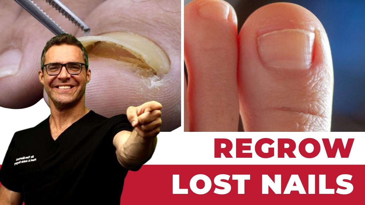
Recovery involves a compressive dressing for 1–2 weeks, protective shoe gear (post-surgical sandal) for 2–3 weeks, and gradual return to regular footwear at 3–4 weeks. The nail regrows over 6–12 months after nail avulsion; nail regrowth quality depends on the extent of nail matrix involvement during excision.
Frequently Asked Questions
How do I know if I have a subungual exostosis or an ingrown toenail?
Key distinguishing features: ingrown toenail pain is at the lateral nail fold (the skin groove beside the nail edge), where the nail has penetrated or compressed soft tissue. Subungual exostosis pain is under the nail plate — at the nail bed or distal hyponychium — and there is typically a hard, fixed lump at the toe tip rather than the nail margin inflammation of an ingrown nail. X-ray taken in the office confirms the presence of bone pathology immediately. Dr. Biernacki takes X-rays at any nail evaluation where subungual exostosis is suspected.
Will the nail grow back normally after excision?
If the nail matrix is undisturbed during surgery, the nail grows back normally over 6–12 months. When the exostosis has eroded extensively into the nail bed or matrix, some permanent nail deformity may persist — but the pain and progressive distortion cease. Dr. Biernacki discusses realistic expectations for nail regrowth at the pre-surgical consultation based on the extent of nail bed involvement.
Is subungual exostosis cancer?
No — subungual exostosis is a benign lesion. It does not transform into bone cancer and is not associated with malignancy. The concern for malignancy arises when subungual lesions are pigmented or ulcerated and could represent subungual melanoma — the X-ray and histological examination of the excised specimen conclusively confirm the benign diagnosis in true exostosis cases.
What is Foot pain?

Foot pain is a common foot/ankle condition that affects mobility and quality of life. Understanding the underlying cause is the first step in successful treatment. Our podiatrists at Balance Foot & Ankle perform a hands-on biomechanical exam, review your activity history, and use diagnostic imaging when appropriate to identify the root cause—not just treat the symptom. Many patients have been told to “rest and ice” without a deeper diagnostic workup; our approach is different.
Symptoms and warning signs
Common signs of foot pain include pain that worsens with activity, morning stiffness, swelling, tenderness when palpated, and difficulty bearing weight. If you experience sudden severe pain, inability to walk, visible deformity, numbness or color change, contact our office the same day or visit urgent care—these can signal a more serious injury such as a fracture, tendon rupture, or vascular compromise. Diabetics with any foot wound should seek same-day care.
Conservative treatment options
Most cases of foot pain respond to non-surgical care: structured rest, supportive footwear changes, custom orthotics, targeted stretching and strengthening protocols, anti-inflammatory medications when medically appropriate, and in-office procedures such as ultrasound-guided injections. We also offer advanced therapies including MLS laser therapy, EPAT/shockwave, regenerative injections, and image-guided procedures. Treatment is sequenced from least invasive to most invasive, and we explain the rationale at every step.
When is surgery considered?
Surgery is reserved for cases that fail 3-6 months of well-structured conservative care, when there is structural pathology (severe deformity, complete tear, advanced arthritis), or when imaging shows damage that will not heal without intervention. Our surgeons have performed 3,000+ foot and ankle procedures and prioritize minimally-invasive techniques whenever appropriate. We discuss recovery timelines, return-to-activity milestones, and realistic outcome expectations before any procedure is scheduled.
Recovery timeline and prevention
Recovery from foot pain varies based on severity and chosen treatment path. Conservative cases often improve within 4-8 weeks with consistent adherence to the protocol. Post-procedural recovery may range from a few days (in-office procedures) to several months (reconstructive surgery). Long-term prevention involves footwear assessment, activity modification, structured strengthening, and regular check-ins with your podiatrist if you have a history of recurrence. We provide written home-exercise plans and digital follow-up support.
Ready to feel better?
Same-week appointments available in Howell and Bloomfield Hills, Michigan.
Book Your VisitVisit Balance Foot & Ankle — Same-Day Appointments Available
Our podiatry team serves patients throughout Michigan including Howell, Brighton, and Bloomfield Hills. Whether you’re dealing with heel pain, ingrown toenails, or a foot injury, we have same-day appointment availability.
Ready to Get Relief?
Same-day appointments available in Howell & Bloomfield Hills, MI
4.9★ | 1,123 Reviews | 3,000+ Surgeries
Or call: (810) 206-1402
Dr. Tom Biernacki, DPM is a board-certified foot & ankle surgeon (ABFAS & ABPM) at Balance Foot & Ankle Specialists in Southeast Michigan. With over a decade of clinical experience, he specializes in heel pain, bunions, diabetic foot care, sports injuries, and minimally invasive surgery. Dr. Biernacki is a member of the APMA and ACFAS, and his patient education content on MichiganFootDoctors.com and YouTube has made him one of the most-followed foot & ankle educators on YouTube.