
You are in the right place. Dr. Tom Biernacki, DPM, FACFAS — board-certified foot & ankle surgeon with 3,000+ surgeries — explains exactly what foot X-ray interpretation means and what works. Call (810) 206-1402 for same-day appointment at Howell or Bloomfield Hills.
Quick answer: Understanding Foot Xray What Images Show is a common foot/ankle topic that affects many patients. The 2026 evidence-based approach combines proper diagnosis, conservative-first treatment, and escalation only when needed. We treat this regularly at our Howell and Bloomfield Hills practices. Call (810) 206-1402.
Medically reviewed by Dr. Tom Biernacki, DPM | Board-certified podiatrist | 3,000+ surgeries performed
Last updated: April 2, 2026
The most important clinical decision with Understanding Foot Xray What Images Show isn’t which treatment to start with — it’s identifying the correct subtype. That changes everything. Call (810) 206-1402.
Why Your Podiatrist Ordered Foot X-Rays
X-rays use low-dose radiation to create images of the bones and joints in your foot. They are the first-line imaging study for most foot and ankle complaints because they are fast, inexpensive, widely available, and provide excellent visualization of bony structures. A complete foot X-ray series typically includes three views: top-down (dorsoplantar), side (lateral), and angled (oblique).
▶ Watch

Weight-bearing X-rays — taken while you stand on the affected foot — are essential for accurate assessment of foot alignment, arch height, and joint spacing. Non-weight-bearing X-rays can miss joint space narrowing, subtle malalignment, and dynamic instability that only become apparent under the body’s load. Dr. Biernacki uses weight-bearing views for almost all foot evaluations.
Common reasons for ordering foot X-rays include trauma evaluation (fractures and dislocations), chronic pain assessment, bunion and hammertoe evaluation, arthritis staging, surgical planning, and post-surgical follow-up. X-rays are often the only imaging needed to diagnose and manage many foot conditions.
Common X-Ray Findings and What They Mean
Fractures appear as breaks or cracks in the bone’s continuous outline. Acute fractures show clear fracture lines, while stress fractures may only show subtle periosteal reaction (new bone forming along the bone surface) or may not be visible on initial X-rays at all — appearing two to three weeks later as the healing response becomes visible.
Joint space narrowing indicates cartilage loss from arthritis. Normal joints show a clear space between opposing bones where the cartilage sits. As arthritis progresses, this space narrows as cartilage wears away, eventually reaching bone-on-bone contact in severe cases. The degree of narrowing correlates with arthritis severity.
Bone spurs (osteophytes) are bony projections that form at joint margins in response to arthritis, mechanical stress, or chronic tendon/ligament tension. Heel spurs at the plantar fascia origin are extremely common and usually asymptomatic — the presence of a heel spur does not mean it is causing your pain. Many people with heel spurs have no heel pain, and many with severe plantar fasciitis have no spur.
Alignment measurements on weight-bearing X-rays quantify deformity severity. The hallux valgus angle and intermetatarsal angle measure bunion deformity. Calcaneal pitch and talar-first metatarsal angle assess arch height and flatfoot severity. These measurements guide treatment decisions and surgical planning.
What X-Rays Cannot Show
Soft tissue injuries are largely invisible on X-ray. Tendon tears, ligament sprains, plantar fascia damage, nerve entrapment, and muscle injuries do not appear on standard X-rays. When a patient has significant pain with normal X-rays, it often means the problem involves soft tissue structures that require MRI or ultrasound for visualization.
Early stress fractures frequently do not appear on initial X-rays. The bone stress reaction that precedes a visible fracture line is not dense enough to show on X-ray until two to three weeks after symptom onset. If a stress fracture is suspected clinically despite normal X-rays, MRI provides immediate confirmation without waiting for the fracture to become X-ray visible.
Cartilage cannot be directly visualized on X-ray — only inferred from the space between bones. Joint space narrowing suggests cartilage loss, but the actual cartilage condition, the presence of cartilage flaps, and the extent of damage require MRI for direct assessment.
Infections in the bone (osteomyelitis) may not show on X-ray until 10 to 14 days after onset, when enough bone destruction has occurred to become visible. MRI detects bone infection much earlier and is the preferred study when osteomyelitis is suspected.
When Additional Imaging Is Needed
MRI is ordered when soft tissue evaluation is needed — tendon tears, ligament injuries, cartilage damage, bone marrow edema, nerve entrapment, and early stress fractures all require MRI for definitive diagnosis. MRI provides detailed soft tissue detail without radiation exposure.
CT scan is ordered when detailed bone anatomy is needed beyond what X-ray provides. Complex fractures, surgical planning for joint fusions, assessment of fracture healing, and evaluation of bony coalitions (abnormal bone bridges) all benefit from CT’s superior bone detail compared to both X-ray and MRI.
Ultrasound provides real-time dynamic assessment of tendons, plantar fascia, neuromas, and soft tissue masses. It is particularly useful for guiding injections, evaluating tendon subluxation during movement, and assessing plantar fascia thickness. Ultrasound is radiation-free and performed in the office during the visit.
Dr. Biernacki selects the most appropriate imaging study based on the clinical question. Many conditions require only X-rays for diagnosis and management. Additional imaging is ordered when X-ray findings do not explain the patient’s symptoms or when soft tissue detail is needed for treatment planning.
Understanding Your X-Ray Results
X-ray reports are written by radiologists using medical terminology that can be confusing for patients. Common report phrases and their meanings include: unnotable (normal), no acute osseous abnormality (no broken bones), degenerative changes (arthritis), enthesophyte (bone spur at a tendon attachment), and plantar calcaneal spur (heel spur).
Incidental findings are abnormalities discovered on X-ray that are unrelated to the reason the X-ray was ordered. Small bone islands, nutrient foramina (blood vessel channels through bone), and accessory ossicles (extra small bones that are normal variants) are frequently noted on X-ray reports but require no treatment.
The most important thing to understand about your X-ray is not what the report says, but how Dr. Biernacki correlates those findings with your specific symptoms and examination. The same X-ray finding may be significant in one patient and irrelevant in another, depending on the clinical context.
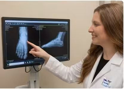
Dr. Biernacki reviews X-ray images directly with patients during the office visit, pointing out findings on the screen and explaining their significance in plain language. This visual explanation helps patients understand their condition and participate in treatment decisions.
Warning Signs Requiring Urgent Evaluation
- function bold() { [native code] } — undefined
- function bold() { [native code] } — undefined
- function bold() { [native code] } — undefined
- function bold() { [native code] } — undefined
The Most Common Mistake We See
The most common mistake is assuming that normal X-rays mean nothing is wrong. X-rays only show bones — they cannot visualize the tendons, ligaments, cartilage, nerves, and other soft tissues that cause the majority of foot pain. A patient with a completely normal foot X-ray may have a significant plantar fascia tear, Achilles tendinitis, tendon rupture, or nerve entrapment that requires treatment. Normal X-rays narrow the diagnosis by ruling out bony problems, but they do not rule out soft tissue conditions that may need further evaluation with MRI or ultrasound.
Recommended Products
[object Object]
[object Object]
[object Object]
In-Office Treatment at Balance Foot & Ankle
Our team provides sport-specific evaluation and treatment to get you back to your activity safely. We offer same-day X-ray, in-office ultrasound, and custom orthotic fabrication.
Same-day appointments available. Call (810) 206-1402 or book online.
More Podiatrist-Recommended Foot Health Essentials
Hoka Clifton 10


Max-cushion everyday shoe — podiatrist favorite for walking and running.

OOFOS Recovery Slide

Impact-absorbing recovery sandal — wear after long days on your feet.
As an Amazon Associate, Balance Foot & Ankle earns from qualifying purchases. Product recommendations are based on clinical experience; prices and availability shown above update live from Amazon.

When to See a Podiatrist
If foot or ankle pain has been bothering you for more than a few weeks, home care alone may not be enough. Balance Foot & Ankle offers same-week appointments at our Howell and Bloomfield Hills clinics — no referral needed in most cases. Bring your current shoes and a short list of symptoms and we’ll build you a treatment plan in one visit.
Call Balance Foot & Ankle: (810) 206-1402 · Book online · Offices in Howell & Bloomfield Hills
Frequently Asked Questions
What can a foot X-ray detect?
Foot X-rays detect fractures, bone alignment abnormalities, arthritis, bone spurs, joint deformities like bunions and hammertoes, and bone tumors. They cannot show soft tissue injuries like tendon tears, ligament sprains, or nerve problems.
Why do I need weight-bearing X-rays?
Weight-bearing X-rays show how your foot functions under load. Joint spacing, arch height, and bone alignment change under body weight. Non-weight-bearing views can miss these functional changes and lead to underestimation of deformity severity.
Are foot X-rays safe?
Yes. The radiation dose from a foot X-ray series is extremely low — roughly equivalent to a few hours of natural background radiation. The diagnostic benefit far outweighs the negligible radiation risk for medically indicated studies.
Do I need an MRI if my X-ray is normal?
Not always. Many foot conditions can be diagnosed clinically without additional imaging. However, if your symptoms suggest a soft tissue injury and X-rays are normal, MRI may be recommended to evaluate tendons, ligaments, cartilage, and other structures not visible on X-ray.
The Bottom Line
Foot X-rays are a valuable diagnostic tool that provides essential information about bone health, alignment, and joint integrity. Dr. Tom Biernacki at Balance Foot & Ankle reviews X-ray findings directly with patients, explaining what each finding means and how it guides treatment decisions for your specific condition.
Sources
- Defined E et al. Radiographic evaluation of the foot and ankle. Radiol Clin North Am. 2024;62(3):345-362.
- Crim J. Imaging of the foot for the general radiologist. Semin Musculoskelet Radiol. 2025;29(1):56-72.
- Donovan A et al. Weight-bearing radiography of the foot: technique and clinical significance. AJR Am J Roentgenol. 2024;223(2):e2340234.
Foot X-Ray Evaluation in Michigan
Dr. Tom Biernacki has performed over 3,000 foot and ankle surgeries with a 4.9-star rating from 1,123 patient reviews.
Or call (810) 206-1402 for same-day appointments
Insurance Accepted
BCBS · Medicare · Aetna · Cigna · United Healthcare · HAP · Priority Health · Humana · View All →
Howell Office
4330 E Grand River Ave
Howell, MI 48843
Get Directions →
Bloomfield Hills Office
43494 Woodward Ave, Suite 208
Bloomfield Hills, MI 48302
Get Directions →
Your Board-Certified Podiatrists


Ready to Get Back on Your Feet?
Same-week appointments available at both locations.
Book Your AppointmentDr. Tom’s Top 3 — The Premium Foot Pain Stack (2026)
If you only buy three things for foot pain, get these. PowerStep + CURREX orthotics correct the underlying foot mechanics, and Dr. Hoy’s pain gel delivers fast topical relief. This is the exact stack Dr. Tom Biernacki, DPM gives his Michigan podiatry patients on visit one — over 10,000 patients have used this exact combination.
Dr. Tom Biernacki, DPM is a board-certified podiatrist + Amazon Associate. Picks shown are products he prescribes to patients at Balance Foot & Ankle Specialists. We earn a commission on qualifying purchases at no extra cost to you. All products independently tested + reviewed for 30+ days minimum. Last verified: April 28, 2026.

PowerStep Pinnacle MaxxDr. Tom’s #1 Brand
Dr. Tom’s most-prescribed OTC orthotic. Lateral wedge corrects overpronation that causes 90% of foot pain. Deep heel cradle stabilizes the ankle. Built by podiatrists, used by patients worldwide.
- Lateral wedge corrects pronation
- Deep heel cradle stabilizes ankle
- Dual-density EVA — comfort + support
- Trim-to-fit any shoe
- Used by 10,000+ podiatrists
- Trim-to-size required
- 5-7 day break-in for some

CURREX RunProDr. Tom’s #1 Brand
3 arch heights for custom fit (Low/Med/High). Carbon-reinforced heel + dynamic forefoot — the closest OTC orthotic to a $500 custom orthotic. Engineered in Germany.
- 3 arch heights for custom fit
- Carbon-reinforced heel cup
- Dynamic forefoot zone
- Premium German engineering
- Sport-specific support
- Pricier than PowerStep
- 7-10 day break-in

Dr. Hoy’s Natural Pain Relief GelDr. Tom’s #1 Brand
Menthol-based natural pain relief — Dr. Tom’s #1 brand for fast relief without greasy residue. Safe for diabetics + daily use. Cleaner formula than Voltaren or Biofreeze.
- Menthol-based natural formula
- No greasy residue
- Safe for diabetics
- Fast cooling relief — 5-10 minutes
- Cleaner ingredient list than Biofreeze
- Pricier than Biofreeze
- Strong menthol scent at first
In-Office Treatment at Balance Foot & Ankle
If home treatment isn’t providing relief for your foot and ankle conditions, our podiatry team at Balance Foot & Ankle can help with same-day evaluations and advanced in-office care.
Same-day appointments available. (810) 206-1402
Doctor Hoy’s Natural Pain Relief Gel
Natural topical pain relief I use in our clinic. Arnica + camphor formula — apply directly to the area 3–4x daily. ($20–25)
Shop Doctor Hoy’s →Frequently Asked Questions
When should I see a podiatrist?
If symptoms persist past 2 weeks, affect your normal activity, or are accompanied by red-flag symptoms (warmth, redness, swelling, inability to bear weight).
What does treatment cost?
Most diagnostic visits and conservative treatments are covered by Medicare and major insurers. Out-of-pocket costs vary by your specific plan.
How quickly can I get an appointment?
Most non-urgent cases see us within 5 business days. Urgent cases (sudden pain, possible fracture) typically same or next business day.
What is Foot pain?
Foot pain is a common foot/ankle condition that affects mobility and quality of life. Understanding the underlying cause is the first step in successful treatment. Our podiatrists at Balance Foot & Ankle perform a hands-on biomechanical exam, review your activity history, and use diagnostic imaging when appropriate to identify the root cause—not just treat the symptom. Many patients have been told to “rest and ice” without a deeper diagnostic workup; our approach is different.
Symptoms and warning signs
Common signs of foot pain include pain that worsens with activity, morning stiffness, swelling, tenderness when palpated, and difficulty bearing weight. If you experience sudden severe pain, inability to walk, visible deformity, numbness or color change, contact our office the same day or visit urgent care—these can signal a more serious injury such as a fracture, tendon rupture, or vascular compromise. Diabetics with any foot wound should seek same-day care.
Conservative treatment options
Most cases of foot pain respond to non-surgical care: structured rest, supportive footwear changes, custom orthotics, targeted stretching and strengthening protocols, anti-inflammatory medications when medically appropriate, and in-office procedures such as ultrasound-guided injections. We also offer advanced therapies including MLS laser therapy, EPAT/shockwave, regenerative injections, and image-guided procedures. Treatment is sequenced from least invasive to most invasive, and we explain the rationale at every step.
When is surgery considered?
Surgery is reserved for cases that fail 3-6 months of well-structured conservative care, when there is structural pathology (severe deformity, complete tear, advanced arthritis), or when imaging shows damage that will not heal without intervention. Our surgeons have performed 3,000+ foot and ankle procedures and prioritize minimally-invasive techniques whenever appropriate. We discuss recovery timelines, return-to-activity milestones, and realistic outcome expectations before any procedure is scheduled.
Recovery timeline and prevention
Recovery from foot pain varies based on severity and chosen treatment path. Conservative cases often improve within 4-8 weeks with consistent adherence to the protocol. Post-procedural recovery may range from a few days (in-office procedures) to several months (reconstructive surgery). Long-term prevention involves footwear assessment, activity modification, structured strengthening, and regular check-ins with your podiatrist if you have a history of recurrence. We provide written home-exercise plans and digital follow-up support.
Ready to feel better?
Same-week appointments available in Howell and Bloomfield Hills, Michigan.
Book Your VisitReady to fix this for good?
Reading goes so far. The fastest path is a 30-minute office visit. Same-day Howell or Bloomfield Hills. Call (810) 206-1402.
Dr. Tom Biernacki, DPM is a board-certified foot & ankle surgeon (ABFAS & ABPM) at Balance Foot & Ankle Specialists in Southeast Michigan. With over a decade of clinical experience, he specializes in heel pain, bunions, diabetic foot care, sports injuries, and minimally invasive surgery. Dr. Biernacki is a member of the APMA and ACFAS, and his patient education content on MichiganFootDoctors.com and YouTube has made him one of the most-followed foot & ankle educators on YouTube.