
Medically reviewed by Dr. Tom Biernacki, DPM — Board-Certified Podiatric Surgeon — Balance Foot & Ankle, Howell & Bloomfield Hills, MI. Last updated April 2026.
▶ Watch

Medically Reviewed by Dr. Tom Biernacki, DPM — Board-Certified Podiatrist, Balance Foot & Ankle Specialists, Michigan. Last updated April 2026.
Introduction to Ankle Arthroscopy
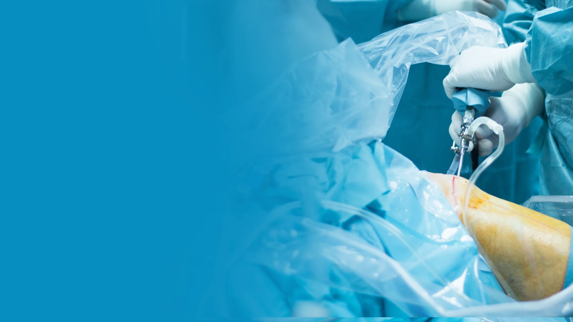
Ankle arthroscopy is a minimally invasive surgical technique that uses a small camera (arthroscope) and specialized instruments inserted through tiny portals (incisions typically 5mm or less) to visualize and treat pathology within the ankle joint. First performed in the 1930s and refined substantially since the 1980s with improvements in small-joint arthroscopic technology, ankle arthroscopy has become the gold standard for diagnosing and treating a many intra-articular ankle conditions — from chronic pain of unclear etiology to specific pathology including osteochondral lesions, impingement syndrome, and synovitis.
The advantages over open ankle surgery are substantial: smaller incisions, reduced perioperative tissue damage, faster recovery, shorter hospital stay (virtually always outpatient), and lower risk of wound complications. The joint is visualized with magnification under direct camera view, allowing detailed assessment of all cartilage surfaces, ligaments, and bone anatomy that X-rays and even MRI cannot fully capture.
Osteochondral Lesions of the Talus (OLT)
What Is an OLT?
An osteochondral lesion of the talus is a defect involving the articular cartilage and the underlying subchondral bone of the talar dome. These lesions are common sequelae of ankle sprains — estimated to occur in up to 50% of acute lateral ankle sprains that involve significant force — though they may also develop spontaneously through avascular mechanisms. The talar dome has two vulnerable zones: the anterolateral portion (most commonly traumatic, following inversion sprains) and the posteromedial portion (more often idiopathic or associated with rotational mechanisms).
Articular cartilage is avascular and has extremely limited intrinsic healing capacity. When cartilage is damaged, the defect persists and — without treatment — typically progresses, exposing the underlying bone to direct mechanical loading and eventually producing early ankle arthrosis.
Classification
OLTs are classified using several systems; the Berndt and Harty classification (modified by Ferkel) describes lesions from Stage I (subchondral compression without fragment) through Stage IV (completely detached, displaced fragment). The International Cartilage Repair Society (ICRS) classification grades articular cartilage damage from Grade I (surface fibrillation) through Grade IV (full-thickness loss to bone). Lesion size — assessed by MRI — is critically important for surgical planning: lesions smaller than 150mm² (approximately 1.5cm² in surface area) respond favorably to marrow stimulation techniques; larger lesions typically require more complex cartilage restoration approaches.
Arthroscopic Treatment of OLTs
Marrow Stimulation (Microfracture/Nanofracture)
The most commonly performed arthroscopic OLT treatment, marrow stimulation involves debriding the unstable cartilage to a stable rim, removing the damaged fibrocartilage and subchondral plate, and creating multiple small perforations (microfractures) in the exposed bone using a specialized pick. These perforations allow marrow elements — including mesenchymal stem cells — to access the defect and form a fibrocartilage repair tissue that fills the void. Microfracture is best indicated for contained lesions smaller than 150mm² in younger patients with good bone quality. Results are good to excellent in 80–85% of appropriately selected patients at intermediate follow-up (5 years); long-term durability is more variable.
Retrograde Drilling
For OLTs with intact cartilage surface but underlying subchondral cyst formation — a finding identified on MRI — retrograde drilling is performed arthroscopically or under fluoroscopic guidance. Drill tracks are created from a remote extra-articular approach through the talus into the cystic cavity, avoiding disruption of the intact cartilage surface. Bone grafting through the drill track fills the subchondral cyst. This technique is particularly elegant for posteromedial lesions where the cartilage surface remains structurally sound.
Ankle Impingement Syndrome
Anterior Ankle Impingement
Anterior ankle impingement — sometimes called ‘footballer’s ankle’ — results from impingement of soft tissue or osteophytes (bone spurs) at the anterior ankle joint during dorsiflexion. Chronic repetitive ankle sprains produce scar tissue in the anterior joint space; years of impact loading produce anterior tibiotalar osteophytes at the anterior tibia lip and talar neck that physically collide during dorsiflexion. The clinical result is a specific dorsiflexion-limited, anterior joint line pain pattern that is distinct from generalized ankle pain.
Arthroscopic management excises the offending soft tissue with a shaver and ablation device, and removes osteophytes with a burr under direct visualization. Postoperative results are excellent — most patients regain full dorsiflexion range and return to sport within 6–8 weeks. Recurrence of osteophytes is possible with continued high-impact activity but is not universal.
Posterior Ankle Impingement
Posterior ankle impingement produces pain at the posterior ankle during plantarflexion, caused by compression of the posterior talar process or os trigonum between the tibia and calcaneus. This condition is particularly common in ballet dancers (en pointe) and soccer players (kicking follow-through). Arthroscopic posterior ankle arthroscopy — performed with the patient prone through posterior portals — excises the os trigonum or prominent posterior talar process, releases the surrounding scar tissue, and decompresses the FHL tendon if it is involved. Recovery is faster than open posterior ankle surgery and produces equivalent outcomes.
Procedure Details and Recovery
Ankle arthroscopy is performed under general, spinal, or regional anesthesia as a day surgery procedure. Noninvasive ankle distraction may be applied to improve visualization of the talar dome. Two to three portals are typically used. Most procedures last 45–90 minutes. Patients are weight-bearing immediately for simple procedures (impingement resection) or non-weight-bearing for 2–6 weeks following cartilage procedures (OLT treatment). Full return to sport typically requires 3–6 months following OLT treatment and 6–12 weeks following impingement resection.
Get Back on Your Feet — Fast
Board-certified podiatrists serving Southeast Michigan. Same-week appointments available.
Ankle Arthroscopy in Michigan
Ankle arthroscopy is a minimally invasive procedure for treating osteochondral lesions (cartilage damage), bone spurs, and impingement. Dr. Tom Biernacki performs advanced ankle arthroscopy with microfracture and cartilage restoration techniques at Balance Foot & Ankle.
Learn About Our Ankle Surgery Options | Book Your Appointment | Call (810) 206-1402
Clinical References
- Zengerink M, et al. “Current concepts: treatment of osteochondral ankle defects.” Foot Ankle Clin. 2006;11(2):331-359.
- Chuckpaiwong B, et al. “Outcome after debridement of osteochondral lesions of the talus.” Foot Ankle Int. 2008;29(5):489-496.
- Ferkel RD, et al. “Arthroscopic treatment of osteochondral lesions of the talus: long-term results.” Am J Sports Med. 2008;36(9):1750-1762.
Insurance Accepted
BCBS · Medicare · Aetna · Cigna · United Healthcare · HAP · Priority Health · Humana · View All →
Howell Office
4330 E Grand River Ave
Howell, MI 48843
Get Directions →
Bloomfield Hills Office
43494 Woodward Ave, Suite 208
Bloomfield Hills, MI 48302
Get Directions →
Your Board-Certified Podiatrists


Ready to Get Back on Your Feet?
Same-week appointments available at both locations.
Book Your AppointmentMore Podiatrist-Recommended Foot Health Essentials
Hoka Clifton 10


Max-cushion everyday shoe — podiatrist favorite for walking and running.

OOFOS Recovery Slide

Impact-absorbing recovery sandal — wear after long days on your feet.
As an Amazon Associate, Balance Foot & Ankle earns from qualifying purchases. Product recommendations are based on clinical experience; prices and availability shown above update live from Amazon.

When to See a Podiatrist
If foot or ankle pain has been bothering you for more than a few weeks, home care alone may not be enough. Balance Foot & Ankle offers same-week appointments at our Howell and Bloomfield Hills clinics — no referral needed in most cases. Bring your current shoes and a short list of symptoms and we’ll build you a treatment plan in one visit.
Call Balance Foot & Ankle: (810) 206-1402 · Book online · Offices in Howell & Bloomfield Hills
In-Office Treatment at Balance Foot & Ankle
When conservative care isn’t enough, Dr. Tom Biernacki and the team at Balance Foot & Ankle offer advanced, same-day options — including Ankle Arthroscopy Michigan at our Howell and Bloomfield Hills clinics.
Same-day appointments available. Call (810) 206-1402 or book online.
What is Foot pain?
Foot pain is a common foot/ankle condition that affects mobility and quality of life. Understanding the underlying cause is the first step in successful treatment. Our podiatrists at Balance Foot & Ankle perform a hands-on biomechanical exam, review your activity history, and use diagnostic imaging when appropriate to identify the root cause—not just treat the symptom. Many patients have been told to “rest and ice” without a deeper diagnostic workup; our approach is different.
Symptoms and warning signs
Common signs of foot pain include pain that worsens with activity, morning stiffness, swelling, tenderness when palpated, and difficulty bearing weight. If you experience sudden severe pain, inability to walk, visible deformity, numbness or color change, contact our office the same day or visit urgent care—these can signal a more serious injury such as a fracture, tendon rupture, or vascular compromise. Diabetics with any foot wound should seek same-day care.
Conservative treatment options
Most cases of foot pain respond to non-surgical care: structured rest, supportive footwear changes, custom orthotics, targeted stretching and strengthening protocols, anti-inflammatory medications when medically appropriate, and in-office procedures such as ultrasound-guided injections. We also offer advanced therapies including MLS laser therapy, EPAT/shockwave, regenerative injections, and image-guided procedures. Treatment is sequenced from least invasive to most invasive, and we explain the rationale at every step.
When is surgery considered?
Surgery is reserved for cases that fail 3-6 months of well-structured conservative care, when there is structural pathology (severe deformity, complete tear, advanced arthritis), or when imaging shows damage that will not heal without intervention. Our surgeons have performed 3,000+ foot and ankle procedures and prioritize minimally-invasive techniques whenever appropriate. We discuss recovery timelines, return-to-activity milestones, and realistic outcome expectations before any procedure is scheduled.
Recovery timeline and prevention
Recovery from foot pain varies based on severity and chosen treatment path. Conservative cases often improve within 4-8 weeks with consistent adherence to the protocol. Post-procedural recovery may range from a few days (in-office procedures) to several months (reconstructive surgery). Long-term prevention involves footwear assessment, activity modification, structured strengthening, and regular check-ins with your podiatrist if you have a history of recurrence. We provide written home-exercise plans and digital follow-up support.
Ready to feel better?
Same-week appointments available in Howell and Bloomfield Hills, Michigan.
Book Your VisitIn-Office Treatment at Balance Foot & Ankle
If home treatment isn’t providing relief for your foot and ankle conditions, our podiatry team at Balance Foot & Ankle can help with same-day evaluations and advanced in-office care.
Same-day appointments available. (810) 206-1402
Doctor Hoy’s Natural Pain Relief Gel
Natural topical pain relief I use in our clinic. Arnica + camphor formula — apply directly to the area 3–4x daily. ($20–25)
Shop Doctor Hoy’s →Dr. Tom Biernacki, DPM is a board-certified foot & ankle surgeon (ABFAS & ABPM) at Balance Foot & Ankle Specialists in Southeast Michigan. With over a decade of clinical experience, he specializes in heel pain, bunions, diabetic foot care, sports injuries, and minimally invasive surgery. Dr. Biernacki is a member of the APMA and ACFAS, and his patient education content on MichiganFootDoctors.com and YouTube has made him one of the most-followed foot & ankle educators on YouTube.