

The most important clinical decision with Fat Pad Atrophy isn’t which treatment to start with — it’s identifying the correct subtype. That changes everything. Call (810) 206-1402.
Products for Fat Pad Atrophy Pain Relief
Medically reviewed by Dr. Tom Biernacki, DPM
Board-certified podiatric surgeon | Balance Foot & Ankle
Last reviewed: May 2026
Fat pad atrophy has no cure — the lost tissue does not regenerate. Treatment is entirely about replacing the cushioning the fat pad no longer provides. These are the products that most effectively compensate for lost plantar padding.
#1 — Maximum Cushion Running Shoes (Non-Negotiable)
For patients with fat pad atrophy, maximum stack height shoes are not a preference — they are a medical necessity. The shoe must replace the cushioning the foot no longer provides internally. HOKA Bondi 8 on Amazon — 4mm heel-to-toe drop, 38mm stack height at heel. Best single shoe recommendation for fat pad atrophy. Also consider the Brooks Ghost 16 for patients who prefer a more traditional ride.
#2 — Heel Cups (For Isolated Heel Fat Pad Atrophy)
When fat pad atrophy is localized to the heel (common in elderly patients and corticosteroid users), a thick silicone heel cup adds back the cushioning the fat pad no longer provides. Silicone Heel Cups for Fat Pad on Amazon — get the 6mm or 12mm thickness version; thinner versions provide insufficient compensation.
#3 — Full-Length Cushioning Insoles
For ball-of-foot fat pad atrophy, full-length cushioning insoles provide metatarsal head offloading that heel cups alone cannot address. Spenco RX Cushioning Insoles on Amazon — Spenco’s polysorb material provides durable forefoot cushioning that maintains its properties through 6+ months of daily use.
#4 — Metatarsal Gel Pads
When metatarsal head fat pad atrophy is the primary complaint, adhesive metatarsal gel pads directly replace the lost tissue at the area of highest pressure. Metatarsal Gel Pads on Amazon — adhesive variety stays positioned correctly during walking; replace every 2–3 weeks.
This page covers the clinical evaluation, evidence-based treatment options, and recovery timeline for fat pad atrophy: heel cushion loss & treatment at Balance Foot & Ankle in Michigan. For same-week appointments at our Howell or Bloomfield Hills offices, call (810) 206-1402.
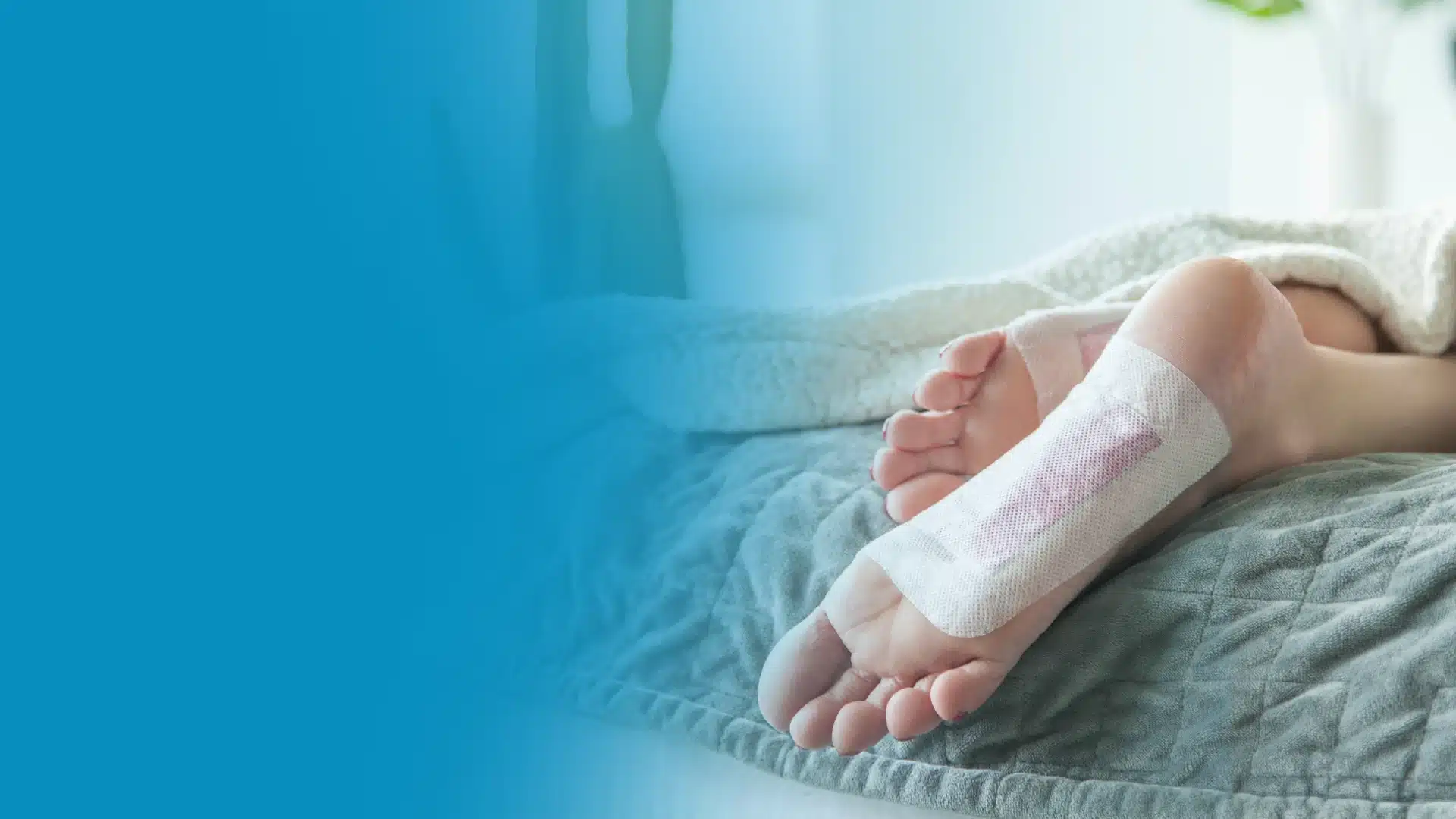
Fat pad atrophy — when the natural cushioning under your heel or ball of foot thins out — makes every step feel like walking on bone. Heel cups and metatarsal pads transform the experience.
You’re in the right place. Dr. Tom Biernacki, DPM, FACFAS — board-certified foot & ankle surgeon with 3,000+ surgeries — explains exactly what fat pad atrophy means and what works. Call (810) 206-1402 for same-day appointment at Howell or Bloomfield Hills.
Foot pain isn’t resolving?
Same-week appointments at Howell & Bloomfield Hills
Board-Certified Podiatric Foot & Ankle Surgeon · Last reviewed: May 5, 2026
Fat pad atrophy occurs when the shock-absorbing fatty tissue under the heel thins and loses its cushioning properties, causing deep, diffuse heel pain that worsens with hard surfaces and barefoot walking. Unlike plantar fasciitis, the pain is maximal directly under the center of the heel rather than at the plantar fascia insertion. In our clinic, treatment focuses on external cushioning replacement — thick gel heel cups, cushioned footwear, and activity modification — since the fat pad itself cannot be regenerated through conservative care alone.
You have been told your heel pain is plantar fasciitis for years, but the stretching, orthotics, and cortisone shots have never fully resolved it. The pain is not in the morning stepping out of bed — it is constant whenever you stand on a hard floor without thick cushioning. This pattern often points to fat pad atrophy, one of the most commonly missed diagnoses in heel pain, particularly in patients over 50 and those who have received multiple cortisone injections to the heel.
What Heel Fat Pad Atrophy Is
The plantar heel fat pad is a specialized adipose structure made of closed fibroelastic cells filled with fat — essentially a biological shock absorber designed to withstand repetitive loading across a lifetime. In younger adults, the fat pad is typically 1.8–2 cm thick and has high viscoelastic properties, absorbing up to 90% of the impact force with each heel strike. As the fat pad atrophies, the heel bone (calcaneus) becomes more directly exposed to ground contact forces, resulting in deep pain that increases with hard surface walking and decreases with thick cushioning.
In our clinic, we measure fat pad thickness with diagnostic ultrasound. A fat pad less than 1.4 cm thick combined with loss of the normal fibro-septal compartment architecture confirms atrophy as a primary pain driver. This measurement matters because it distinguishes fat pad atrophy from plantar fasciitis — two conditions that can coexist but require different treatment priorities.
Causes and Risk Factors for Heel Fat Pad Atrophy
| Cause | Mechanism |
|---|---|
| Age (primary cause) | Fat cell count decreases, fibrous septa degrade — normal aging process accelerating after age 40 |
| Repeated cortisone injections | Corticosteroids cause fat cell lysis (lipoatrophy) — the most preventable cause in clinical practice |
| High-impact activity without cushioning | Chronic compression degrades fat cell integrity over decades |
| Prolonged barefoot walking on hard floors | Direct calcaneal loading without fat pad protection accelerates atrophy |
| Systemic conditions | Rheumatoid arthritis, lupus, and diabetes alter fat pad composition |
| Cavus (high arch) foot | Concentrated heel loading from rigid high-arch mechanics |
The iatrogenic cause — cortisone injection-related fat pad atrophy — is the most important to understand clinically. In our practice, we limit heel injections to a maximum of 2–3 per heel per year and always use ultrasound guidance to inject into the plantar fascia sheath rather than the fat pad itself. Injecting directly into the fat pad is a technical error that accelerates atrophy and should be avoided.
Fat Pad Atrophy vs. Plantar Fasciitis — How to Tell the Difference
These two conditions are frequently confused — and frequently coexist. Here is how to distinguish them clinically.
| Feature | Fat Pad Atrophy | Plantar Fasciitis |
|---|---|---|
| Pain location | Central plantar heel (directly under calcaneus) | Medial plantar heel (plantar fascia insertion) |
| Worst time | Throughout standing — no “first step” pattern | Morning first steps, after sitting |
| Relief with cushioning | Significant — thick heel cup dramatically reduces pain | Partial — orthotics help but not primarily through cushioning |
| Barefoot on hard floor | Very painful — often the most reliable trigger | Less specific to surface hardness |
| Age group | Primarily 50+ and athletes with many injections | Any adult, peak 40–60 |
| Windlass test | Negative or mildly positive | Positive (toe dorsiflexion reproduces heel pain) |
Treatment Protocol for Heel Fat Pad Atrophy
Fat pad atrophy treatment has one primary goal: replace externally what the body can no longer provide internally. Unlike plantar fasciitis where stretching and strengthening produce meaningful tissue-level change, fat pad atrophy does not respond to these interventions — the fat cells are gone and cannot be regenerated through conservative means.
Silicone Heel Cups (first-line): Thick, high-density silicone heel cups that cradle the calcaneus are the most effective conservative intervention. The key distinction is thickness — standard thin heel cushions are insufficient. Look for cups with at least 1 cm of silicone thickness under the heel center. Wear in all footwear including house slippers. Barefoot walking on hard floors must be eliminated completely.
Cushioned Footwear (mandatory): Maximum-cushion running shoes (Hoka, Brooks Glycerin, ASICS Gel-Nimbus category) dramatically reduce calcaneal impact stress. These are not optional accessories — they are functional medical devices for fat pad atrophy. Dress shoes and hard-soled footwear without cushioning insoles should be avoided entirely.
Activity Modification: Hard surface walking, running, and prolonged standing on concrete or tile are the primary aggravating factors. Switching to softer surfaces for exercise (grass, tracks, treadmills) and using anti-fatigue mats at standing workstations produce meaningful symptom reduction.
Emerging Treatments (in-office): Platelet-rich plasma (PRP) injection into the fat pad has shown early promise in regenerating fat pad tissue architecture in small studies. It is not yet first-line but offers a potential option for patients with confirmed severe atrophy and failed conservative care. Autologous fat transfer (using the patient’s own fat cells) is a surgical option in severe cases performed by specialized foot and ankle surgeons.
- Additional cortisone injections directly into the fat pad (worsens atrophy)
- Barefoot walking on tile, hardwood, or concrete floors
- Minimalist shoes, thin-soled shoes, or ballet flats
- Standing for prolonged periods without cushioned matting
- Night splints if they increase plantar pressure on an already compromised heel
See a podiatrist if: heel pain is not responding to thick cushioning after 6 weeks, pain is increasing despite cushion use, or you notice skin breakdown over the bony heel (indicates severe atrophy with no protective tissue remaining).
PowerStep Pinnacle Maxx insoles provide the combination that fat pad atrophy patients need: a deep heel cup that centralizes and maximizes the remaining fat pad tissue under the calcaneus (preventing lateral spread), plus firm arch support that reduces overall calcaneal loading by distributing weight across the entire foot. In our clinic, we recommend these as the OTC insole of choice for fat pad atrophy — the heel cup geometry specifically addresses the lateral fat pad displacement that occurs as the pad thins. Part of our Foundation Wellness portfolio.
Not Ideal For: Patients needing maximum-thickness cushioning alone — add a silicone heel cup underneath for severe atrophy. Size down half a size for best fit.
Shop PowerStep at MFDFat Pad Atrophy Treatment at Balance Foot & Ankle
Accurate diagnosis of fat pad atrophy changes the treatment plan completely — avoiding further cortisone injections, selecting the right footwear and insole, and in appropriate cases evaluating for PRP or surgical fat grafting. At Balance Foot & Ankle, Dr. Tom Biernacki uses diagnostic ultrasound to measure fat pad thickness, distinguishes fat pad atrophy from coexisting plantar fasciitis, and creates a personalized cushioning and activity plan. For patients with severe atrophy who are not responding to conservative measures, we discuss all available options including regenerative approaches.
Same-day appointments. Dr. Biernacki — 3,000+ surgeries, 4.9 stars, 1,123 reviews.
Book Online (810) 206-1402Howell: 4330 E Grand River Ave · Bloomfield Hills: 43494 Woodward Ave #208
Frequently Asked Questions
Can heel fat pad atrophy be reversed?
Fat pad atrophy from normal aging cannot be reversed through conservative treatment — the fat cells that have degraded do not regenerate. However, PRP injections and autologous fat grafting are emerging options that may restore some pad volume in appropriate candidates. Conservative management focuses on replacing the cushioning externally.
How is fat pad atrophy different from plantar fasciitis?
Fat pad atrophy causes central heel pain directly under the calcaneus that is worse with hard surfaces and dramatically relieved by cushioning. Plantar fasciitis causes medial heel pain at the fascia insertion that is worst with morning first steps and responds to stretching. The windlass test (toe dorsiflexion reproducing heel pain) is positive in plantar fasciitis and negative in isolated fat pad atrophy.
Does cortisone cause fat pad atrophy?
Yes — cortisone injections directly into the heel fat pad can cause fat cell lysis (lipoatrophy), permanently thinning the pad. The risk increases with multiple injections, large volumes, and non-ultrasound-guided technique that misses the fascia and deposits steroid in the fat pad. This is why we limit injections and use ultrasound guidance for all heel injections in our practice.
What shoes are best for fat pad atrophy?
Maximum-cushion shoes with thick midsoles (Hoka One One, Brooks Glycerin, ASICS Gel-Nimbus) are ideal. Avoid minimalist shoes, thin-soled casual shoes, and any footwear without substantial heel cushioning. Add a silicone heel cup inside the shoe for additional cushion and to centralize the remaining fat pad under the calcaneus.
When should I see a podiatrist about fat pad atrophy?
See a podiatrist if heel pain persists despite thick cushioning and shoe modification, if cortisone injections have been previously administered to the heel, if pain is affecting daily function, or if you want a diagnostic ultrasound to confirm fat pad thickness and rule out coexisting plantar fasciitis or calcaneal stress fracture.
Sources
- Jahss MH, et al. Investigations into the fat pads of the sole of the foot: anatomy and histology. Foot Ankle. 1992;13(5):233–242.
- Baan H, et al. The relationship between fat pad atrophy and chronic heel pain. Foot Ankle Int. 2011;32(7):673–678.
- Cunha M, et al. Clinical and ultrasonographic evaluation of fat pad syndrome in patients with rheumatoid arthritis. Rev Bras Reumatol. 2012;52(4):581–591.
- Uzel AP, et al. Plantar fat pad augmentation using autologous fat grafting. J Foot Ankle Surg. 2019;58(2):247–252.
- Balance Foot & Ankle. Heel Pain Treatment — Dr. Tom Biernacki DPM.
Dr. Tom’s Clinic-Recommended Products
The OTC orthotic I recommend most. Medical-grade arch support at a fraction of custom orthotic cost. Holds shape 12+ months.
View on Amazon →
Natural topical pain relief — arnica + menthol + magnesium. Used in our clinic. No greasy residue. FSA-eligible.
View on Amazon →
As an Amazon Associate and Foundation Wellness affiliate I earn from qualifying purchases at no extra cost to you.
Ready to fix this for good?
Reading goes so far. The fastest path is a 30-minute office visit. Same-day Howell or Bloomfield Hills. Call (810) 206-1402.
In This Article
- What is heel fat pad atrophy?
- What Heel Fat Pad Atrophy Is
- Causes and Risk Factors for Heel Fat Pad Atrophy
- Fat Pad Atrophy vs. Plantar Fasciitis — How to Tell the Difference
- Treatment Protocol for Heel Fat Pad Atrophy
- Fat Pad Atrophy Treatment at Balance Foot & Ankle
- Frequently Asked Questions
- Sources
Related Conditions
In-Office Treatment at Balance Foot & Ankle
If home treatment isn’t providing relief for your foot and ankle conditions, our podiatry team at Balance Foot & Ankle can help with same-day evaluations and advanced in-office care.
Same-day appointments available. (810) 206-1402
What is heel fat pad atrophy and what causes it?
The heel fat pad is a specialized shock-absorbing structure — made of fibro-fatty tissue compartmentalized by fibrous septa — that cushions the calcaneus during weight-bearing. Fat pad atrophy is the thinning and deterioration of this cushioning layer, leaving the heel bone with inadequate protection from ground impact forces. Causes include aging (the fat pad naturally thins after age 40-50), repeated corticosteroid injections into the heel (which cause fat cell atrophy — a known complication), chronic plantar fasciitis, high-arch feet (cavus foot) that concentrate central heel loading, and prolonged walking barefoot on hard surfaces.
How is heel fat pad atrophy diagnosed?
Clinical diagnosis is confirmed by direct palpation — the heel feels hard and bony with reduced compressibility, and diffuse tenderness over the central heel (distinct from the localized tenderness of plantar fasciitis at the calcaneal insertion). Ultrasound measures fat pad thickness — a measurement below 10mm indicates significant atrophy. MRI provides the most detailed assessment when the diagnosis is uncertain or when associated bursitis or stress reaction needs evaluation. The key clinical distinction from plantar fasciitis: fat pad pain is central (under the heel bone), worse on hard surfaces, present throughout the day rather than only with first steps, and not improved by stretching.
What treatments help heel fat pad atrophy?
Treatment focuses on external cushioning to replace the lost natural cushioning. Silicone heel cups provide the best mechanical protection and significantly reduce plantar pressure under the calcaneus — far superior to standard foam insoles. Custom accommodative orthotics with a deep heel cup and soft EVA or PPT material offload the central heel. Maximally cushioned shoes (HOKA Bondi, Brooks Ghost, New Balance 1080) reduce impact forces. Avoid hard-soled shoes, barefoot walking, and further corticosteroid heel injections. Platelet-rich plasma (PRP) injection has emerging evidence for stimulating fat pad restoration in selected patients. Autologous fat grafting is an option in severe refractory cases.
Can heel fat pad atrophy be prevented?
Some prevention is possible. Limiting heel corticosteroid injections to the minimum number necessary — and using ultrasound guidance to place the injection precisely into the plantar fascia rather than the fat pad — reduces iatrogenic fat atrophy. Wearing well-cushioned footwear (rather than hard-soled dress shoes or flip flops) throughout adulthood slows the mechanical deterioration of the fat pad. Patients with high-arch feet who experience concentrated central heel loading benefit from custom orthotics with heel cushioning to reduce cumulative impact. Once significant fat pad atrophy has occurred, management is focused on symptom relief rather than restoration.
Ready to get relief? Book an appointment at Balance Foot & Ankle or call (810) 206-1402. Same-day appointments available in Howell & Bloomfield Hills, MI.
Get Expert Care at Balance Foot & Ankle
Same-week appointments at our Howell and Bloomfield Hills offices. Board-certified podiatric surgeons. Most insurance accepted.
Same-Week Appointments in Howell & Bloomfield Hills
Three board-certified podiatric surgeons. 1,123+ five-star reviews. Most insurance accepted.
Ready for Expert Care?
Same-day appointments in Howell & Bloomfield Hills, MI.
4.9★ | 1,123 Reviews | 3,000+ Surgeries
Or call: (810) 206-1402
📋 Dr. Tom Biernacki, DPM, FACFAS answers:
Heel fat pad atrophy — thinning of the specialized adipose cushion under the calcaneus — is one of the more difficult podiatric conditions to treat because the fat pad cannot regenerate once significantly lost. Causes include aging (natural fat pad thinning begins after age 40), repeated corticosteroid injections into the heel (which can accelerate fat cell destruction), prolonged high-impact activity, and certain inflammatory conditions. Conservative management focuses on substituting for the lost cushioning: gel heel cups with 10–15mm of cushioning absorb impact that the atrophied fat pad cannot. Custom orthotics with full contact and extra cushioning at the heel distribute load. Rocker-bottom shoes reduce peak heel pressure. Avoid thin-soled shoes and barefoot walking on hard floors entirely. Experimental treatments including autologous fat grafting (injecting fat harvested from the patient’s own body into the heel pad) show promising results in research settings but are not yet widely available.
Feel like you’re walking on the bones of your feet?
Thinning fat pads make every step painful — but offloading and cushioning help a lot. In Michigan? our podiatrists can build a plan. Same-week visits in Howell & Bloomfield Hills, most insurance accepted.
📅 Book an Appointment 📞 (810) 206-1402★ 4.9 from 1,123+ Google reviews · Same-week visits · We verify your insurance free — most visits are just a copay.
Dr. Tom Biernacki, DPM is a board-certified foot & ankle surgeon (ABFAS & ABPM) at Balance Foot & Ankle Specialists in Southeast Michigan. With over a decade of clinical experience, he specializes in heel pain, bunions, diabetic foot care, sports injuries, and minimally invasive surgery. Dr. Biernacki is a member of the APMA and ACFAS, and his patient education content on MichiganFootDoctors.com and YouTube has made him one of the most-followed foot & ankle educators on YouTube.