
Quick answer: Flexor Digitorum Longus Tendon Transfer Flatfoot is a common foot/ankle topic that affects many patients. The 2026 evidence-based approach combines proper diagnosis, conservative-first treatment, and escalation only when needed. We treat this regularly at our Howell and Bloomfield Hills practices. Call (810) 206-1402.
▶ Watch
Why the PTT Fails and Why Reconstruction Is Needed
Medically Reviewed by Dr. Tom Biernacki, DPM — Board-Certified Podiatrist, Balance Foot & Ankle Specialists, Michigan. Last updated April 2026.
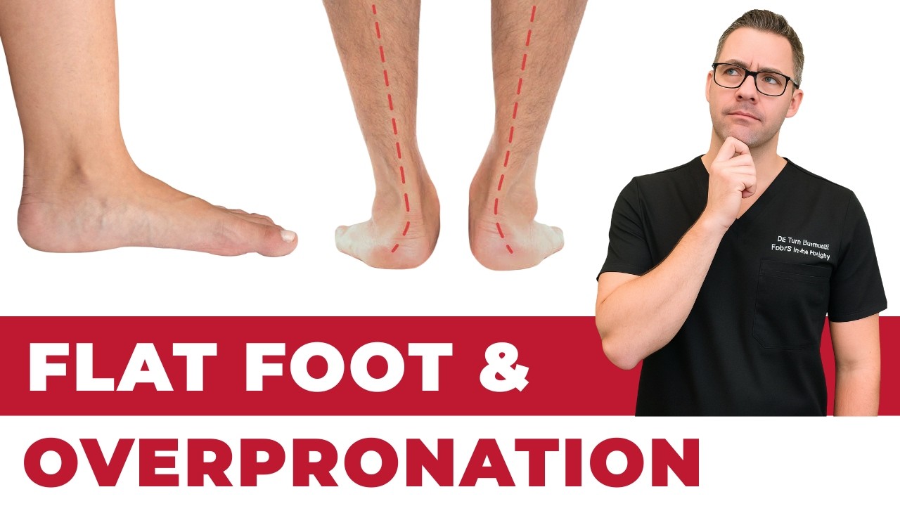
The posterior tibial tendon (PTT) is the primary dynamic stabilizer of the medial longitudinal arch. When it fails through progressive degeneration, partial tearing, or complete rupture — a condition called posterior tibial tendon dysfunction (PTTD) — the medial arch collapses, the hindfoot tips into valgus alignment, and the forefoot abducts. The clinical result is adult acquired flatfoot deformity (AAFD), a painful, progressive condition that ultimately prevents normal walking if left untreated.
Conservative management with custom ankle-foot orthoses (AFOs) can stabilize early PTTD and prevent progression. However, once Stage II deformity is established — with flexible but structurally significant flatfoot — surgical reconstruction offers more reliable and durable outcomes for active patients. The cornerstone of surgical reconstruction is tendon transfer: replacing the failed PTT with a functional tendon harvest from an adjacent structure capable of performing a similar biomechanical role.
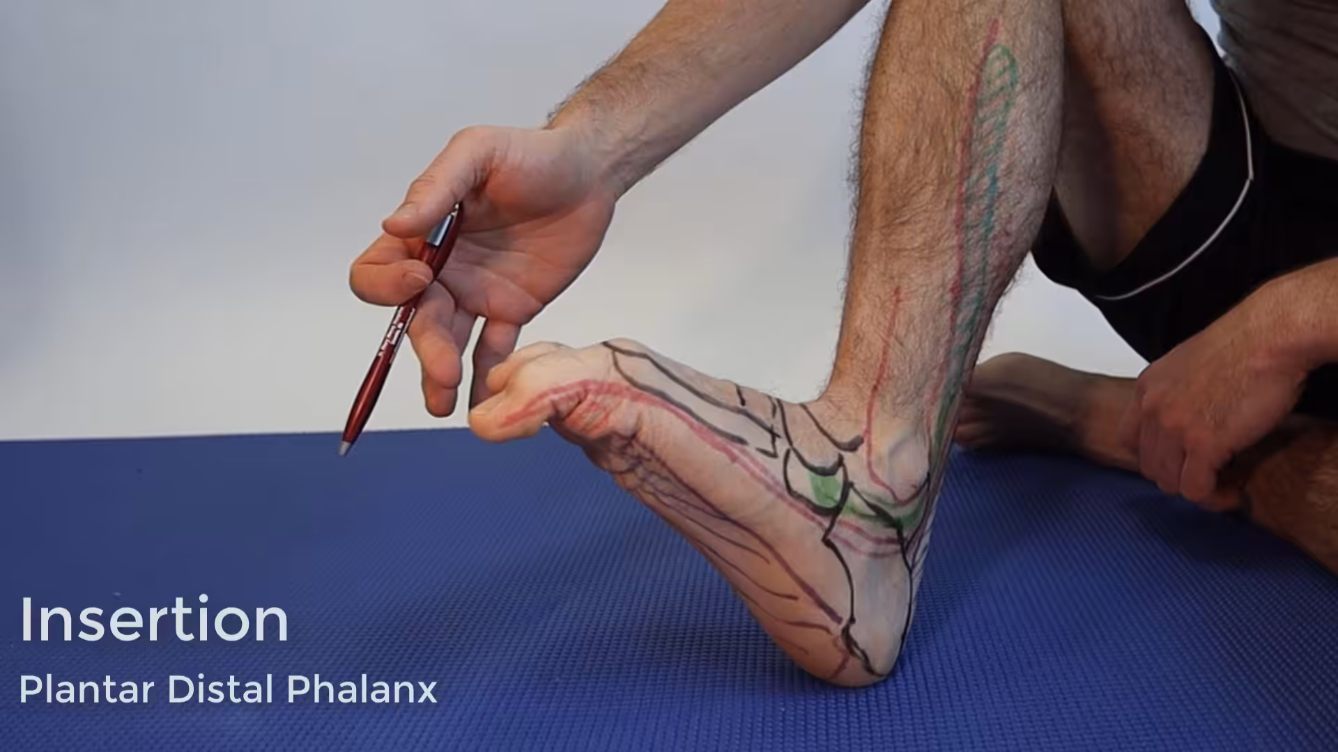
Why the FDL Is the Ideal Transfer Tendon
The flexor digitorum longus (FDL) is the preferred transfer tendon for PTT reconstruction for several reasons. It runs in close proximity to the PTT throughout its course — sharing the tarsal tunnel — simplifying the surgical approach. Its muscle-tendon unit is expendable: the great majority of toe flexion is performed by the intrinsic foot muscles, and sacrificing FDL power produces minimal functional deficit (mild weakness in lesser toe curl that most patients do not notice in daily activity). The FDL provides a strong, well-vascularized tendon of appropriate size and length for transfer to the navicular, the PTT’s primary insertion site. Its direction of pull — supination, inversion, and arch support — closely mimics the biomechanical role of the PTT.
Surgical Technique
Approach and Tendon Harvest
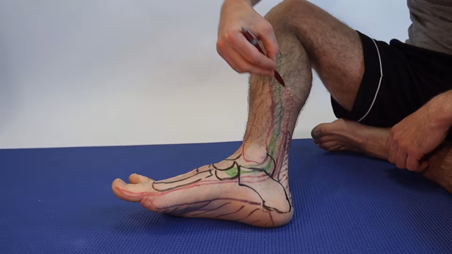
The FDL tendon transfer is performed through a longitudinal incision along the posteromedial aspect of the foot from the medial malleolus to the navicular. The native PTT is identified and assessed; even partially intact degenerated tendon may be incorporated into the repair as augmentation. The FDL is identified running parallel and posterior to the PTT, then harvested at the Master Knot of Henry — the point near the mid-arch where the FDL and FHL tendons cross — maximizing harvest length while preserving distal toe flexion through the FHL and intrinsic muscles.
Tendon Fixation
The harvested FDL is routed through a bone tunnel drilled through the navicular — the bone into which the PTT normally inserts — from the dorsal to plantar cortex, or from medial to lateral. The foot is held in a position of maximal inversion and plantarflexion (corrected alignment) while the FDL is tensioned and secured within the bone tunnel using an interference screw, a suture anchor, or button-pull-through technique. The goal is to set the transfer under appropriate tension so that it actively supports the arch in the corrected position during weight-bearing.
Adjunct Procedures
FDL transfer alone is rarely sufficient to correct all components of PTTD at Stage II. It is almost universally combined with one or more bony procedures:
- Medializing calcaneal osteotomy: corrects hindfoot valgus by shifting the heel medially
- Evans lateral column lengthening osteotomy: corrects forefoot abduction in Stage IIb
- Spring ligament repair or reconstruction: addresses the primary passive arch stabilizer that has typically been compromised alongside the PTT
- Gastrocnemius recession or Achilles lengthening: addresses equinus contracture — the almost-universal concomitant finding in PTTD — that would otherwise defeat the reconstruction by generating excessive arch-flattening tension through the Achilles
Postoperative Protocol
The reconstructed foot is immobilized in a non-weight-bearing splint for 2 weeks for wound healing, then transitioned to a non-weight-bearing fiberglass cast for an additional 4 weeks while the osteotomies and tendon transfer begin to heal. At 6 weeks, progressive weight-bearing in a CAM boot begins — typically starting with 50% weight-bearing and advancing weekly. Transition to a supportive shoe with a custom AFO occurs at 10–12 weeks. Physical therapy focusing on posterior tibial muscle activation, calf stretching, and proprioceptive retraining begins at 10–12 weeks and continues for 4–6 months. Return to full unrestricted activity — including sports — typically requires 9–12 months from the date of surgery.
Outcomes and Evidence
FDL transfer combined with calcaneal osteotomy and adjunct procedures is the most evidence-supported surgical strategy for Stage IIa and IIb PTTD. Long-term outcomes studies demonstrate significant improvement in pain, function, and radiographic alignment compared to pre-operative baselines. American Orthopaedic Foot and Ankle Society (AOFAS) hindfoot scores improve by an average of 40–50 points. Patient satisfaction rates are 80–90% in well-selected series. Predictors of less favorable outcomes include obesity (BMI >35), advanced age, severity of preoperative deformity, and incomplete adherence to postoperative rehabilitation protocols.
Consultation for PTTD
Patients with medial ankle pain, progressive flatfoot deformity, or difficulty rising onto one tiptoe should seek podiatric evaluation promptly. Early diagnosis preserves more treatment options and generally produces better outcomes. Our podiatric surgeons at Balance Foot & Ankle perform comprehensive PTTD staging, conservative management, and surgical reconstruction for all stages of adult acquired flatfoot deformity.
Expert Podiatric Care Across Southeast Michigan
Board-certified podiatrists — same-week appointments, most insurance accepted.
Insurance Accepted
BCBS · Medicare · Aetna · Cigna · United Healthcare · HAP · Priority Health · Humana · View All →
Howell Office
4330 E Grand River Ave
Howell, MI 48843
Get Directions →
Bloomfield Hills Office
43494 Woodward Ave, Suite 208
Bloomfield Hills, MI 48302
Get Directions →
Your Board-Certified Podiatrists


Ready to Get Back on Your Feet?
Same-week appointments available at both locations.
More Podiatrist-Recommended Flat Feet Essentials
PowerStep Pinnacle Insole


![How to Fix Flat Feet? [Collapsing Arch Pain & Flat Foot Correction!]](https://www.michiganfootdoctors.com/wp-content/cache/flying-press/7d7817e5895b520e184d5a23725fccc6.jpg)
Watch: How to Fix Flat Feet? [Collapsing Arch Pain & Flat Foot Correction!] — MichiganFootDoctors YouTube
Top orthotic for flat feet — lifts the collapsed arch and controls pronation.
Stability Running Shoe
New Balance Fresh Foam X 860 — designed for overpronators with flat feet.
Supportive Stability Shoe
Brooks Adrenaline GTS 25 — gold-standard stability shoe for flat feet.
As an Amazon Associate, Balance Foot & Ankle earns from qualifying purchases. Product recommendations are based on clinical experience; prices and availability shown above update live from Amazon.
When to See a Podiatrist
Painful flat feet in adults can signal posterior tibial tendon dysfunction — a progressive condition that needs early intervention to avoid surgery. Balance Foot & Ankle evaluates adult flatfoot with weight-bearing imaging and custom orthotic prescriptions. Catching PTTD at stage 1-2 makes the difference between a brace and a reconstruction.
Call Balance Foot & Ankle: (810) 206-1402 · Book online · Offices in Howell & Bloomfield Hills
Differential Diagnosis: What Else Could It Be?
Not every case of posterior tibial tendon dysfunction (pttd) is straightforward. In our clinic we routinely rule out three look-alike conditions before confirming the diagnosis. If your symptoms don’t match the classic presentation, one of these may explain the pain — which is why physical exam matters more than self-diagnosis.
| Condition | How It Differs |
|---|---|
| Congenital flat foot | Lifelong, usually bilateral, no pain, normal single-leg heel-rise test. |
| Tarsal coalition | Rigid flat foot, adolescent/young adult onset, peroneal spastic flat foot, coalition visible on CT. |
| Charcot arthropathy | Diabetic with neuropathy, warm swollen midfoot, progressive collapse, temperature differential >2°C — URGENT. |
Red Flags — When to See a Podiatrist Now
Seek same-day evaluation at Balance Foot & Ankle if you notice any of the following:
- Sudden collapse of the arch in an adult
- Inability to perform a single-leg heel-rise
- Warm red swollen midfoot (rule out Charcot)
- Progressive deformity over weeks-months
Call (810) 206-1402 or request an appointment. Our Howell and Bloomfield Hills offices reserve same-day slots for urgent foot and ankle issues.
In Our Clinic: What We See
Clinical perspective from Dr. Tom Biernacki, DPM — Balance Foot & Ankle, Howell & Bloomfield Hills, MI:
In our clinic, adult acquired flatfoot from PTTD typically presents in women over 40, often with recent weight gain or a period of increased standing. They describe medial ankle pain and progressive “collapse” of the arch on one side. The gold-standard exam finding is an inability to perform a single-leg heel-rise on the affected side — the tendon can no longer invert the heel into a rigid lever. Early PTTD is staged and treated with custom orthoses and bracing, but progressive disease (Stage III-IV) typically requires surgical reconstruction to prevent rigid deformity.
In-Office Treatment at Balance Foot & Ankle
When conservative care isn’t enough, Dr. Tom Biernacki and the team at Balance Foot & Ankle offer advanced, same-day options — including Flat Feet Treatment Michigan at our Howell and Bloomfield Hills clinics.
Same-day appointments available. Call (810) 206-1402 or book online.
In-Office Treatment at Balance Foot & Ankle
If home treatment isn’t providing relief for your tendon pain, our podiatry team at Balance Foot & Ankle can help with same-day evaluations and advanced in-office care.
Same-day appointments available. (810) 206-1402
Doctor Hoy’s Natural Pain Relief Gel
Natural topical pain relief I use in our clinic. Arnica + camphor formula — apply directly to the area 3–4x daily. ($20–25)
Frequently Asked Questions
When should I see a podiatrist?
If symptoms persist past 2 weeks, affect your normal activity, or are accompanied by red-flag symptoms (warmth, redness, swelling, inability to bear weight).
What does treatment cost?
Most diagnostic visits and conservative treatments are covered by Medicare and major insurers. Out-of-pocket costs vary by your specific plan.
How quickly can I get an appointment?
Most non-urgent cases see us within 5 business days. Urgent cases (sudden pain, possible fracture) typically same or next business day.
Dr. Tom Biernacki, DPM is a board-certified foot & ankle surgeon (ABFAS & ABPM) at Balance Foot & Ankle Specialists in Southeast Michigan. With over a decade of clinical experience, he specializes in heel pain, bunions, diabetic foot care, sports injuries, and minimally invasive surgery. Dr. Biernacki is a member of the APMA and ACFAS, and his patient education content on MichiganFootDoctors.com and YouTube has made him one of the most-followed foot & ankle educators on YouTube.