
Quick answer: Lisfranc Injury Midfoot Fracture Dislocation 3 is a common foot/ankle topic that affects many patients. The 2026 evidence-based approach combines proper diagnosis, conservative-first treatment, and escalation only when needed. We treat this regularly at our Howell and Bloomfield Hills practices. Call (810) 206-1402.
▶ Watch
What Is the Lisfranc Joint Complex?
Medically Reviewed by Dr. Tom Biernacki, DPM — Board-Certified Podiatrist, Balance Foot & Ankle Specialists, Michigan. Last updated April 2026.
The Lisfranc joint complex — formally the tarsometatarsal (TMT) joint complex — is the articulation between the five metatarsal bases and the three cuneiform bones plus the cuboid, forming the junction between the midfoot and forefoot. Named after French surgeon Jacques Lisfranc de St. Martin who described amputations at this level during the Napoleonic Wars, injuries at this anatomical site are among the most frequently missed diagnoses in acute traumatic foot care. The Lisfranc ligament — a strong dorsal, interosseous, and plantar ligament complex connecting the medial cuneiform to the second metatarsal base — is the anatomical keystone. Its disruption destabilizes the entire TMT complex.
Mechanisms of Injury
High-Energy Trauma
Motor vehicle accidents, falls from height, and direct crush injuries produce high-energy Lisfranc injuries with significant displacement, multiple fractures, and obvious deformity. These injuries present acutely in emergency department settings and are typically diagnosed promptly with plain radiographs.
Low-Energy Indirect Mechanism
The most commonly missed Lisfranc injuries occur through low-energy indirect mechanisms: a stumbling fall on a plantarflexed foot, a sports injury when a running athlete’s foot is fixed on the ground while the body rotates, or the ‘fall off a step’ injury. These injuries may produce purely ligamentous disruption without fracture — which is more difficult to diagnose radiographically and carries greater risk of missed diagnosis. Athletes often initially describe a midfoot sprain; the true severity is not recognized until X-rays are obtained (often days later) or the patient fails to improve on the trajectory expected for a simple sprain.
Clinical Presentation
Acute Lisfranc injury produces midfoot pain, swelling, and ecchymosis (bruising) across the dorsal foot. A classic finding is plantar ecchymosis visible across the arch — when midfoot ligaments rupture, blood tracks to the plantar surface and produces bruising in the arch that is pathognomonic (highly specific) for significant ligamentous disruption at the TMT complex. Inability to bear weight, or severe pain with any forefoot loading, should prompt X-ray evaluation.
Radiographic Evaluation
Weight-Bearing X-Rays
Standard non-weight-bearing X-rays frequently appear normal in ligamentous Lisfranc injuries because the partially disrupted ligaments can maintain near-normal joint alignment when the joint is unloaded. Weight-bearing X-rays — obtained with the patient standing, which is often severely painful and may require modification — are essential and frequently reveal diastasis (widening) between the first and second metatarsal bases at the TMT level that is not visible on non-weight-bearing films. A diastasis greater than 2mm is considered diagnostic.
CT Scan
CT provides far superior osseous detail compared to plain X-rays, identifying small avulsion fractures at the second metatarsal base (the ‘fleck sign’ — a pathognomonic finding for Lisfranc ligament avulsion), cuneonavicular fractures, and the full extent of displacement. CT is routinely obtained in all suspected Lisfranc injuries to guide surgical planning.
MRI
MRI directly visualizes ligamentous integrity, grading injuries from partial to complete disruption. It is particularly valuable for purely ligamentous Lisfranc injuries without fracture where CT is negative, providing definitive confirmation of ligamentous instability and guiding the decision between conservative and surgical management.
Classification
Myerson’s modification of Hardcastle’s classification describes Lisfranc injuries in three types: Type A (total incongruity — all five TMT joints displaced), Type B (partial incongruity — medial column or lateral column displacement), and Type C (divergent — the first metatarsal displaces medially while the second through fifth displace laterally). This classification guides surgical approach selection. The Nunley-Vertullo classification specifically addresses athletic (low-energy) Lisfranc injuries in a clinically useful staging system from Stage I (sprain without diastasis) through Stage III (diastasis with arch height loss).
Non-Surgical Treatment
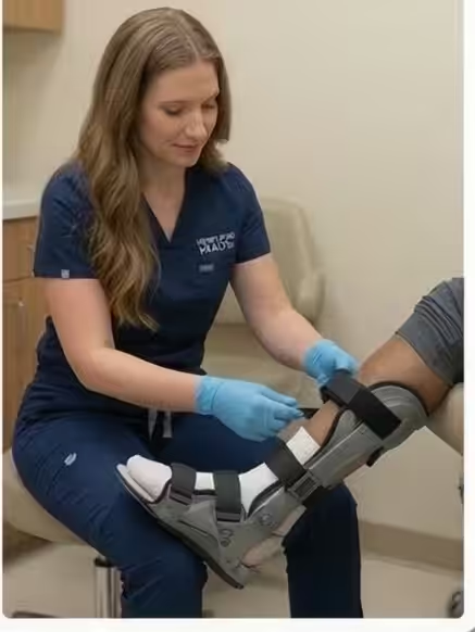
Purely ligamentous, non-displaced Lisfranc injuries (Nunley-Vertullo Stage I) — confirmed by normal weight-bearing X-rays and MRI showing partial ligamentous injury — may be managed non-surgically with non-weight-bearing casting for 6 weeks, followed by progressive rehabilitation in a stiff-soled shoe or boot. However, this approach requires close radiographic follow-up to confirm maintained alignment, and any displacement on follow-up imaging mandates surgical revision of management. Athletes with Stage I injuries treated conservatively show variable outcomes — a significant proportion develop chronic midfoot pain and require eventual surgical stabilization.
Surgical Treatment: ORIF vs. Primary Arthrodesis
Open Reduction and Internal Fixation (ORIF)
Displaced Lisfranc injuries require surgical open reduction and internal fixation. Surgical exposure identifies the disrupted TMT joints; the medial column (first TMT, medial and intermediate cuneiform joints) is anatomically reduced and fixed with transarticular screws or bridge plating. The lateral column (fourth and fifth TMT joints) — which requires flexible motion for normal gait — is provisionally fixed with flexible suture button devices rather than rigid screws to preserve motion. Hardware is typically removed at 3–4 months to restore joint motion before definitive healing.
Primary Arthrodesis
Growing evidence supports primary arthrodesis (immediate fusion) of the medial column TMT joints for purely ligamentous Lisfranc injuries and for high-energy injuries with comminuted articular damage. Fusion eliminates the risk of post-traumatic arthrosis of the medial column — the most common long-term complication of Lisfranc injury — while achieving stability without requiring hardware removal. Multiple prospective studies demonstrate superior long-term functional outcomes with primary arthrodesis compared to ORIF for medial column ligamentous injuries.
Recovery and Prognosis
Recovery from Lisfranc injury — regardless of treatment method — requires patience. Non-weight-bearing for 6–8 weeks is followed by gradual progressive loading over 4–6 weeks. Full return to athletic activity typically requires 6–9 months for ORIF and 9–12 months for arthrodesis. Prognosis depends critically on time to diagnosis: injuries identified and stabilized within the first 48–72 hours have substantially better outcomes than those diagnosed weeks later after progressive displacement and articular cartilage damage. Any athlete with significant midfoot pain after a stumble or twisting injury who is not improving as expected for a routine sprain should have weight-bearing X-rays and orthopaedic or podiatric surgical evaluation.
Trusted Podiatric Care Across Southeast Michigan
Board-certified podiatrists — same-week appointments, most insurance accepted including Medicare.
Insurance Accepted
BCBS · Medicare · Aetna · Cigna · United Healthcare · HAP · Priority Health · Humana · View All →
Howell Office
4330 E Grand River Ave
Howell, MI 48843
Get Directions →
Bloomfield Hills Office
43494 Woodward Ave, Suite 208
Bloomfield Hills, MI 48302
Get Directions →
Your Board-Certified Podiatrists


Ready to Get Back on Your Feet?
Same-week appointments available at both locations.
Differential Diagnosis: What Else Could It Be?
Not every case of lisfranc (midfoot) injury is straightforward. In our clinic we routinely rule out three look-alike conditions before confirming the diagnosis. If your symptoms don’t match the classic presentation, one of these may explain the pain — which is why physical exam matters more than self-diagnosis.
| Condition | How It Differs |
|---|---|
| Midfoot sprain | No diastasis on X-ray; able to bear weight after initial pain. |
| Navicular stress fracture | Dorsal midfoot pain with impact loading; stress fx confirmed on MRI. |
| Cuboid syndrome | Lateral midfoot pain, often following ankle inversion; relieved by cuboid whip. |
Red Flags — When to See a Podiatrist Now
Seek same-day evaluation at Balance Foot & Ankle if you notice any of the following:
- Pain out of proportion to injury severity
- Plantar bruising across the arch (classic Lisfranc sign)
- Inability to bear weight for >24 hours
- Widening of tarsometatarsal joints on weight-bearing X-ray
Call (810) 206-1402 or request an appointment. Our Howell and Bloomfield Hills offices reserve same-day slots for urgent foot and ankle issues.
In Our Clinic: What We See
Clinical perspective from Dr. Tom Biernacki, DPM — Balance Foot & Ankle, Howell & Bloomfield Hills, MI:
Lisfranc injury is the most-missed foot injury in primary care and emergency rooms. Patients walk in weeks after a misstep complaining of midfoot pain that never resolves. In our clinic the first clue is often the bruising pattern — plantar bruising across the arch is pathognomonic. Weight-bearing X-rays comparing both feet reveal the widening that non-weight-bearing films miss. Non-displaced Lisfranc sprains can heal in a boot; any displacement requires surgery. Dr. Biernacki has handled dozens of missed Lisfranc injuries and always comments: if a midfoot sprain isn’t significantly better at 3 weeks, get weight-bearing films — don’t wait.
More Podiatrist-Recommended Foot Health Essentials
Hoka Clifton 10


Max-cushion everyday shoe — podiatrist favorite for walking and running.

OOFOS Recovery Slide

Impact-absorbing recovery sandal — wear after long days on your feet.
As an Amazon Associate, Balance Foot & Ankle earns from qualifying purchases. Product recommendations are based on clinical experience; prices and availability shown above update live from Amazon.
When to See a Podiatrist
If foot or ankle pain has been bothering you for more than a few weeks, home care alone may not be enough. Balance Foot & Ankle offers same-week appointments at our Howell and Bloomfield Hills clinics — no referral needed in most cases. Bring your current shoes and a short list of symptoms and we’ll build you a treatment plan in one visit.
Call Balance Foot & Ankle: (810) 206-1402 · Book online · Offices in Howell & Bloomfield Hills
In-Office Treatment at Balance Foot & Ankle
When conservative care isn’t enough, Dr. Tom Biernacki and the team at Balance Foot & Ankle offer advanced, same-day options — including Foot & Ankle Fracture Repair Michigan at our Howell and Bloomfield Hills clinics.
Same-day appointments available. Call (810) 206-1402 or book online.
In-Office Treatment at Balance Foot & Ankle
If home treatment isn’t providing relief for your foot and ankle conditions, our podiatry team at Balance Foot & Ankle can help with same-day evaluations and advanced in-office care.
Same-day appointments available. (810) 206-1402
Doctor Hoy’s Natural Pain Relief Gel
Natural topical pain relief I use in our clinic. Arnica + camphor formula — apply directly to the area 3–4x daily. ($20–25)
Frequently Asked Questions
When should I see a podiatrist?
If symptoms persist past 2 weeks, affect your normal activity, or are accompanied by red-flag symptoms (warmth, redness, swelling, inability to bear weight).
What does treatment cost?
Most diagnostic visits and conservative treatments are covered by Medicare and major insurers. Out-of-pocket costs vary by your specific plan.
How quickly can I get an appointment?
Most non-urgent cases see us within 5 business days. Urgent cases (sudden pain, possible fracture) typically same or next business day.
What is Stress fracture?
Stress fracture is a common foot/ankle condition that affects mobility and quality of life. Understanding the underlying cause is the first step in successful treatment. Our podiatrists at Balance Foot & Ankle perform a hands-on biomechanical exam, review your activity history, and use diagnostic imaging when appropriate to identify the root cause—not just treat the symptom. Many patients have been told to “rest and ice” without a deeper diagnostic workup; our approach is different.
Symptoms and warning signs
Common signs of stress fracture include pain that worsens with activity, morning stiffness, swelling, tenderness when palpated, and difficulty bearing weight. If you experience sudden severe pain, inability to walk, visible deformity, numbness or color change, contact our office the same day or visit urgent care—these can signal a more serious injury such as a fracture, tendon rupture, or vascular compromise. Diabetics with any foot wound should seek same-day care.
Conservative treatment options
Most cases of stress fracture respond to non-surgical care: structured rest, supportive footwear changes, custom orthotics, targeted stretching and strengthening protocols, anti-inflammatory medications when medically appropriate, and in-office procedures such as ultrasound-guided injections. We also offer advanced therapies including MLS laser therapy, EPAT/shockwave, regenerative injections, and image-guided procedures. Treatment is sequenced from least invasive to most invasive, and we explain the rationale at every step.
When is surgery considered?
Surgery is reserved for cases that fail 3-6 months of well-structured conservative care, when there is structural pathology (severe deformity, complete tear, advanced arthritis), or when imaging shows damage that will not heal without intervention. Our surgeons have performed 3,000+ foot and ankle procedures and prioritize minimally-invasive techniques whenever appropriate. We discuss recovery timelines, return-to-activity milestones, and realistic outcome expectations before any procedure is scheduled.
Recovery timeline and prevention
Recovery from stress fracture varies based on severity and chosen treatment path. Conservative cases often improve within 4-8 weeks with consistent adherence to the protocol. Post-procedural recovery may range from a few days (in-office procedures) to several months (reconstructive surgery). Long-term prevention involves footwear assessment, activity modification, structured strengthening, and regular check-ins with your podiatrist if you have a history of recurrence. We provide written home-exercise plans and digital follow-up support.
Ready to feel better?
Same-week appointments available in Howell and Bloomfield Hills, Michigan.
Dr. Tom Biernacki, DPM is a board-certified foot & ankle surgeon (ABFAS & ABPM) at Balance Foot & Ankle Specialists in Southeast Michigan. With over a decade of clinical experience, he specializes in heel pain, bunions, diabetic foot care, sports injuries, and minimally invasive surgery. Dr. Biernacki is a member of the APMA and ACFAS, and his patient education content on MichiganFootDoctors.com and YouTube has made him one of the most-followed foot & ankle educators on YouTube.