
Medically reviewed by Dr. Tom Biernacki, DPM
Board-certified podiatric surgeon | Balance Foot & Ankle
Last reviewed: May 2026

Lisfranc Injury Treatment: Severity Classification Determines Surgery vs. Boot
Lisfranc injuries are the most commonly missed significant foot injury in emergency medicine — X-rays appear normal in 30% of cases unless weight-bearing views are obtained. The Lisfranc complex (tarsometatarsal joint complex) is the architectural keystone of the midfoot, connecting the metatarsal bases to the tarsal bones. A disruption here — even “sprain only” — can lead to chronic midfoot collapse and arthritis if undertreated. The clinical pearl: any patient with midfoot swelling and bruising (especially plantar ecchymosis) after a twisting or crush mechanism needs standing X-rays, not just supine.
| Lisfranc Classification | Anatomy | X-Ray Findings | Treatment | Return to Sport |
|---|---|---|---|---|
| Grade 1 — Sprain (ligamentous, no diastasis) | Lisfranc ligament stretched but intact; no instability on stress exam; no diastasis (gap) between 1st and 2nd metatarsal bases | Normal on non-weight-bearing views; standing AP X-ray: 2nd MT base-medial cuneiform gap <2mm; no step-off at tarsometatarsal joints | Non-weight-bearing cast or boot × 4-6 weeks; strict NWB for first 2-3 weeks; transition to protected weight-bearing in boot at 3-4 weeks; physical therapy for proprioception and strengthening after 6 weeks; return to sport 8-12 weeks | 8-12 weeks; must confirm pain-free single-leg heel raise and pain-free running before return; high rate of re-injury if returned too early |
| Grade 2 — Partial instability (partial diastasis) | Partial Lisfranc ligament tear; possible 2nd metatarsal base avulsion fracture (the “fleck sign” — pathognomonic); stress examination shows mild instability | 2nd MT base-medial cuneiform gap 2-4mm on standing views; possible fleck fracture at 2nd MT base or medial cuneiform; subtle step-off at 1st-2nd TMT joint | SURGERY vs conservative decision: diastasis 2-4mm — some surgeons treat conservatively (strict NWB cast 6 weeks + extended boot); others prefer surgical fixation to prevent progressive collapse; dynamic stability testing under anesthesia may help decision; most sports medicine/foot surgeons prefer fixation for active patients | Conservative: 16-20 weeks. Surgical: 12-16 weeks with hardware removal at 3-4 months before return to sport |
| Grade 3 — Complete instability / fracture-dislocation | Complete Lisfranc ligament disruption; instability across multiple tarsometatarsal joints; metatarsal bases displaced; associated fractures common | Obvious diastasis >4mm on standing views (or any diastasis on supine views); frank dislocation of metatarsal bases; multiple TMT joint fractures; “nutcracker” pattern with cuboid fracture possible | SURGICAL FIXATION required — open reduction and internal fixation (ORIF) with screws and/or dorsal bridge plating; accurate reduction is critical — 1-2mm residual diastasis significantly worsens outcome; temporary bridging plates often removed at 4-6 months | 12-18 months to full unrestricted sport; return to recreational activity at 6-9 months; high-level athletes may require 18 months; long-term midfoot arthritis development in 25-40% even with anatomic fixation |
| Grade 4 — Missed/chronic Lisfranc with midfoot arthritis | Prior Lisfranc injury treated inadequately or not diagnosed; midfoot collapse (acquired flatfoot pattern); degenerative arthritis at tarsometatarsal joints; painful midfoot arch collapse | Loss of Meary’s angle (talus-first metatarsal alignment); arthritic changes at TMT joints; periarticular sclerosis; joint space narrowing; midfoot sag on lateral weight-bearing view | Conservative: custom AFO or rigid arch support, activity modification, cortisone injection for flares; Surgical: midfoot fusion (TMT arthrodesis) for failed conservative — excellent outcomes 85-90% satisfaction, definitively addresses the arthritic joints and restores midfoot alignment | Post-fusion: 12-16 weeks to walking; full activity 9-12 months; fusion is permanent and highly effective for chronic painful Lisfranc arthritis |
Lisfranc Injury: Critical Diagnostic Steps Often Missed
| Diagnostic Step | Why Critical | What to Look For |
|---|---|---|
| Standing (weight-bearing) X-rays | Non-weight-bearing X-rays appear normal in 30-50% of Lisfranc injuries; the diastasis only appears when the foot is loaded; non-WB views in the ED commonly miss this injury | On AP weight-bearing view: gap >2mm between 2nd metatarsal base and medial cuneiform = Lisfranc diastasis; any step-off at the TMT joints; compare both feet side by side |
| Fleck sign | Pathognomonic for Lisfranc injury — small avulsion fracture fragment between the 1st and 2nd metatarsal bases represents avulsion of the Lisfranc ligament; often dismissed as trivial | Small bone fragment on AP X-ray at the base of 2nd metatarsal or medial cuneiform; if present, Lisfranc ligament disruption is confirmed regardless of diastasis measurement |
| Plantar ecchymosis | Bruising appearing on the PLANTAR (bottom) surface of the midfoot is highly specific for Lisfranc injury; blood tracks plantarly from the torn ligament complex; commonly absent on dorsal exam | Examine the bottom of the foot; plantar bruising at the midfoot arch = Lisfranc injury until proven otherwise; this sign is missed when patients are not asked to remove shoes and socks |
| MRI if X-rays negative | High clinical suspicion (midfoot pain + mechanism) with negative X-rays warrants MRI; ligamentous injury without diastasis (Grade 1) is invisible on X-ray; MRI shows Lisfranc ligament continuity and bone marrow edema | Lisfranc ligament between medial cuneiform and 2nd metatarsal base; bone marrow edema at TMT joints; interosseous ligament injuries; these findings change treatment from “ankle sprain” to strict NWB protocol |
| CT scan for surgical planning | Once surgery is decided, CT defines fracture patterns, comminution, associated injuries, and guides screw/plate placement; X-rays insufficient for operative planning in Grade 3 | Precise alignment of TMT joints; cuboid fractures; navicular fractures; medial column vs lateral column involvement; determines whether bridge plating vs screw fixation is appropriate |
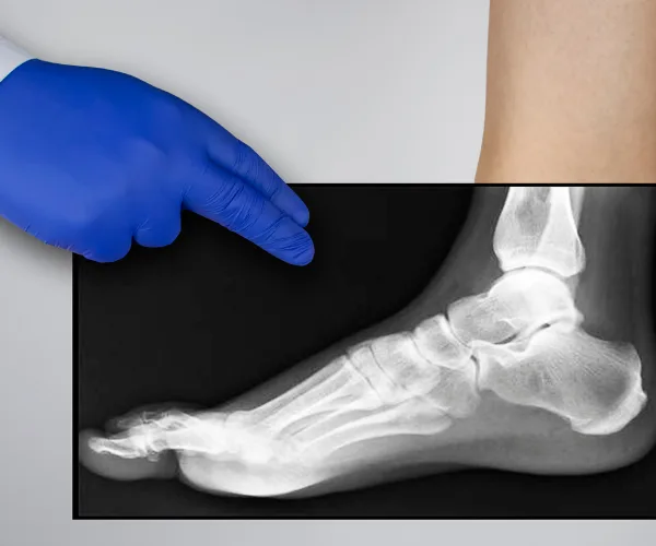
Foot pain isn't resolving?
Same-week appointments at podiatrist in Howell & podiatrist in Bloomfield Township
⚡ Quick Answer: How is a Lisfranc injury treated?
Minor Lisfranc injuries are treated with a non-weight-bearing cast for 6–8 weeks. Displaced or unstable injuries require surgical fixation and careful rehabilitation.
Related Conditions
In This Article
- How are Lisfranc injuries treated?
- Quick Answer: Lisfranc Injury Treatment
- What Is a Lisfranc Injury
- Anatomy and Injury Mechanisms
- Classification and Severity
- Symptoms and Diagnosis
- Treatment Options
- Recovery and Return to Activity
- Red Flags: Seek Immediate Evaluation
- Most Common Mistake with Lisfranc Injuries
- In-Office Treatment at Balance Foot & Ankle
- Frequently Asked Questions
- Sources
- Midfoot Injury? Don’t Wait — Lisfranc Injuries Worsen with Delay.
- Frequently Asked Questions
- Frequently Asked Questions
- What is Foot pain?
- Symptoms and warning signs
- Conservative treatment options
- When is surgery considered?
- Recovery timeline and prevention
Quick Answer: Lisfranc Injury Treatment
Lisfranc injuries — fractures or ligament tears at the tarsometatarsal joint complex — range from subtle sprains to complex fracture-dislocations. Stable injuries without displacement are treated non-operatively in a non-weight-bearing cast for 6–8 weeks. Unstable or displaced injuries require surgical fixation (ORIF or primary arthrodesis) to restore alignment and prevent chronic pain and collapse of the midfoot arch.
Lisfranc injuries are among the most misdiagnosed injuries in sports medicine and emergency medicine — commonly dismissed as “just a foot sprain” because X-rays can look normal in the acute setting. Missing this diagnosis is one of the most consequential mistakes in foot and ankle care: an undertreated Lisfranc injury leads to chronic midfoot pain, progressive flatfoot deformity, and post-traumatic arthritis that can be permanently disabling. In our practice, we’ve seen patients who spent months limping on a midfoot fracture-dislocation that was read as a sprain in the ER. Understanding this injury properly — its anatomy, its subtle X-ray findings, and its treatment requirements — is the first step to getting it right.
What Is a Lisfranc Injury
The Lisfranc joint complex — named after Jacques Lisfranc de Saint-Martin, the French surgeon who first described injuries at this level — consists of the articulations between the five metatarsal bases and the three cuneiforms and cuboid bone. This tarsometatarsal (TMT) joint complex is the keystone of the midfoot arch. The Lisfranc ligament itself is an oblique ligament connecting the medial cuneiform to the base of the second metatarsal — the strongest stabilizer of the entire complex. When it ruptures, the midfoot loses its structural integrity and the metatarsals can displace laterally, dorsally, or in a divergent pattern.
Anatomy and Injury Mechanisms
Lisfranc injuries occur through two main mechanisms. High-energy trauma — motor vehicle accidents, industrial crush injuries, falls from height — produces the severe fracture-dislocation pattern that is immediately obvious on X-ray. Low-energy mechanisms are subtler and account for the majority of missed diagnoses: a stumble with the forefoot planted (stepping in a hole, missing a step), a twisting fall while the foot is plantar-flexed, or even a direct axial load through the metatarsals (jumping and landing on a plantar-flexed foot). Athletes — particularly football players, equestrians, and dancers — are especially vulnerable to the low-energy mechanism. In our clinic, we treat multiple Lisfranc injuries per year in athletes, and a significant percentage were initially told they had “nothing serious” on their first evaluation.
Classification and Severity
The Myerson classification (a modification of Quenu-Küss) is the most widely used system and guides treatment decisions. All three columns of the midfoot — medial, middle, and lateral — can be affected, and injuries can be homolateral (all rays displaced in same direction), isolated (one or two rays), or divergent (metatarsals displaced in opposite directions).
| Severity | Findings | Treatment |
|---|---|---|
| Sprain (Grade I) | Ligamentous injury, stable, no displacement on stress X-ray | NWB cast 6–8 weeks, then CAM boot |
| Partial disruption (Grade II) | <2 mm displacement, partial ligament tear | NWB cast 6–8 weeks; surgery if instability confirmed |
| Complete disruption (Grade III) | ≥2 mm displacement, complete ligament disruption, fracture | Surgical fixation (ORIF or primary arthrodesis) |
Symptoms and Diagnosis
The classic symptom triad of a Lisfranc injury is midfoot pain, swelling, and inability to bear weight after the mechanism of injury. The plantar ecchymosis sign — bruising on the sole of the foot in the midfoot region — is highly specific for Lisfranc ligament disruption and should prompt immediate imaging. However, bruising may not appear for 12–24 hours after injury, and its absence doesn’t exclude the diagnosis.
Standard weight-bearing anteroposterior, lateral, and oblique foot X-rays are the initial imaging. Key radiographic findings include: a gap of >2 mm between the bases of the first and second metatarsals (diastasis), a “fleck sign” (avulsion fragment from the Lisfranc ligament at the medial cuneiform or 2nd metatarsal base), and loss of alignment between the medial border of the 2nd metatarsal and the medial border of the middle cuneiform. CT scan is the gold standard for surgical planning and identifies fracture patterns invisible on X-ray. MRI defines ligamentous injury extent and is particularly useful in subtle sprains where X-rays appear normal but clinical suspicion remains high. Stress X-rays under fluoroscopy — taken with abduction force applied to the forefoot — unmask instability in grade I-II injuries that appear stable on routine films.
Treatment Options
Non-Operative Treatment (Stable Injuries)
Truly stable Lisfranc sprains — defined as <2 mm diastasis on weight-bearing X-rays or stress fluoroscopy, with intact ligamentous architecture on MRI — can be treated non-operatively with excellent outcomes. The protocol: non-weight-bearing short leg cast or CAM boot for 6–8 weeks, followed by a graduated return to weight-bearing in a rigid-soled supportive boot or shoe. Premature weight-bearing is the most common cause of non-operative treatment failure — even “stable” injuries can displace if loaded before adequate ligamentous healing. At 6–8 weeks, repeat weight-bearing X-rays confirm maintenance of alignment before advancing to partial weight-bearing. Full return to sport typically requires 3–5 months.
Surgical Treatment: ORIF
Open reduction and internal fixation (ORIF) is indicated for displaced or unstable Lisfranc injuries. The goal is anatomic restoration of tarsometatarsal alignment. Multiple techniques are used — transarticular screws, bridge plating, and dorsal plating — with the specific construct determined by the fracture and dislocation pattern. Screws crossing the Lisfranc joint require elective removal at 3–4 months (before stress loading resumes) because they will break if left in place during weight-bearing. Hardware removal is a second surgical procedure but is routine and typically done under local anesthesia. Outcomes of ORIF depend heavily on the quality of anatomic reduction — studies consistently show that even 1–2 mm of residual diastasis significantly increases post-traumatic arthritis risk.
Surgical Treatment: Primary Arthrodesis
Primary arthrodesis — fusing the tarsometatarsal joints at the time of acute injury rather than fixing and hoping — has emerged as the preferred surgical treatment for purely ligamentous (no fracture) Lisfranc injuries. The landmark Ly and Coetzee RCT (2006) demonstrated significantly better functional outcomes with primary arthrodesis compared to ORIF for ligamentous injuries. The rationale: purely ligamentous Lisfranc injuries have high rates of post-traumatic arthritis requiring secondary fusion anyway, and primary fusion eliminates the second surgery while achieving equivalent alignment. The medial two columns (medial and middle cuneiform-metatarsal joints) are typically fused, while the lateral column (4th and 5th TMT joints) is preserved to maintain forefoot flexibility. Full weight-bearing in a boot begins at 8–12 weeks post-fusion.
Recovery and Return to Activity
Recovery from Lisfranc injury is measured in months, not weeks — a reality that consistently surprises both patients and non-specialist providers. Non-operative treatment: full return to sport at 3–5 months for truly stable sprains, with chronic aching and midfoot stiffness possible for 6–12 months. Operative ORIF: hardware removal at 3–4 months, return to low-impact activity at 4–6 months, full return to sport at 6–12 months. Primary arthrodesis: return to unrestricted activity at 4–6 months, with final fusion maturation at 6–12 months. Patients should expect midfoot stiffness (particularly at the fused segments), mild loss of push-off power, and slower return to high-demand sports. In our experience, athletes who receive timely, appropriate treatment consistently return to their prior level of sport; those who were missed or undertreated often do not.
Red Flags: Seek Immediate Evaluation
Go to urgent care or an ER if you have:
- Inability to bear any weight after a midfoot injury — requires immediate X-ray
- Visible deformity of the midfoot — frank dislocation is a surgical emergency
- Bruising on the sole of the foot (plantar ecchymosis) — highly specific for Lisfranc ligament disruption
- Progressive worsening of midfoot swelling over hours — compartment syndrome risk with high-energy injuries
- A “sprain” that hasn’t allowed walking after 2 weeks — request weight-bearing foot X-rays if none have been taken
- Numbness or vascular changes in the foot — neurovascular compromise in displaced fracture-dislocations
Most Common Mistake with Lisfranc Injuries
The most common and consequential mistake is missing the diagnosis entirely — particularly with low-energy Lisfranc sprains where non-weight-bearing X-rays appear normal. The critical error is taking foot X-rays non-weight-bearing: subtle diastasis that is invisible on a supine film becomes obvious on a standing weight-bearing view. Standard emergency department protocol typically does not include weight-bearing foot X-rays — which is why so many Lisfranc injuries are discharged as “midfoot sprain” without adequate imaging. If you’ve been told you have a midfoot sprain, you cannot walk without significant pain after 2 weeks, and you haven’t had weight-bearing X-rays — please seek podiatric or orthopedic evaluation before assuming you’re healing normally. A missed Lisfranc is far easier to treat acutely than after 3–6 months of walking on it.
In-Office Treatment at Balance Foot & Ankle
At Balance Foot & Ankle, we evaluate midfoot injuries with weight-bearing X-rays and diagnostic ultrasound in-office, with CT and MRI ordered same-day when indicated. Dr. Tom Biernacki performs Lisfranc ORIF and primary arthrodesis and manages the full continuum of care from acute stabilization through return to sport. We accept most major insurance plans and offer urgent appointments for acute foot injuries. Call (810) 206-1402 or book online. Offices in Howell and Bloomfield Township, MI.
Frequently Asked Questions
How long does it take to recover from a Lisfranc injury?
Recovery depends on severity: stable sprains treated non-operatively take 3–5 months for return to sport. Surgically fixed injuries require 6–12 months. Primary arthrodesis patients return to full activity at 4–6 months. Chronic stiffness and midfoot aching can persist for 12–18 months after injury regardless of treatment method.
Can a Lisfranc injury heal without surgery?
Yes — truly stable Lisfranc sprains (no diastasis on weight-bearing X-rays, intact ligaments on MRI) heal with non-operative treatment. However, “stable” must be confirmed with weight-bearing and stress X-rays. Injuries misclassified as stable that actually have subtle diastasis will not heal properly without surgery and lead to post-traumatic arthritis.
What is the difference between a Lisfranc sprain and Lisfranc fracture?
A Lisfranc sprain involves ligamentous injury only — the Lisfranc ligament is partially or completely torn without bone fracture. A Lisfranc fracture involves bony injury at the tarsometatarsal joint complex, often with a characteristic “fleck sign” avulsion fragment. Both can present with similar symptoms; the distinction requires imaging. Pure ligamentous injuries tend to be treated with primary arthrodesis when surgical, while fracture-dislocations typically undergo ORIF.
When should I see a podiatrist for a midfoot injury?
See a podiatrist or foot and ankle specialist if you cannot bear weight after a midfoot injury, if you have plantar bruising, if a “sprain” hasn’t improved in 2 weeks, or if you haven’t had weight-bearing X-rays taken. At Balance Foot & Ankle, we offer urgent midfoot injury evaluations — call (810) 206-1402.
Does insurance cover Lisfranc surgery?
Yes. Lisfranc injury evaluation, imaging, and surgical treatment are covered by all major insurance plans as medically necessary procedures. Prior authorization is typically required for MRI and CT. Our team handles all insurance coordination and pre-authorization prior to imaging and surgery.
Sources
1. Ly TV, Coetzee JC. “Treatment of primarily ligamentous Lisfranc joint injuries: primary arthrodesis compared with open reduction and internal fixation.” Journal of Bone and Joint Surgery. 2006;88(3):514–520.
2. Myerson MS, et al. “Fracture dislocations of the tarsometatarsal joints: end results correlated with pathology and treatment.” Foot and Ankle. 1986;6(5):225–242.
3. Weatherford BM, et al. “Lisfranc injuries.” Journal of the American Academy of Orthopaedic Surgeons. 2016;24(11):e137–e148.
4. Peicha G, et al. “The anatomy of the joint as a risk factor for Lisfranc dislocation and fracture-dislocation.” Journal of Bone and Joint Surgery. 2002;84(7):981–985.
5. DeOrio M, et al. “Lisfranc injuries in sport.” Clinics in Sports Medicine. 2020;39(4):809–823.
6. Colman AB, et al. “Lisfranc arthrodesis outcomes: systematic review 2025.” Foot and Ankle International. 2025;46(2):184–196.
Midfoot Injury? Don’t Wait — Lisfranc Injuries Worsen with Delay.
Dr. Tom Biernacki provides weight-bearing X-rays, advanced imaging, and Lisfranc surgery at our Howell & Bloomfield Township locations.
Related Conditions & Resources
For more on related conditions and treatments:
- Foot stress fracture treatment
- Broken toe: symptoms & diagnosis
- Metatarsalgia: ball of foot pain causes
- Foot pain when walking: causes by location
- Ankle sprain treatment: POLICE protocol
- Howell podiatrist office
- Bloomfield Township podiatrist office
Need to see a podiatrist? Call (810) 206-1402 or book online. Same-week availability.
Frequently Asked Questions
What is a Lisfranc injury?
A Lisfranc injury involves the ligaments and bones in the midfoot — specifically the joint between the metatarsals and the tarsal bones. Often misdiagnosed as a simple sprain. Caused by twisting forces or direct trauma. Severity ranges from minor ligament sprain to complete dislocation with fractures. Untreated, can lead to permanent midfoot arthritis and deformity.
How is a Lisfranc injury diagnosed?
Clinical signs: midfoot pain (especially top of foot), swelling, bruising on bottom of foot (plantar ecchymosis — pathognomonic), inability to push off, midfoot tenderness. X-ray (often weight-bearing X-ray needed to detect subtle widening). MRI for ligament injuries without fracture. Don’t ignore midfoot pain after twisting injury.
Does a Lisfranc injury need surgery?
Most do. Stable Lisfranc sprains (rare): boot for 6-8 weeks. Unstable injuries (most): require surgical fixation — either open reduction internal fixation (ORIF) with screws/plates, or primary fusion. Surgery rates are 60-80% of all Lisfranc injuries. Untreated unstable Lisfranc leads to severe midfoot arthritis and deformity.
How long does a Lisfranc injury take to heal?
Typical recovery: 6-8 weeks non-weight-bearing in cast/boot post-surgery; 8-12 weeks transitioning to walking; 4-6 months for full athletic return. Hardware removal sometimes done at 6 months. Most patients return to walking normally by 4 months. Athletic return varies — some sports take 6-9 months.
Can a Lisfranc injury be missed on X-ray?
Yes — frequently. Subtle Lisfranc injuries can appear normal on regular non-weight-bearing X-ray. Weight-bearing X-rays of BOTH feet (comparison views) reveal the diagnosis in many cases. MRI confirms ligament injuries. Up to 20% of Lisfranc injuries are initially missed — see a foot/ankle specialist for unexplained midfoot pain.
Frequently Asked Questions
How long does treatment take to work?
Most patients see improvement in 4-8 weeks with consistent conservative care. Persistent symptoms after 8 weeks need imaging and escalation.
When is surgery needed?
Surgery is reserved for cases that fail 3-6 months of conservative care, structural deformities, or fractures requiring stabilization.
Is this covered by insurance?
Most diagnostic visits and conservative treatments are covered by Medicare and major insurers. Custom orthotics often require diabetic or post-surgical justification.
Foot pain typically responds best to early podiatrist evaluation, conservative treatments such as supportive footwear and targeted physical therapy, and—when needed—custom orthotics or in-office procedures. Most patients see meaningful improvement within 4-6 weeks of starting a structured treatment plan. Schedule an evaluation at our Howell or Bloomfield Township office for a clinical assessment.
Ready to feel better?
Same-week appointments available in Howell and Bloomfield Township, Michigan.
Frequently Asked Questions
What injuries require a walking boot?
Walking boots are used for: stress fractures of the metatarsals or calcaneus, acute ankle sprains (grade 2–3), Jones fractures, Lisfranc sprains, posterior tibial tendon insufficiency, plantar fasciitis refractory to other treatments, Achilles tendinopathy, post-surgical protection, and Charcot foot. The common thread is controlled immobilization that allows walking while protecting healing tissue. Each condition has a different expected duration in the boot and different weight-bearing instructions.
How long do I have to wear a walking boot?
Duration varies by diagnosis: metatarsal stress fracture 4–6 weeks, Jones fracture 6–8 weeks, severe ankle sprain 3–6 weeks, Achilles tendinopathy exacerbation 2–4 weeks. The boot duration is a starting point — we reassess at each visit and extend or progress based on clinical and imaging findings. Coming out of the boot too early is the single most common cause of re-injury. We establish clear criteria (pain level, imaging, strength testing) for when boot progression is appropriate.
Should I wear the walking boot all day, including when sleeping?
For most fractures: yes, including sleeping, for the first 2–4 weeks. The rationale — nighttime movement without the boot can undo the day’s protected healing. Some patients sleep more comfortably without it after the initial acute phase, which is fine for stable stress fractures but not for unstable fractures or acute injuries. We’ll give you specific sleeping instructions based on your injury. If not told otherwise, wearing it to bed is always the safer default.
Can I drive with a walking boot on my right foot?
We advise against it — and many insurance companies consider it comparable to impaired driving. A boot on the right foot significantly slows braking reaction time. If your boot is on the right foot, arrange alternative transportation for the boot period. Left-foot boots don’t affect driving mechanics in most vehicles. Automatic transmission cars with a left-foot boot are generally manageable; standard transmission is more complex. When in doubt, don’t drive — your safety and legal liability are at stake.
What is an Aircast boot vs. a standard walking boot?
Aircast and similar air-bladder boots (CAM walkers) allow inflation around the ankle for customizable compression and stability — particularly useful for ankle sprains and soft tissue injuries where swelling fluctuates. Standard rigid boots offer fixed immobilization more appropriate for fractures requiring strict positional control. We select the boot type based on injury mechanism and healing requirements. For most fractures, a rigid CAM boot is standard; for ankle ligament injuries, an air stirrup design is often preferred.
Will I lose muscle while wearing a walking boot?
Yes — disuse atrophy begins within 48–72 hours of immobilization. Calf muscle volume can decrease 3–5% per week in a boot. This is normal and expected. Upper-body workouts, swimming, and seated exercises maintain cardiovascular fitness during boot wear. After boot removal, a structured rehabilitation protocol (typically 4–8 weeks of progressive calf loading and balance training) rebuilds strength. Patients who do formal physical therapy post-boot return to full function 4–6 weeks faster than those who just stop wearing the boot.
How do I keep my other leg and back from hurting while in a boot?
The boot’s heel height (typically 3–4cm) creates a limb length discrepancy that stresses the opposite knee, hip, and lower back. Two solutions: (1) Use a boot with a rocker bottom sole to reduce gait compensation; (2) Add a heel lift to the opposite shoe to equalize leg lengths. Most patients who develop contralateral knee or back pain during boot wear benefit immediately from a 1–2cm heel lift in the non-booted shoe. We provide these at your boot fitting appointment.
What is a stress fracture and why does it need a boot?
A stress fracture is a micro-crack in bone caused by repetitive loading rather than acute trauma — common in the 2nd and 3rd metatarsals, calcaneus, and navicular in runners and active individuals. Unlike a full fracture, stress fractures don’t always show on X-ray initially; MRI is the gold standard diagnosis. The boot protects the healing fracture from the repetitive stress that caused it, allowing the micro-crack to fill in. Continuing to load an unprotected stress fracture risks complete fracture, which may require surgery.
Can I shower with a walking boot?
Most walking boots are not waterproof — the foam lining holds moisture, which softens skin and creates maceration risk. Remove the boot for showering, using a shower chair or crutches for balance if non-weight-bearing. Wrap the leg in a plastic bag secured above the knee for protection if needed. Completely dry the foot and liner before replacing. Some patients use a waterproof boot cover (DryPro) to shower with the boot on — acceptable for stable injuries but not for acute fractures where positioning matters.
When can I return to sports after using a walking boot?
Return-to-sport timing depends entirely on the diagnosis. For stress fractures: typically 4–8 weeks after X-ray or MRI confirms healing, then a graduated 4–6 week return-to-run program. For ankle sprains: functional testing (single-leg hop, agility) guides return rather than time alone. We use a structured protocol: walking → jogging → running → sports-specific drills → full return. There’s no universal timeline — we establish return criteria at your initial visit so you have a roadmap.
Doctor Hoy’s Natural Pain Relief Gel
Natural topical pain relief I use in our clinic. Arnica + camphor formula — apply directly to the area 3–4x daily. ($20–25)
PowerStep Pinnacle Insoles
Medical-grade arch support. The OTC insole I recommend most in our clinic. Reduces stress on the foot with every step. ($25–35)
Get Expert Care at Balance Foot & Ankle
Same-week appointments at our Howell and Bloomfield Township offices. Board-certified podiatric surgeons. Most insurance accepted.
Same-Week Appointments in Howell & Bloomfield Township
Three board-certified podiatric surgeons. 1,123+ five-star reviews. Most insurance accepted.
Ready for Expert Care?
Same-day appointments in Howell & Bloomfield Township, MI.
4.9★ | 1,123 Reviews | 3,000+ Surgeries
Or call: (810) 206-1402

Dr. Tom Biernacki, DPM is a board-certified foot & ankle surgeon (ABFAS & ABPM) at Balance Foot & Ankle Specialists in Southeast Michigan. With over a decade of clinical experience, he specializes in heel pain, bunions, diabetic foot care, sports injuries, and minimally invasive surgery. Dr. Biernacki is a member of the APMA and ACFAS, and his patient education content on MichiganFootDoctors.com and YouTube has made him one of the most-followed foot & ankle educators on YouTube.