
Quick answer: Osteochondral Lesion Talus Cartilage Repair Recovery is a common foot/ankle topic that affects many patients. The 2026 evidence-based approach combines proper diagnosis, conservative-first treatment, and escalation only when needed. We treat this regularly at our Howell and Bloomfield Hills practices. Call (810) 206-1402.
Medically reviewed by Dr. Tom Biernacki, DPM — Board-Certified Podiatric Surgeon — Balance Foot & Ankle, Howell & Bloomfield Hills, MI. Last updated April 2026.
▶ Watch

Medically reviewed by Dr. Tom Biernacki, DPM | Board-certified podiatrist | 3,000+ surgeries performed
Last updated: April 2, 2026
The most important clinical decision with Osteochondral Lesion Talus Cartilage Repair Recovery isn’t which treatment to start with — it’s identifying the correct subtype. That changes everything. Call (810) 206-1402.
How Osteochondral Lesions Develop
The talar dome is covered by articular cartilage that is among the thinnest in the body (1-1.7mm) yet bears some of the highest loads per unit area of any joint. When an ankle sprain or fracture impacts the talar dome against the tibial plafond, the cartilage and underlying subchondral bone can be compressed, sheared, or avulsed, creating a focal area of damage.
OLTs typically occur in two characteristic locations: the medial (inner) talar dome (60% of cases) and the lateral (outer) talar dome (40% of cases). Medial lesions tend to be deeper, cup-shaped, and caused by repetitive microtrauma or inversion-plantarflexion mechanisms. Lateral lesions are typically shallower, wafer-shaped, and result from acute inversion-dorsiflexion injuries.
The poor blood supply to the talar dome — particularly in its central and lateral regions — means that these lesions have limited capacity for spontaneous healing. Unlike cartilage in the knee, which has some access to bone marrow blood supply through subchondral channels, talar cartilage is largely dependent on synovial fluid nutrition, making natural repair slow and often incomplete.
Recognizing OLT Symptoms
The classic presentation is persistent deep ankle pain following a significant ankle sprain that fails to improve with standard rehabilitation. Patients describe the pain as deep within the joint rather than over the lateral ligaments, often with catching, clicking, or locking sensations during ankle motion.
Swelling with OLT is typically mild to moderate and intermittent — worse after activity and better with rest. This differs from ligament sprains where swelling is more dramatic initially and resolves progressively. The chronic, activity-related nature of OLT swelling is an important diagnostic clue.
Many patients report that their ankle ‘never fully recovered’ from a sprain that occurred months or years earlier. This persistent dysfunction despite adequate ligament healing strongly suggests an underlying OLT that was not identified at the time of the original injury. Up to 50% of OLTs are missed on initial evaluation because attention focuses on the more obvious ligament injury.
Diagnostic Imaging for OLT
Standard ankle X-rays detect only 50-60% of OLTs because the lesion must be relatively large and involve significant subchondral bone change to be visible on plain films. Weight-bearing X-rays may show a subtle lucency or irregularity on the talar dome that prompts further imaging.
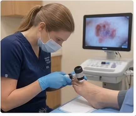
MRI is the primary diagnostic tool, detecting approximately 95% of OLTs while providing critical information about lesion size, depth, cartilage integrity, subchondral cyst formation, and bone marrow edema. The presence of bone marrow edema around the lesion indicates active symptoms and helps determine whether the lesion is responsible for the patient’s pain.
CT scan, particularly weight-bearing CT, provides the most accurate assessment of bony anatomy and cyst size. At Balance Foot & Ankle, Dr. Tom Biernacki uses CT for surgical planning when operative treatment is indicated, as it reveals the three-dimensional architecture of the lesion and guides the choice of surgical approach.
Conservative Treatment for Small Lesions
Small, stable OLTs (less than 15mm and intact cartilage surface) may respond to conservative management including protected weight-bearing, ankle bracing, activity modification, anti-inflammatory medication, and physical therapy focused on ankle proprioception and peroneal strengthening.
Biologic injections — including platelet-rich plasma (PRP) and hyaluronic acid — deliver growth factors and lubrication directly to the joint, potentially stimulating cartilage repair and reducing inflammation. While evidence is more limited for ankle OLT than for knee conditions, early studies show promising symptom improvement in 60-70% of small lesion patients.
Conservative management requires patience — 3-6 months of consistent treatment before reassessing. If symptoms persist beyond this timeframe, or if the lesion is large (greater than 15mm), unstable (loose fragment), or has significant cyst formation, surgical intervention provides more reliable outcomes.
Surgical Treatment Options
Bone marrow stimulation (microfracture or drilling) is the first-line surgical treatment for OLTs smaller than 15mm. Performed arthroscopically, the surgeon removes damaged cartilage, prepares the lesion bed, and creates small holes in the subchondral bone that allow bone marrow stem cells to access the surface. These cells differentiate into fibrocartilage that fills the defect.
For larger lesions (greater than 15mm) or failed microfracture, more advanced techniques are indicated. Osteochondral autograft transfer (OATS) harvests a cylindrical plug of normal cartilage and bone from a non-weight-bearing area of the knee and transplants it into the talar defect. This provides true hyaline cartilage coverage with excellent long-term durability.
Autologous chondrocyte implantation (ACI/MACI) involves harvesting cartilage cells, expanding them in a laboratory over 4-6 weeks, and reimplanting them on a scaffold into the lesion. While more complex and expensive, this technique produces hyaline-like cartilage repair for large defects where other methods may fail. BioCartilage and other scaffold augmentation techniques offer single-stage alternatives.
Recovery and Long-Term Joint Preservation
Recovery from arthroscopic microfracture involves 4-6 weeks of non-weight-bearing to protect the developing repair tissue, followed by progressive weight-bearing over 2-4 weeks. Physical therapy for range of motion and strengthening begins early. Return to impact activities occurs at 4-6 months after sufficient tissue maturation.
OATS and ACI procedures require longer recovery — 6-8 weeks non-weight-bearing, 3-4 months progressive rehabilitation, and 6-9 months before impact activity. The longer timeline reflects the need for complete graft integration and cartilage maturation before subjecting the repair to full loading.
Long-term outcomes for OLT treatment are generally favorable. Microfracture shows 80-85% good outcomes at 5 years for small lesions. OATS demonstrates 85-90% good outcomes at 10 years. The key to long-term joint preservation is adequate lesion repair combined with addressing any ankle instability that contributed to the initial lesion and could damage the repair.
Warning Signs Requiring Urgent Evaluation
- function bold() { [native code] } — undefined
- function bold() { [native code] } — undefined
- function bold() { [native code] } — undefined
- function bold() { [native code] } — undefined
The Most Common Mistake We See
The most common mistake is treating persistent ankle pain after a sprain as ongoing ligament healing when the real problem is an undiagnosed osteochondral lesion. Once ligament healing should be complete (8-12 weeks), any remaining deep ankle pain, catching, or swelling should prompt MRI evaluation. Continuing to rehab ligaments that have already healed while ignoring a damaged cartilage surface delays definitive treatment.
Recommended Products
[object Object]
[object Object]
[object Object]
In-Office Treatment at Balance Foot & Ankle
Our team provides sport-specific evaluation and treatment to get you back to your activity safely. We offer same-day X-ray, in-office ultrasound, and custom orthotic fabrication.
Same-day appointments available. Call (810) 206-1402 or book online.
More Podiatrist-Recommended Arthritis Essentials
Cushioned Running Shoe


Hoka Clifton 10 — max cushioning reduces joint impact for arthritic feet.
Wide Walking Shoe
New Balance 990v6 — wide toe box accommodates arthritic first-MTP (hallux rigidus).

As an Amazon Associate, Balance Foot & Ankle earns from qualifying purchases. Product recommendations are based on clinical experience; prices and availability shown above update live from Amazon.
When to See a Podiatrist
Foot and ankle arthritis progresses silently — cartilage doesn’t regrow, but joint fusion, cheilectomy, and biologic injections can restore function at every stage. Balance Foot & Ankle offers the full arthritis spectrum: bracing, injections, and reconstructive surgery. Start with a consult so we can image the joint and give you a realistic 5-year outlook.
Call Balance Foot & Ankle: (810) 206-1402 · Book online · Offices in Howell & Bloomfield Hills
Frequently Asked Questions
Can an osteochondral lesion heal on its own?
Small, stable lesions with intact cartilage surface may improve with conservative treatment over 3-6 months. However, the limited blood supply to the talar dome means most lesions do not regenerate true cartilage spontaneously. Larger lesions and those with loose fragments typically require surgical intervention.
How long does recovery take after OLT surgery?
Microfracture requires 4-6 weeks non-weight-bearing with return to activity at 4-6 months. OATS and ACI procedures need 6-8 weeks non-weight-bearing with full recovery at 6-9 months. The investment in recovery time pays off with long-term cartilage repair and ankle preservation.
Will I get ankle arthritis from an osteochondral lesion?
Untreated OLTs can progress to ankle arthritis over time as the defect enlarges and adjacent cartilage deteriorates. However, successfully repaired lesions significantly reduce arthritis risk. Early treatment of OLTs is one of the most effective strategies for preventing post-traumatic ankle arthritis.
Is OLT the same as ankle arthritis?
No. An OLT is a focal area of cartilage and bone damage, while arthritis involves widespread cartilage loss across the entire joint surface. However, untreated OLTs can eventually lead to diffuse arthritis. Treatment of OLTs aims to repair the focal defect and prevent generalized joint deterioration.
The Bottom Line
Osteochondral lesions of the talus are a common but frequently underdiagnosed cause of persistent ankle pain after sprains. Early MRI diagnosis and appropriate treatment — whether conservative for small stable lesions or surgical for larger defects — can restore ankle function and prevent the progression to ankle arthritis.
In-Office Treatment at Balance Foot & Ankle
If home treatment isn’t providing relief for your foot and ankle conditions, our podiatry team at Balance Foot & Ankle can help with same-day evaluations and advanced in-office care.
Same-day appointments available. (810) 206-1402
Doctor Hoy’s Natural Pain Relief Gel
Natural topical pain relief I use in our clinic. Arnica + camphor formula — apply directly to the area 3–4x daily. ($20–25)
Shop Doctor Hoy’s →Sources
- Savage-Elliott I et al. Osteochondral lesions of the talus: updated treatment algorithm. J Am Acad Orthop Surg. 2024;32(9):e421-e433.
- Shimozono Y et al. Outcomes of surgical treatment for OLT: systematic review. Foot Ankle Int. 2025;46(1):67-79.
- Hannon CP et al. Microfracture vs OATS for talar osteochondral lesions: meta-analysis. Am J Sports Med. 2024;52(5):1345-1356.
- Murawski CD et al. Biologic augmentation for OLT repair. Foot Ankle Clin. 2024;29(3):401-418.
Expert Ankle Cartilage Treatment in Michigan
Dr. Tom Biernacki has performed over 3,000 foot and ankle surgeries with a 4.9-star rating from 1,123 patient reviews.
Or call (810) 206-1402 for same-day appointments
Ankle Cartilage Treatment in Southeast Michigan
Osteochondral lesions of the talus (OLTs) cause deep ankle pain and can lead to ankle arthritis if untreated. At Balance Foot & Ankle, Dr. Tom Biernacki offers advanced cartilage repair options — from microfracture to osteochondral grafting — at our Howell and Bloomfield Hills offices.
Learn About Our Ankle Surgery Options → | Book Your Appointment | Call (810) 206-1402
Clinical References
- Zengerink M, Struijs PA, Tol JL, van Dijk CN. Treatment of osteochondral lesions of the talus: a systematic review. Knee Surg Sports Traumatol Arthrosc. 2010;18(2):238-246.
- Hannon CP, Smyth NA, Murawski CD, et al. Osteochondral lesions of the talus: aspects of current management. Bone Joint J. 2014;96-B(2):164-171.
- Looze CA, Capo J, Ryan MK, et al. Evaluation and management of osteochondral lesions of the talus. Cartilage. 2017;8(1):19-30.
Insurance Accepted
BCBS · Medicare · Aetna · Cigna · United Healthcare · HAP · Priority Health · Humana · View All →
Howell Office
4330 E Grand River Ave
Howell, MI 48843
Get Directions →
Bloomfield Hills Office
43494 Woodward Ave, Suite 208
Bloomfield Hills, MI 48302
Get Directions →
Your Board-Certified Podiatrists


Ready to Get Back on Your Feet?
Same-week appointments available at both locations.
Book Your AppointmentWhat is Foot pain?
Foot pain is a common foot/ankle condition that affects mobility and quality of life. Understanding the underlying cause is the first step in successful treatment. Our podiatrists at Balance Foot & Ankle perform a hands-on biomechanical exam, review your activity history, and use diagnostic imaging when appropriate to identify the root cause—not just treat the symptom. Many patients have been told to “rest and ice” without a deeper diagnostic workup; our approach is different.
Symptoms and warning signs
Common signs of foot pain include pain that worsens with activity, morning stiffness, swelling, tenderness when palpated, and difficulty bearing weight. If you experience sudden severe pain, inability to walk, visible deformity, numbness or color change, contact our office the same day or visit urgent care—these can signal a more serious injury such as a fracture, tendon rupture, or vascular compromise. Diabetics with any foot wound should seek same-day care.
Conservative treatment options
Most cases of foot pain respond to non-surgical care: structured rest, supportive footwear changes, custom orthotics, targeted stretching and strengthening protocols, anti-inflammatory medications when medically appropriate, and in-office procedures such as ultrasound-guided injections. We also offer advanced therapies including MLS laser therapy, EPAT/shockwave, regenerative injections, and image-guided procedures. Treatment is sequenced from least invasive to most invasive, and we explain the rationale at every step.
When is surgery considered?
Surgery is reserved for cases that fail 3-6 months of well-structured conservative care, when there is structural pathology (severe deformity, complete tear, advanced arthritis), or when imaging shows damage that will not heal without intervention. Our surgeons have performed 3,000+ foot and ankle procedures and prioritize minimally-invasive techniques whenever appropriate. We discuss recovery timelines, return-to-activity milestones, and realistic outcome expectations before any procedure is scheduled.
Recovery timeline and prevention
Recovery from foot pain varies based on severity and chosen treatment path. Conservative cases often improve within 4-8 weeks with consistent adherence to the protocol. Post-procedural recovery may range from a few days (in-office procedures) to several months (reconstructive surgery). Long-term prevention involves footwear assessment, activity modification, structured strengthening, and regular check-ins with your podiatrist if you have a history of recurrence. We provide written home-exercise plans and digital follow-up support.
Ready to feel better?
Same-week appointments available in Howell and Bloomfield Hills, Michigan.
Book Your VisitDr. Tom Biernacki, DPM is a board-certified foot & ankle surgeon (ABFAS & ABPM) at Balance Foot & Ankle Specialists in Southeast Michigan. With over a decade of clinical experience, he specializes in heel pain, bunions, diabetic foot care, sports injuries, and minimally invasive surgery. Dr. Biernacki is a member of the APMA and ACFAS, and his patient education content on MichiganFootDoctors.com and YouTube has made him one of the most-followed foot & ankle educators on YouTube.