

Quick answer: Total Ankle Replacement Revision Surgery Managing Failed Replacements is a common foot/ankle topic that affects many patients. The 2026 evidence-based approach combines proper diagnosis, conservative-first treatment, and escalation only when needed. We treat this regularly at our Howell and Bloomfield Hills practices. Call (810) 206-1402.
Medically reviewed by Dr. Tom Biernacki, DPM, FACFAS — Board-Certified Podiatric Surgeon at Balance Foot & Ankle PLLC. Last updated April 3, 2026.
Quick Answer: Total ankle replacement revision surgery addresses a failed or failing ankle replacement by exchanging worn components, correcting alignment, managing bone loss, or converting to an ankle fusion when the implant cannot be salvaged. Modern revision techniques using custom implants and bone grafting achieve 70–85% patient satisfaction at 5 years, though outcomes are less predictable than primary replacement. This guide explains why ankle replacements fail, what revision options exist, and what to expect from recovery.
Table of Contents
Medically Reviewed by Dr. Tom Biernacki, DPM — Board-Certified Podiatrist, Balance Foot & Ankle Specialists, Michigan. Last updated April 2026.
- Why Total Ankle Replacements Fail
- Signs Your Ankle Replacement Is Failing
- Diagnostic Workup for Failed Ankle Replacement
- Revision Surgery Options
- Component Exchange Revision
- Conversion to Ankle Fusion (Arthrodesis)
- Bone Grafting Techniques in Revision
- Recovery Timeline After Revision Surgery
- Best Products for Revision Recovery
- Outcomes and Success Rates
- Most Common Mistake
- Warning Signs After Revision Surgery
- Frequently Asked Questions
- Sources
Why Total Ankle Replacements Fail
Learning that your ankle replacement may need revision surgery is understandably concerning. You went through a significant procedure expecting long-term relief, and the possibility of another surgery can feel overwhelming. Understanding why ankle replacements fail helps you make informed decisions about your treatment options and feel more confident in the revision process.
Total ankle replacement (TAR) has improved dramatically over the past two decades, but even modern implants have a finite lifespan. Published data shows that 85–90% of ankle replacements survive 10 years, meaning 10–15% will require some form of revision during that timeframe. Several mechanisms contribute to implant failure.
Polyethylene wear. The plastic (polyethylene) bearing surface that sits between the metal tibial and talar components gradually wears down with use. As the bearing thins, it changes the biomechanics of the ankle joint, creates wear debris that triggers inflammation (osteolysis), and can eventually allow metal-on-metal contact. Wear is accelerated in younger, more active patients and those with higher body weight.
Aseptic loosening. The most common cause of revision, aseptic loosening occurs when the bond between the metal implant and the surrounding bone weakens over time. Microscopic motion at the bone-implant interface creates a fibrous membrane rather than a solid bony ingrowth, leading to progressive loosening, subsidence (sinking), and pain. Wear particle-induced osteolysis (bone resorption) around the implant accelerates this process.
Component malalignment. If the original implant was positioned with even slight malalignment, abnormal loading patterns develop that accelerate wear on one side of the bearing, cause edge loading, and create asymmetric stress on the bone-implant interface. Over time, this uneven loading leads to progressive deformity and component failure.
Periprosthetic fracture. Fractures of the tibia or talus around the implant components can destabilize the replacement. These fractures may occur from trauma (falls, ankle sprains) or from bone weakened by osteolysis around a loosening implant. Periprosthetic fractures frequently require revision surgery to address both the fracture and the compromised implant.
Deep infection. Infection around an ankle replacement (periprosthetic joint infection) occurs in 1–4% of cases and represents one of the most challenging complications. Bacteria form biofilms on the implant surface that are resistant to antibiotics alone, typically requiring implant removal, a period of antibiotic treatment, and staged reconstruction.
Progressive cyst formation. Osteolytic cysts — pockets of bone destruction caused by wear debris — can develop around the implant components without initially causing symptoms. As cysts enlarge, they weaken the bone’s ability to support the implant, eventually leading to subsidence or fracture. Regular surveillance imaging can detect cysts before they reach a critical size.
Signs Your Ankle Replacement May Be Failing
Recognizing the warning signs of a failing ankle replacement early gives you the best chance for a successful revision with the most treatment options available. Not all of these signs mean your replacement has failed — but each warrants evaluation by your surgeon.
Increasing pain. The most common sign of implant failure. If ankle pain that was well-controlled after your replacement begins gradually returning — particularly deep, aching pain with weight bearing that worsens over months — this suggests component loosening, wear, or osteolysis. Sudden sharp pain after a specific incident may indicate periprosthetic fracture.
Progressive swelling. Persistent or worsening ankle swelling that does not respond to elevation and compression may indicate wear-particle synovitis (inflammation caused by polyethylene debris), infection, or component loosening with increased joint fluid production.
Loss of motion. Gradually decreasing ankle dorsiflexion or plantarflexion can signal heterotopic ossification (abnormal bone growth), component subsidence that changes the joint mechanics, or gutter impingement from bone overgrowth around the implant edges.
Visible deformity. Progressive tilting of the foot into varus (inward lean) or valgus (outward lean) suggests asymmetric component wear, loosening, or bone collapse on one side of the implant. This deformity may be subtle initially but becomes more apparent over time.
Clicking, grinding, or instability. Mechanical symptoms like clicking or grinding during ankle motion suggest polyethylene wear, bearing dislocation, or component loosening. A feeling of the ankle “giving way” indicates ligamentous insufficiency or component instability that may require surgical correction.
Diagnostic Workup for Failed Ankle Replacement
A thorough diagnostic workup is essential before planning revision surgery. The cause of failure determines the best revision strategy, and missing an underlying infection before operating can lead to catastrophic outcomes.
Weight-bearing X-rays. Standing ankle X-rays (AP, lateral, and mortise views) reveal component position, alignment, subsidence (sinking), and lucencies (dark lines around the implant indicating loosening). Serial comparison with prior X-rays identifies progressive changes that confirm failure. Stress views may demonstrate instability not apparent on standard films.
CT scan with metal artifact reduction. Modern CT protocols using metal artifact reduction software (MARS) provide detailed assessment of bone stock around the implant, periprosthetic cyst size and location, and component positioning in three dimensions. CT is essential for surgical planning because it maps the exact bone deficiency that must be addressed during revision.
Laboratory studies. ESR (erythrocyte sedimentation rate), CRP (C-reactive protein), and CBC (complete blood count) screen for infection. Elevated inflammatory markers require further investigation with joint aspiration before any revision surgery is planned. Missing a low-grade infection and proceeding with component exchange leads to revision failure.
Joint aspiration. Needle aspiration of the ankle joint under fluoroscopic guidance retrieves fluid for cell count, differential, culture, and crystal analysis. This is the most important test for ruling out periprosthetic joint infection. Cultures should be held for a minimum of 14 days to detect slow-growing organisms.
Nuclear imaging (bone scan or PET-CT). In equivocal cases where X-rays and labs are inconclusive, nuclear imaging can differentiate between aseptic loosening and infection. Combined white blood cell/bone marrow scans provide the highest specificity for diagnosing periprosthetic joint infection when other tests are borderline.
Revision Surgery Options for Failed Ankle Replacement
The revision strategy depends on the cause of failure, the remaining bone stock, the patient’s activity level and health status, and the condition of the surrounding soft tissues. Your surgeon will discuss the options most appropriate for your specific situation.
Polyethylene exchange (bearing swap). When the metal components are well-fixed and properly aligned but the polyethylene bearing is worn, a simple bearing exchange may be sufficient. This is the least invasive revision option with the quickest recovery, but it is only appropriate when the underlying implant fixation and alignment are satisfactory.
Component revision (partial or total). When one or both metal components have loosened, subsided, or become malaligned, they must be removed and replaced. Modern revision systems include larger, offset, and custom components designed to address the bone deficiency created by removing the failed implant. This is a more complex procedure than primary replacement but can restore excellent function when adequate bone stock remains.
Conversion to ankle fusion (arthrodesis). When bone loss is too severe for reliable component revision, or when the patient’s anatomy or health status makes a revision replacement inadvisable, conversion to a solid ankle fusion provides predictable pain relief and stability. While fusion eliminates ankle motion, it creates a stable, pain-free limb that allows walking, working, and many recreational activities.
Staged revision for infection. When periprosthetic joint infection is confirmed, a two-stage revision is typically necessary. Stage 1 involves implant removal, thorough debridement, and placement of an antibiotic-loaded cement spacer. After 6–12 weeks of IV antibiotics and confirmation that the infection is eradicated, Stage 2 involves reimplantation of a new ankle replacement or conversion to fusion.
Component Exchange Revision: What to Expect
Component exchange — removing and replacing the failed metal and/or polyethylene components — is the preferred revision option when adequate bone stock remains and infection has been excluded. Modern revision implant systems have significantly improved the options available for complex reconstructions.
Custom and modular implants. Revision-specific implant systems include thicker polyethylene bearings, longer-stemmed tibial and talar components for enhanced fixation, and custom 3D-printed titanium components manufactured from the patient’s CT scan. These custom implants precisely match the patient’s unique bone deficiency, providing optimal fit and fixation where standard revision components may not suffice.
Surgical technique. The revision is typically performed through the same anterior approach used for the primary replacement. The failed components are carefully removed while preserving as much bone as possible. The revision bed is prepared, bone defects are grafted, and the new components are implanted with or without cement depending on the bone quality and implant design. The procedure typically takes 2–3 hours, longer than primary replacement due to the complexity of component removal and reconstruction.
Conversion to Ankle Fusion (Arthrodesis): When Revision Replacement Is Not Possible
Conversion to ankle fusion is not a failure — it is a well-established salvage procedure that provides reliable pain relief and functional stability when revision replacement is not feasible or advisable. Many patients who undergo conversion to fusion report significant improvement in quality of life compared to living with a painful, failing replacement.
Bone loss management. The primary challenge in conversion to fusion is the bone deficit left after implant removal. Structural bone grafts — including femoral head allografts (cadaver bone), iliac crest autografts, or bulk tibial allografts — fill the gap and provide biological substrate for fusion healing. Modern fixation with compression screws, locking plates, and intramedullary nails maintains alignment while the graft incorporates.
Fusion position. The ankle is fused in a neutral position (90 degrees to the tibia) with slight external rotation and 5 degrees of hindfoot valgus. This position optimizes gait mechanics and allows the surrounding joints (subtalar, talonavicular, calcaneocuboid) to compensate partially for the lost ankle motion.
Functional expectations. After fusion, patients lose the up-and-down motion of the ankle but retain side-to-side motion through the subtalar joint. Most patients walk with a mild limp that is often less noticeable than the limp caused by a painful failing replacement. Walking on flat surfaces, driving, and many daily activities proceed normally. Stairs, inclines, and uneven terrain require adaptation but are manageable for most patients.
Bone Grafting Techniques in Ankle Revision Surgery
Bone grafting is a central element of most ankle revision procedures because failed implants invariably leave bone deficits that must be filled for the revision to succeed. The choice of graft material depends on the size and location of the defect.
Structural allografts. Large bone defects (greater than 1 cm) typically require structural allografts — prepared cadaver bone shaped to fill the specific deficiency. Femoral head allografts are the most commonly used structural graft in ankle revision because they provide adequate volume and structural integrity while healing predictably with modern fixation techniques.
Cancellous autograft. Bone harvested from the patient’s own iliac crest (hip bone) or proximal tibia provides living cells with the highest biological potency for healing. Autograft is preferred for smaller defects and is often mixed with allograft for larger reconstructions to enhance healing rates.
Bone graft substitutes. Synthetic materials including calcium phosphate, tricalcium phosphate, and demineralized bone matrix provide osteoconductive scaffolding that supports new bone growth. These are used to fill smaller cavitary defects and to augment structural grafts. Biologics including platelet-rich plasma (PRP) and bone morphogenetic proteins (BMP) may be added to enhance healing in select cases.
Recovery Timeline After Ankle Revision Surgery
Recovery from ankle revision surgery is typically longer and more demanding than primary ankle replacement recovery. Setting realistic expectations from the outset helps you prepare mentally and physically for each phase.
Weeks 0–6: Non-weight-bearing. Strict non-weight-bearing in a splint or cast is essential to protect the revision construct and allow bone graft incorporation. A knee scooter or wheelchair is used for mobility. The foot is elevated above heart level as much as possible to control swelling.
Weeks 6–10: Protected weight bearing. Transition to a walking boot with gradual weight bearing. X-rays confirm early healing and appropriate component position before weight bearing progresses. DASS medical grade compression socks become essential during this phase to manage the significant swelling that accompanies increased activity after prolonged non-weight-bearing.
Weeks 10–16: Progressive weight bearing and physical therapy. Full weight bearing in the boot progresses to walking in supportive shoes with orthotic insoles. Physical therapy focuses on ankle range of motion (for revision replacement) or gait optimization (for conversion to fusion), strengthening, and balance training.
Months 4–6: Functional rehabilitation. Transition to regular activities with continued physical therapy. PowerStep orthotic insoles in supportive shoes protect the reconstruction and optimize alignment during the ongoing healing process. Maximum improvement typically occurs between 6–12 months post-revision.
Months 6–12: Continued improvement. Swelling gradually resolves, strength improves, and confidence in the ankle increases. Final functional outcome may not be apparent until 12 months post-revision. Continued use of compression socks for activity and orthotics in all shoes is recommended long-term.
Best Products for Ankle Revision Recovery
Recovery from ankle revision surgery is a marathon, not a sprint. These products help manage the prolonged recovery and optimize your long-term outcome.
Affiliate disclosure: Some links below are affiliate links, meaning we may earn a small commission if you purchase through them — at no extra cost to you. We only recommend products we use in our own practice.
PowerStep Orthotic Insoles for Post-Revision Support
Once you transition from the boot to regular shoes (typically 3–4 months post-revision), a quality orthotic insole is essential for protecting the reconstruction and optimizing alignment. PowerStep orthotic insoles provide the semi-rigid arch support and heel cushioning that distribute pressure evenly across the foot, reducing stress on the revision construct. For patients who have undergone conversion to fusion, PowerStep orthotics help the surrounding joints compensate for the lost ankle motion by controlling midfoot and hindfoot alignment. We recommend long-term orthotic use — ideally indefinitely — for all ankle revision patients.
DASS Medical Grade Compression Socks
Swelling management is arguably the single biggest challenge in ankle revision recovery. The ankle and lower leg can remain significantly swollen for 6–12 months post-revision, and uncontrolled edema delays healing, increases stiffness, and impairs rehabilitation. DASS medical grade compression socks provide true 20–30 mmHg graduated compression that effectively controls post-surgical edema. Begin wearing compression daily as soon as the surgical incision is healed (typically 2–3 weeks post-op) and continue for a minimum of 6 months. Many revision patients find that long-term compression wear significantly improves comfort and function.
Doctor Hoy’s Natural Pain Relief
Managing pain during the prolonged revision recovery without excessive reliance on opioids is a priority. Doctor Hoy’s Natural Pain Relief gel provides effective topical analgesic relief that can supplement (and eventually replace) oral pain medications. Apply around the ankle joint (avoiding the incision until fully healed) for localized anti-inflammatory and pain-relieving benefits. The cooling menthol sensation provides immediate comfort during physical therapy sessions and after increased activity.
Ankle Revision Surgery Outcomes and Success Rates
Ankle revision surgery outcomes are less predictable than primary ankle replacement, and honest expectations are important for patient satisfaction. That said, modern revision techniques have improved results significantly.
Revision replacement outcomes: Component exchange and revision replacement achieve 70–85% patient satisfaction at 5-year follow-up. Pain improvement averages 60–75% compared to the pre-revision state. Range of motion is typically preserved at 75–85% of the primary replacement level. Second revision rates are approximately 15–20% at 10 years.
Conversion to fusion outcomes: Ankle fusion after failed replacement achieves 80–90% union rates, though healing may take longer than primary fusion due to the bone deficiency. Pain relief is reliable once fusion is achieved, and patient satisfaction averages 75–85%. The trade-off of lost motion for predictable pain relief is one most patients find acceptable.
Factors affecting outcomes: Younger, healthier patients with smaller bone defects and well-aligned limbs have the best revision outcomes. Infection history, large bone loss, severe malalignment, and peripheral vascular disease are negative prognostic factors. Smoking significantly impairs bone healing and increases fusion nonunion rates — smoking cessation before revision surgery is strongly recommended.
Most Common Mistake With Failing Ankle Replacements
🔑 Key Takeaway: The most common mistake patients make with a failing ankle replacement is waiting too long to seek evaluation. Many patients tolerate gradually increasing pain for months or years, hoping it will improve on its own. During this delay, bone loss from osteolysis progresses, making eventual revision more complex and less predictable. Cysts enlarge, components subside further, and what could have been a straightforward bearing exchange becomes a major revision with structural bone grafting. If your ankle replacement pain is increasing, your range of motion is decreasing, or your ankle is progressively swelling — do not wait. Earlier evaluation preserves more options and improves outcomes.
Warning Signs After Ankle Revision Surgery
⚠️ Contact your surgeon immediately if you experience any of the following after ankle revision surgery:
- Increasing wound redness, warmth, or drainage especially after the first week — infection risk is higher in revision surgery than primary replacement
- Fever above 101°F (38.3°C) with ankle pain or swelling
- Sudden severe pain or a cracking sensation during weight bearing — may indicate periprosthetic fracture or graft failure
- Progressive numbness or weakness in the foot that was not present before surgery
- Calf pain, redness, or swelling unrelated to the ankle — potential deep vein thrombosis requiring urgent evaluation
- Shortness of breath or chest pain — rare but potentially life-threatening signs of pulmonary embolism requiring emergency care
- Wound breakdown or skin necrosis around the incision, which may require wound care intervention to prevent deep infection
Contact Balance Foot & Ankle at (586) 207-4540 or go to your nearest emergency room for any of these warning signs.
Ankle Revision Surgery at Balance Foot & Ankle
At Balance Foot & Ankle, Dr. Biernacki evaluates patients with failed or failing ankle replacements and provides comprehensive treatment planning. Our approach includes thorough diagnostic workup with advanced imaging, infection screening, and honest discussion of all revision options — from bearing exchange to component revision to conversion to fusion. We collaborate with orthopedic ankle specialists when complex revision reconstruction is indicated, ensuring you receive the most appropriate care for your specific situation.
Ankle Surgery: Understanding Your Options [Video]
Dr. Biernacki discusses ankle surgery options in this educational video from Balance Foot & Ankle:
More Podiatrist-Recommended Surgery Essentials
OOFOS Recovery Slide


Post-op approved — impact-absorbing slide for early recovery.
HOKA Ora 3 Recovery Slide
Max-cushion recovery sandal — comfort for post-surgical swelling.
Hoka Bondi 9
Max-cushion walking shoe — ease into return-to-walking post-surgery.
As an Amazon Associate, Balance Foot & Ankle earns from qualifying purchases. Product recommendations are based on clinical experience; prices and availability shown above update live from Amazon.

When to See a Podiatrist
Foot and ankle surgery in 2026 is dramatically different than a decade ago — most procedures are now minimally-invasive, outpatient, and allow weight-bearing within days. Balance Foot & Ankle surgeons have performed 3,000+ foot/ankle surgeries with modern techniques. If another surgeon has recommended a traditional open procedure, a second opinion may reveal a faster, less-invasive option.
Call Balance Foot & Ankle: (810) 206-1402 · Book online · Offices in Howell & Bloomfield Hills
Frequently Asked Questions About Ankle Revision Surgery
How long does a revision ankle replacement last?
Modern revision ankle replacements using contemporary implant systems have published 5-year survival rates of 70–85% and 10-year survival rates of 55–70%. Factors that influence revision longevity include the extent of bone loss at the time of revision, component alignment, patient activity level, body weight, and compliance with orthotic and footwear recommendations. Using PowerStep orthotic insoles long-term helps optimize implant loading and may extend the life of the revision.
Is ankle fusion a good option after failed replacement?
Yes — ankle fusion (arthrodesis) after failed replacement is a well-established salvage procedure with reliable pain relief and high patient satisfaction rates (75–85%). While fusion eliminates ankle dorsiflexion and plantarflexion, it creates a stable, pain-free limb that allows walking, driving, and many activities. Most patients report that the loss of motion is a worthwhile trade-off for predictable pain relief after living with a painful failing replacement. Fusion union rates are 80–90% with modern fixation and bone grafting techniques.
Can a failed ankle replacement be replaced again?
Yes, in many cases a failed ankle replacement can be revised with new components rather than converted to fusion. The feasibility depends primarily on the remaining bone stock — if adequate bone remains or can be reconstructed with grafting, component revision is often possible. Custom 3D-printed implants have expanded the range of cases amenable to revision replacement by precisely matching the patient’s unique bone deficiency. Your surgeon will determine whether revision replacement or conversion to fusion is more appropriate based on CT imaging, bone quality assessment, and your specific goals.
How long is recovery from ankle revision surgery?
Recovery from ankle revision surgery is typically 6–12 months to maximum improvement, significantly longer than primary replacement (3–6 months). Expect 6 weeks of non-weight-bearing, 4–6 weeks of protected weight bearing in a boot, then a gradual return to regular shoes and activities. Swelling can persist for 6–12 months. Wearing DASS compression socks daily throughout recovery significantly improves comfort and reduces the swelling that delays rehabilitation.
What causes an ankle replacement to fail?
The most common causes of ankle replacement failure are aseptic loosening (weakening of the bone-implant bond, 40–50% of failures), polyethylene bearing wear (20–30%), component malalignment (10–15%), periprosthetic fracture (5–10%), and deep infection (5–10%). Risk factors include higher body weight, high activity level, component malalignment during the original surgery, poor bone quality (osteoporosis), and smoking. Regular follow-up with surveillance X-rays helps detect early signs of failure before bone loss becomes severe.
In-Office Treatment at Balance Foot & Ankle
If home treatment isn’t providing relief for your foot and ankle conditions, our podiatry team at Balance Foot & Ankle can help with same-day evaluations and advanced in-office care.
Same-day appointments available. (810) 206-1402
Sources
- Hintermann B, Zwicky L, Knupp M, et al. “HINTEGRA revision arthroplasty for failed total ankle prostheses.” J Bone Joint Surg Am. 2013;95(13):1166-1174.
- Prissel MA, Roukis TS. “Management of extensive tibial osteolysis with the Agility total ankle replacement systems using geometric metal-reinforced polymethylmethacrylate cement augmentation.” J Foot Ankle Surg. 2014;53(1):101-107.
- Ellington JK, Gupta S, Myerson MS. “Management of failures of total ankle replacement with the Agility total ankle arthroplasty.” J Bone Joint Surg Am. 2013;95(23):2112-2118.
- Gross CE, Lewis JS, Adams SB, et al. “Secondary arthrodesis after total ankle arthroplasty.” Foot Ankle Int. 2016;37(7):709-714.
- Palanca A, Mann RA, Mann JA, et al. “Scandinavian total ankle replacement: 15-year follow-up.” Foot Ankle Int. 2018;39(2):135-142.
Failed Ankle Replacement? Expert Evaluation Available
If your ankle replacement is causing increasing pain, swelling, or instability, Dr. Biernacki at Balance Foot & Ankle provides thorough evaluation and honest discussion of your revision options. Early assessment preserves the most treatment options and leads to better outcomes.
Balance Foot & Ankle | (586) 207-4540 | Serving Southeast Michigan
Related Ankle Surgery Guides
- Ankle Arthroscopy: Minimally Invasive Diagnosis & Treatment
- Ankle Fusion Surgery: Complete Guide
- Achilles Tendon Repair Surgery Guide
- Foot Health After 65: Complete Senior Care Guide
- Podiatrist Recommended Foot Care Products 2026
Insurance Accepted
BCBS · Medicare · Aetna · Cigna · United Healthcare · HAP · Priority Health · Humana · View All →
Howell Office
4330 E Grand River Ave
Howell, MI 48843
Get Directions →
Bloomfield Hills Office
43494 Woodward Ave, Suite 208
Bloomfield Hills, MI 48302
Get Directions →
Your Board-Certified Podiatrists



Ready to Get Back on Your Feet?
Same-week appointments available at both locations.
When Shoes Aren’t Enough — Dr. Tom’s Top 9 Orthotics
About 30% of patients I see for foot pain need MORE than a great shoe — they need a structured insole. Below: my complete 2026 orthotic ranking with pros, cons, and the specific patient I’d give each one to.
★ DR. TOM’S COMPLETE 2026 ORTHOTIC RANKING
9 Best Prefab Orthotics by Use Case
PowerStep, Currex, Spenco, Vionic, and PowerStep Pinnacle — every orthotic I’ve fitted to thousands of patients across both Michigan offices. Each card includes pros, cons, and the specific patient I’d give it to. Real Amazon ratings, review counts, and prices below.
Best All-Purpose Orthotic for Most Patients
Semi-rigid arch shell + dual-layer cushion + deep heel cup. The orthotic I’ve fitted to more patients than any other for 15 years. APMA-accepted. Trim-to-fit design works in athletic shoes, casual shoes, and most work boots.

✓ Pros
- Semi-rigid arch shell provides true biomechanical correction
- Deep heel cup centers the heel and reduces lateral instability
- Dual-layer cushion (top + bottom) lasts 9-12 months daily wear
- Available in 8 sizes for precise fit
- APMA-accepted and clinically validated
- Lower price than PowerStep Pinnacle Green for equivalent function
✗ Cons
- Too thick for most dress shoes (use ProTech Slim instead)
- Some break-in period required (3-7 days for arch tolerance)
- Not enough correction for severe pes planus or rigid pes cavus
Dr. Tom’s Recommendation: If a patient has run-of-the-mill plantar fasciitis, mild flat feet, or arch fatigue, this is the first orthotic I try. Better value than PowerStep Pinnacle for 90% of patients, which is why I swapped it into our clinic kits three years ago. Sub-$50 typically.
Maximum Motion Control · Flat Feet & Severe Over-Pronation
PowerStep’s most aggressive stability orthotic. Adds a 2°-7° medial heel post on top of the standard PowerStep platform — designed specifically for flat-footed patients and severe pronators who need real corrective force.

✓ Pros
- 2°-7° medial heel post adds aggressive pronation control
- Same trusted PowerStep arch shell, more correction
- Built specifically for flat-foot biomechanics
- Excellent for posterior tibial tendon dysfunction (PTTD)
- Removable top cover for cleaning
✗ Cons
- Too aggressive for neutral-arch patients
- Needs longer break-in (10-14 days) due to stronger correction
- Adds 2-3 mm of stack height — won’t fit slim dress shoes
Dr. Tom’s Recommendation: When a patient comes in with significant flat feet AND symptoms (heel pain, arch pain, knee pain), the Original PowerStep isn’t aggressive enough. The Maxx is what gets prescribed. About 25% of my flat-footed patients end up here.
Low-Profile · Fits Dress Shoes & Narrow Casuals
3 mm slim profile with podiatrist-designed tri-planar arch technology. Engineered specifically to fit inside dress shoes, oxfords, loafers, and women’s flats without crowding the toe box. Vionic was founded by an Australian podiatrist.
✓ Pros
- 3 mm slim profile (vs 7-10 mm for standard orthotics)
- Tri-planar arch technology adds support without bulk
- Built-in deep heel cup despite slim design
- Fits dress shoes WITHOUT having to remove the factory insole
- Trim-to-fit · APMA-accepted
✗ Cons
- Less arch support than full-volume orthotics
- Top cover wears faster than thicker alternatives
- Not enough correction for severe foot deformities
Dr. Tom’s Recommendation: My default when a patient says ‘I need orthotics but I have to wear dress shoes for work.’ Slim enough to fit in oxfords and pumps without the heel sliding out. The single highest-impact change you can make for office workers with foot pain.
Built-In Metatarsal Pad · Morton’s Neuroma · Ball-of-Foot Pain
Standard Pinnacle orthotic with a built-in metatarsal pad positioned proximal to the metatarsal heads — the exact location that offloads neuromas and metatarsalgia. No need for separate met pads or pad placement guesswork.

✓ Pros
- Built-in met pad eliminates DIY pad placement errors
- Specifically designed for Morton’s neuroma + metatarsalgia
- Same trusted PowerStep arch + heel cup platform
- Top cover protects sensitive forefoot skin
- Faster relief than orthotics + add-on met pads
✗ Cons
- Met pad position is fixed (can’t fine-tune individual placement)
- Some patients with very small or very large feet need custom
- Slightly thicker than the standard Pinnacle
Dr. Tom’s Recommendation: If a patient has Morton’s neuroma, sesamoiditis, or generalized ball-of-foot pain (metatarsalgia), this saves a clinic visit and a prescription. The built-in pad placement is anatomically correct for 80% of feet. Way better than DIY met pads.
Adaptive Dynamic Arch · Athletic & Daily Wear
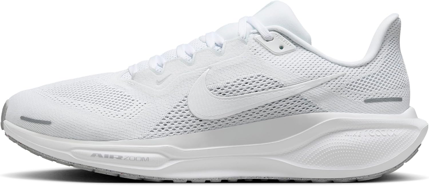
Currex’s flagship adaptive arch technology — the orthotic flexes with your gait instead of fighting it. Different stiffness zones along the length give you targeted support at the heel, midfoot, and forefoot. Available in three arch heights (low/medium/high).
✓ Pros
- Dynamic flex zones adapt to natural gait cycle
- Three arch heights ensure precise fit
- Lighter than rigid orthotics (no ‘heavy foot’ feel)
- Excellent for runners and athletic walkers
- European podiatric design (German engineering)
✗ Cons
- More expensive than PowerStep Original ($55-65 typically)
- Less aggressive correction than Pinnacle Maxx for severe cases
- Three arch heights means you must self-select correctly
Dr. Tom’s Recommendation: I started recommending Currex three years ago for runners who said PowerStep felt ‘too rigid.’ The dynamic flex zones respect natural gait. Best for active patients who walk 8K+ steps daily and don’t need maximum motion control.
Running-Specific · Heel Strike + Forefoot Strike Compatible
Currex’s purpose-built running orthotic. The midfoot flex zone is positioned for runner’s gait mechanics, with a flared heel cushion for heel strikers and a forefoot rocker for midfoot/forefoot strikers. Tested on 1000+ runners during product development.

✓ Pros
- Designed by German biomechanics lab specifically for runners
- Dynamic arch flexes with running gait (not static like PowerStep)
- Three arch heights (low/medium/high)
- Reduces overuse injury risk in mid-distance runners
- Lightweight (no impact on cadence)
✗ Cons
- Premium price ($60-75)
- Not aggressive enough for severe over-pronators (use Pinnacle Maxx)
- Runner-specific design = less ideal for daily walking shoes
Dr. Tom’s Recommendation: If a patient runs 20+ miles per week and has plantar fasciitis or shin splints, this is the orthotic I prescribe. The dynamic flex zones respect running biomechanics in a way that no rigid PowerStep can match. Pricier but worth it for serious runners.
Cavus Foot & High-Arch Patients
Polyurethane base with a deeper heel cup and higher arch profile than PowerStep — built for cavus (high-arched) feet that need maximum cushion and support. The 5-zone cushioning system addresses the unique pressure points of high-arch feet.

✓ Pros
- Deeper heel cup centers the heel for cavus foot stability
- Higher arch profile fills the void under high arches
- 5-zone cushioning addresses cavus foot pressure points
- Polyurethane base lasts 12+ months
- Available in Wide width
✗ Cons
- Too tall/aggressive for normal or low arches
- Won’t fit slim dress shoes
- Pricier than PowerStep Original
- Some patients find the arch height uncomfortable initially
Dr. Tom’s Recommendation: Cavus foot patients are often misdiagnosed and given low-arch orthotics — that makes everything worse. Spenco’s Total Support has the arch profile that high-arch feet actually need. About 15% of my patients have cavus feet; this is what they wear.
Cushion Layer · Standing All Day · Gel Pressure Relief
NOT a true biomechanical orthotic — this is a cushion insole. But for patients who want gel pressure relief instead of arch correction (or to add ON TOP of factory insoles in work boots), this is the best gel option on Amazon.

✓ Pros
- Genuine gel cushioning (not foam pretending to be gel)
- Targeted gel waves under heel and ball of foot
- Trim-to-fit · works in most shoe types
- Sub-$15 price (most affordable option in this list)
- Massaging texture is genuinely soothing
✗ Cons
- ZERO arch support — this is cushion only
- Won’t fix plantar fasciitis or flat-foot issues
- Compresses faster than PowerStep (4-6 months)
- Top cover wears through in high-mileage applications
Dr. Tom’s Recommendation: I recommend these to patients who tell me ‘I just want my feet to stop hurting at the end of my shift’ and who don’t have a biomechanical issue. Construction workers, factory workers, retail. Pure cushion does the job for them.
Tight-Fitting Shoes · Cycling Shoes · Hockey Skates
PowerStep Pinnacle’s slim version of their famous Green insole. The trademark stabilizer cap is preserved but the overall thickness is reduced — works in cycling shoes, hockey skates, ski boots, and other tight-fitting footwear that the standard PowerStep Pinnacle Green can’t fit into.

✓ Pros
- Stabilizer cap centers the heel (PowerStep Pinnacle’s signature feature)
- Slim profile fits tight athletic footwear
- Lasts 12+ months daily wear
- Excellent for cycling shoes specifically
- Built-in odor-control treatment
✗ Cons
- Premium price ($45-55)
- Less cushion than PowerStep equivalents
- Not as aggressive correction as Pinnacle Maxx for flat feet
- The signature ‘heel cup feel’ takes 1-2 weeks to adapt to
Dr. Tom’s Recommendation: If you’re a cyclist with foot numbness, hot spots, or knee pain — this is the orthotic. The stabilizer cap solves cycling-specific biomechanical issues that no other orthotic addresses. Worth the premium for athletes.
None of these solving your foot pain?
Some patients (about 30%) need custom-molded prescription orthotics. We make 3D-scanned custom orthotics in our Howell and Bloomfield Hills offices — specifically built for your foot mechanics.
Schedule a Custom Orthotic Fitting →
FSA/HSA eligible · Most insurance accepted · (810) 206-1402
What is Foot pain?
Foot pain is a common foot/ankle condition that affects mobility and quality of life. Understanding the underlying cause is the first step in successful treatment. Our podiatrists at Balance Foot & Ankle perform a hands-on biomechanical exam, review your activity history, and use diagnostic imaging when appropriate to identify the root cause—not just treat the symptom. Many patients have been told to “rest and ice” without a deeper diagnostic workup; our approach is different.
Symptoms and warning signs
Common signs of foot pain include pain that worsens with activity, morning stiffness, swelling, tenderness when palpated, and difficulty bearing weight. If you experience sudden severe pain, inability to walk, visible deformity, numbness or color change, contact our office the same day or visit urgent care—these can signal a more serious injury such as a fracture, tendon rupture, or vascular compromise. Diabetics with any foot wound should seek same-day care.
Conservative treatment options
Most cases of foot pain respond to non-surgical care: structured rest, supportive footwear changes, custom orthotics, targeted stretching and strengthening protocols, anti-inflammatory medications when medically appropriate, and in-office procedures such as ultrasound-guided injections. We also offer advanced therapies including MLS laser therapy, EPAT/shockwave, regenerative injections, and image-guided procedures. Treatment is sequenced from least invasive to most invasive, and we explain the rationale at every step.
When is surgery considered?
Surgery is reserved for cases that fail 3-6 months of well-structured conservative care, when there is structural pathology (severe deformity, complete tear, advanced arthritis), or when imaging shows damage that will not heal without intervention. Our surgeons have performed 3,000+ foot and ankle procedures and prioritize minimally-invasive techniques whenever appropriate. We discuss recovery timelines, return-to-activity milestones, and realistic outcome expectations before any procedure is scheduled.
OrthoInfo – AAOS: Total Ankle Replacement
Recovery timeline and prevention
Recovery from foot pain varies based on severity and chosen treatment path. Conservative cases often improve within 4-8 weeks with consistent adherence to the protocol. Post-procedural recovery may range from a few days (in-office procedures) to several months (reconstructive surgery). Long-term prevention involves footwear assessment, activity modification, structured strengthening, and regular check-ins with your podiatrist if you have a history of recurrence. We provide written home-exercise plans and digital follow-up support.
Ready to feel better?
Same-week appointments available in Howell and Bloomfield Hills, Michigan.

Dr. Tom Biernacki, DPM is a board-certified foot & ankle surgeon (ABFAS & ABPM) at Balance Foot & Ankle Specialists in Southeast Michigan. With over a decade of clinical experience, he specializes in heel pain, bunions, diabetic foot care, sports injuries, and minimally invasive surgery. Dr. Biernacki is a member of the APMA and ACFAS, and his patient education content on MichiganFootDoctors.com and YouTube has made him one of the most-followed foot & ankle educators on YouTube.