

Medically reviewed by Dr. Tom Biernacki, DPM
Board-certified podiatric surgeon | Balance Foot & Ankle, Howell & Bloomfield Township, MI
Last reviewed: May 2026
| Feature | Anterior Ankle Impingement | Posterior Ankle Impingement |
|---|---|---|
| Location | Anterior ankle joint (talar neck / distal tibia) | Posterior ankle (between talus and calcaneus / tibia) |
| Pain with motion | Dorsiflexion — pain at end range (squatting, uphill) | Plantarflexion — pain at end range (ballet, downhill, push-off) |
| Common Patient | Soccer players; footballers; runners; jumping athletes | Ballet dancers; gymnasts; soccer players (repetitive plantarflexion) |
| Cause | Anterior osteophytes (tibia + talar neck); soft tissue (AITFL, synovium) | Os trigonum; Stieda process; FHL tenosynovitis; posterior capsule |
| Imaging | Lateral X-ray: anterior tibial + talar osteophytes; MRI: synovitis, AITFL tear | Lateral X-ray: os trigonum or large posterior talus process; MRI: bone edema, FHL fluid |
| Examination | Anterior drawer pain; forced dorsiflexion pain; anterior joint line tenderness | Passive plantarflexion pain; Tinel’s FHL posteromedial; posterior ankle compression test |
| Surgical Treatment | Ankle arthroscopy: anterior osteophyte excision + synovectomy | Endoscopic posterior ankle surgery: os trigonum excision + FHL release |
| Treatment | Type | Indication | Success Rate | Return to Sport |
|---|---|---|---|---|
| Activity Modification + PT | Both | First-line for mild-moderate impingement | 50–65% | 6–10 weeks |
| Corticosteroid Injection (joint) | Both | Synovitis predominant; diagnostic utility | 50–70% short-term | Symptom dependent |
| Ankle Arthroscopy (anterior impingement) | Anterior | Failed 3–6 months conservative; confirmed osteophytes | 85–92% good/excellent | 6–12 weeks; dancers 3–4 months |
| Endoscopic Posterior Surgery | Posterior | Os trigonum; FHL release; failed conservative | 85–95%; excellent for os trigonum excision | 6–10 weeks; dancers 3–4 months en pointe |
| Open Posterior Surgery | Posterior | Large Stieda process; revision; complex anatomy | 80–90% | 3–4 months |
Medically Reviewed | Dr. Tom Biernacki, DPM | Board-Certified Podiatric Surgeon | Balance Foot & Ankle, Michigan
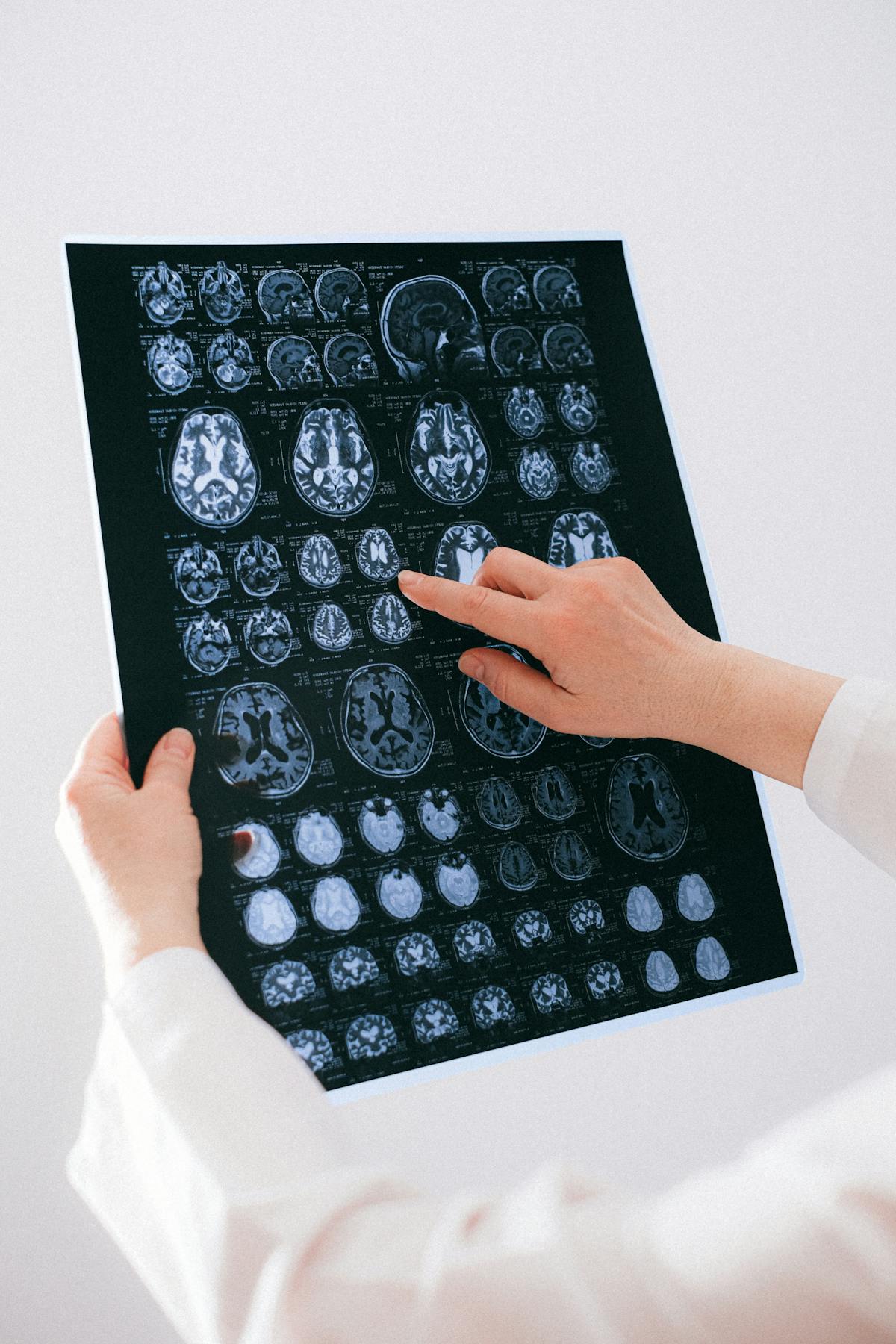
The most important clinical decision with Ankle Impingement Syndrome Anterior Posterior Michigan Podiatrist isn’t which treatment to start with — it’s identifying the correct subtype. That changes everything. Call (810) 206-1402.
The most important clinical decision with Ankle Impingement Syndrome Anterior Posterior Michigan Podiatrist isn’t which treatment to start with — it’s identifying the correct subtype. That changes everything. Call (810) 206-1402.
What Is Ankle Impingement?
Ankle impingement is pain caused by abnormal contact between anatomical structures at the extremes of ankle movement. Anterior ankle impingement produces pain with ankle dorsiflexion (foot flexion upward), as bone spurs or inflamed soft tissue are pinched between the front of the tibia and talus. Posterior ankle impingement causes pain with plantarflexion (foot pointing down), as the os trigonum accessory bone or enlarged posterior talar process is compressed between the tibia and calcaneus. Both forms are common in athletes—particularly football linemen, dancers, gymnasts, and runners.
Anterior Ankle Impingement
Anterior impingement develops from repeated ankle sprains that trigger talar dome osteophyte formation, tibial lip spurring, and anterior joint capsule thickening. Patients report deep anterior ankle pain and stiffness with squat, climb stairs, or sport-specific deep dorsiflexion. The classic “footballer’s ankle” results from decades of ball-kicking impacts driving the anterior joint capsule into the tibial rim. Clinical examination demonstrates anterior joint line tenderness, limited dorsiflexion compared to the contralateral ankle, and pain reproduced by forced dorsiflexion. Weight-bearing X-rays demonstrate the characteristic tibial and talar osteophytes.
Posterior Ankle Impingement
Posterior impingement involves the os trigonum (an accessory bone present in 5–10% of the population), the enlarged posterior talar process (Stieda process), or thickened posterior ankle ligaments. It produces pain with ankle plantarflexion and is particularly disabling for ballet dancers (en pointe), soccer players, and downhill runners. The forced plantarflexion test—passive hyperplantarflexion producing posterior ankle pain—is the diagnostic hallmark. CT scan best delineates bony anatomy; MRI identifies soft-tissue pathology including flexor hallucis longus tendinopathy in the os trigonum tunnel.
Conservative Treatment for Ankle Impingement
Initial management includes activity modification to avoid provocative positions, NSAID anti-inflammatory therapy, and physical therapy focusing on eccentric calf strengthening, proprioception training, and ankle mobility work within the pain-free range. Ultrasound-guided corticosteroid injection into the anterior or posterior joint space reduces acute inflammation and provides diagnostic confirmation when pain relief correlates with the injection site. Custom orthotics can off-load impingement-prone positions. Most cases of soft-tissue impingement respond to conservative care; bony impingement with established osteophytes rarely resolves without surgical debridement.
Arthroscopic Ankle Impingement Surgery
Arthroscopic debridement is the definitive treatment for impingement that fails conservative care. Dr. Biernacki performs ankle arthroscopy through two or three small incisions (portals), visualizing the joint interior with a small camera and removing offending osteophytes, synovial tissue, and bony fragments with motorized shavers and burrs. Posterior impingement with os trigonum is addressed via posterior ankle arthroscopy or endoscopic os trigonum excision in the prone position. Arthroscopic impingement surgery is outpatient, recovery to regular footwear takes two to four weeks, and return to full sport is typically six to ten weeks. Outcomes are excellent with over 85% of patients reporting significant pain reduction and functional improvement.
Dr. Tom's Product Recommendations
Lace-Up Ankle Brace (McDavid 195)
⭐ Highly Rated
Lace-up ankle brace limits extreme dorsiflexion and plantarflexion that provoke anterior and posterior impingement, allowing athletes to continue participating while managing conservative treatment.
Dr. Tom says: “A lace-up ankle brace is the most effective bracing option for controlling the end-range ankle positions that trigger impingement pain during sport.”
Athletes with ankle impingement who need to continue sport during conservative treatment
Those with confirmed bony impingement requiring arthroscopic surgery for definitive relief
Disclosure: We earn a commission at no extra cost to you.
Vive Ankle Ice Wrap
⭐ Highly Rated
Contoured ice wrap provides targeted cold therapy to the anterior or posterior ankle following activity, reducing post-exercise inflammation in impingement patients.
Dr. Tom says: “Ice after activity is simple and effective for controlling impingement flare-ups during conservative management.”
Ankle impingement patients managing symptoms conservatively with activity modification
Those post-arthroscopy needing proper wound care protocol
Disclosure: We earn a commission at no extra cost to you.
✅ Pros / Benefits
- A
- r
- t
- h
- r
- o
- s
- c
- o
- p
- i
- c
- s
- u
- r
- g
- e
- r
- y
- i
- s
- m
- i
- n
- i
- m
- a
- l
- l
- y
- i
- n
- v
- a
- s
- i
- v
- e
- w
- i
- t
- h
- r
- a
- p
- i
- d
- r
- e
- c
- o
- v
- e
- r
- y
- ;
- m
- o
- s
- t
- a
- t
- h
- l
- e
- t
- e
- s
- r
- e
- t
- u
- r
- n
- t
- o
- f
- u
- l
- l
- s
- p
- o
- r
- t
- w
- i
- t
- h
- i
- n
- s
- i
- x
- t
- o
- t
- e
- n
- w
- e
- e
- k
- s
- p
- o
- s
- t
- –
- o
- p
- e
- r
- a
- t
- i
- v
- e
- l
- y
- .
❌ Cons / Risks
- B
- o
- n
- y
- i
- m
- p
- i
- n
- g
- e
- m
- e
- n
- t
- r
- a
- r
- e
- l
- y
- r
- e
- s
- o
- l
- v
- e
- s
- w
- i
- t
- h
- o
- u
- t
- s
- u
- r
- g
- e
- r
- y
- ;
- s
- o
- f
- t
- –
- t
- i
- s
- s
- u
- e
- i
- m
- p
- i
- n
- g
- e
- m
- e
- n
- t
- m
- a
- y
- r
- e
- c
- u
- r
- w
- i
- t
- h
- o
- n
- g
- o
- i
- n
- g
- a
- t
- h
- l
- e
- t
- i
- c
- e
- x
- p
- o
- s
- u
- r
- e
- .
Dr. Tom Biernacki’s Recommendation
Ankle impingement is a very satisfying condition to treat surgically because the results are so consistently good. Athletes who have struggled with deep ankle pain on squatting, running, or dancing for years are often back to full activity within two months of an arthroscopic procedure. If ankle pain limits your range of motion, let’s image it properly and see if impingement is the culprit.
— Dr. Tom Biernacki, DPM | Board-Certified Podiatric Surgeon | Balance Foot & Ankle
Frequently Asked Questions
Can ankle impingement resolve without surgery?
Soft-tissue impingement without established bone spurs frequently responds to conservative care including injections and physical therapy. Bony impingement with osteophytes rarely resolves without arthroscopic removal.
How is ankle arthroscopy different from open surgery?
Arthroscopy uses small camera portals instead of a large incision, reducing recovery time, infection risk, and surgical trauma significantly. Most ankle impingement procedures are now performed arthroscopically with outpatient same-day discharge.
Can I walk after ankle impingement surgery?
Most patients walk in a boot or regular shoe within a few days of arthroscopic impingement surgery. Full return to sport is typically six to ten weeks depending on the extent of the procedure.
Does ankle impingement come back after surgery?
Recurrence is uncommon after thorough arthroscopic debridement. However, continued high-impact sport activity can produce new osteophyte formation over years. Proper rehabilitation and footwear optimization reduce recurrence risk.
Michigan Foot Pain? See Dr. Biernacki In Person
4.9★ rated | 1,123 Reviews | 3,000+ Surgeries
Same-week appointments · Howell & Bloomfield Township
📞 (810) 206-1402 Book Online →Frequently Asked Questions
What causes this condition?
Causes include mechanical stress, biomechanical imbalance, age-related changes, and sometimes systemic disease. Our clinical exam plus imaging identifies the specific driver.
Can it go away on its own?
Mild cases sometimes resolve with rest and supportive footwear. Persistent symptoms past 4-6 weeks rarely resolve without active treatment.
Is surgery required?
Most patients resolve with non-surgical care. Surgery is reserved for refractory cases or structural deformity.
Visit Balance Foot & Ankle — Same-Day Appointments Available
Our podiatry team serves patients throughout Michigan including Howell, Brighton, and Bloomfield Township. If you’re dealing with heel pain, ingrown toenails, or a foot injury, we have same-day appointment availability.
American Podiatric Medical Association: Find a Podiatrist
Ready to Get Relief?
Same-day appointments available in Howell & Bloomfield Township, MI
4.9★ | 1,123 Reviews | 3,000+ Surgeries
Or call: (810) 206-1402

Dr. Tom Biernacki, DPM is a board-certified foot & ankle surgeon (ABFAS & ABPM) at Balance Foot & Ankle Specialists in Southeast Michigan. With over a decade of clinical experience, he specializes in heel pain, bunions, diabetic foot care, sports injuries, and minimally invasive surgery. Dr. Biernacki is a member of the APMA and ACFAS, and his patient education content on MichiganFootDoctors.com and YouTube has made him one of the most-followed foot & ankle educators on YouTube.
More questions patients ask
What is ankle impingement syndrome and what causes anterior vs posterior ankle pain with motion?
Ankle impingement syndrome is a condition in which soft tissue or bony structures are mechanically compressed (impinged) within the ankle joint during motion, producing pain that limits dorsiflexion (anterior impingement) or plantarflexion (posterior impingement); it is a leading cause of chronic ankle pain in athletes and active individuals and is frequently underdiagnosed because standard X-rays may appear normal in soft tissue impingement cases. Anterior ankle impingement: caused by compression of soft tissue or bone at the anterior ankle joint during dorsiflexion; sources of anterior impingement: anterolateral soft tissue impingement: the most common form; scar tissue or a thickened synovial fold (a meniscoid lesion) forms in the anterolateral gutter of the ankle after a lateral ankle sprain; the scar tissue is trapped between the fibula and the talus during dorsiflexion; anterior osteophytes: bone spurs on the anterior tibial lip and the talar neck (talar beak); these osteophytes contact each other during dorsiflexion, blocking motion and causing pain; common in athletes who repeatedly jam the ankle in flexion (soccer, football); Symptoms of anterior impingement: anterior ankle pain that worsens with dorsiflexion activities (squatting, ascending stairs, running uphill); restricted ankle dorsiflexion range of motion; point tenderness at the anterior ankle joint line; pain reproduced by the forced dorsiflexion-eversion test (the examiner forces the ankle into dorsiflexion while the patient reports pain); Posterior ankle impingement: caused by compression of structures at the posterior ankle during plantarflexion; sources of posterior impingement: os trigonum: an accessory ossicle posterior to the talus that is compressed between the calcaneus and the posterior tibial plafond during plantarflexion; posterior talar process fracture (Shepherd's fracture): fracture of the posterior talar process that becomes an impingement lesion; soft tissue posterior impingement: thickened posterior capsule or synovitis; Symptoms of posterior impingement: posterior ankle pain that worsens with plantarflexion (pointing the foot, descending stairs, ballet en pointe, soccer kicking); pain directly posterior to the Achilles tendon between the tendons on deep palpation; both types are confirmed by MRI.
How is ankle impingement syndrome treated and when does it require arthroscopic surgery?
Ankle impingement treatment follows a conservative-first approach for soft tissue impingement, while bony impingement with osteophytes or os trigonum typically requires arthroscopic removal for definitive resolution; the good news is that ankle arthroscopy for impingement produces excellent outcomes with rapid recovery. Conservative treatment: Physical therapy: the cornerstone of non-surgical management; ankle joint mobilization to restore dorsiflexion range of motion; soft tissue mobilization around the ankle; peroneal and calf strengthening; proprioception training; activity modification: temporarily avoiding the impingement-provoking activities (dorsiflexion loading for anterior, plantarflexion for posterior); Corticosteroid injection: ultrasound or fluoroscopy-guided injection into the anterolateral gutter (for soft tissue anterolateral impingement) or the posterior ankle (for posterior impingement); highly effective for soft tissue impingement; less effective for bony impingement where the mechanical obstruction cannot be resolved by reducing inflammation; provides diagnostic information -- significant pain relief after injection confirms the impingement is the pain source; NSAIDs: for the inflammatory component; Footwear: for anterior impingement, a mild heel lift reduces the depth of ankle dorsiflexion; for posterior impingement, avoiding high heels and deep plantarflexion positions; Surgical treatment -- ankle arthroscopy: indications: failure of 3-6 months of conservative management; bony impingement (osteophytes, os trigonum, talar beak) -- surgery is typically the most efficient treatment; anterior impingement arthroscopy: two small portals (anterolateral and anteromedial) are made at the ankle joint; the arthroscope visualizes the anterior compartment; soft tissue impingement lesions (meniscoid scar, thickened synovium) are debrided with a motorized shaver; anterior osteophytes on the tibial plafond and talar neck are removed with a burr until full dorsiflexion is restored under direct visualization; Posterior impingement arthroscopy (2-portal endoscopic approach): the patient is prone; two portals are made posterior to the Achilles; the posterior ankle is visualized; the os trigonum is excised; posterior osteophytes are removed; the FHL tendon is inspected and released if concurrent tendinopathy is present; Recovery: anterior arthroscopy: weight-bearing in a boot immediately; out of boot in 2 weeks; return to sports in 4-8 weeks; posterior endoscopy: similar timeline; return to full ballet or soccer: 6-12 weeks; outcomes: 85-95% excellent results for both anterior and posterior arthroscopic ankle impingement debridement.
Still have a question about coverage or cost? Book online and we will check your benefits before your visit: Book in Howell · Book in Bloomfield Township. Prefer to talk it through first? Call (810) 206-1402.