
You are in the right place. Dr. Tom Biernacki, DPM, FACFAS — board-certified foot & ankle surgeon with 3,000+ surgeries — explains exactly what ankle X-ray vs MRI means and what actually works. Call (810) 206-1402 for a same-day appointment at our Howell or Bloomfield Hills office.
Medically reviewed by Dr. Tom Biernacki, DPM · Board-Certified Podiatric Surgeon · Last reviewed: April 2026 · Editorial Policy
The most important clinical decision with Ankle Xray Vs Mri Michigan isn’t which treatment to start with — it’s identifying the correct subtype. That changes everything. Call (810) 206-1402.
Quick Answer
Ankle X-Ray vs. MRI: Which Do You Need in Michigan? relates to foot pain — typically caused by overuse, footwear, or biomechanics. Most patients improve in 6-12 weeks with conservative care. Same-week appointments in Howell + Bloomfield Hills: (810) 206-1402.

Quick Answer
Most foot and ankle problems respond to conservative care — proper footwear, supportive inserts, activity modification, and targeted stretching — within 4-8 weeks. Persistent pain beyond that window, or any symptom that prevents walking, warrants a podiatric evaluation to rule out fracture, tendon tear, or systemic cause.
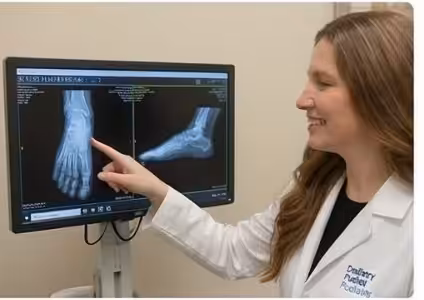
Watch: Dr. Tom Biernacki, DPM

Medically reviewed by Dr. Tom Biernacki, DPM — Board-Certified Podiatric Surgeon — Balance Foot & Ankle, Howell & Bloomfield Hills, MI. Last updated April 2026.
▶ Watch

When you come to a podiatrist with ankle or foot pain, one of the first decisions is what imaging — if any — is needed. X-rays and MRI are the two most commonly ordered studies, but they provide very different information and are appropriate for different clinical situations. At Balance Foot & Ankle in Southeast Michigan, Dr. Tom Biernacki uses targeted imaging to make accurate diagnoses efficiently, without unnecessary cost or radiation exposure.
Medically reviewed by Dr. Tom Biernacki, DPM
Board-certified podiatric surgeon | Balance Foot & Ankle
Last reviewed: April 2, 2026


The most important clinical decision with Ankle Xray Vs Mri Michigan isn't which treatment to start with — it's which subtype or underlying cause you actually have. Our podiatrists regularly see patients who've been treated for months for the wrong diagnosis. The correct identification changes the entire treatment path. Call (810) 206-1402 — Dr. Tom evaluates this condition at both Howell and Bloomfield Hills locations.
What X-Rays Show
X-rays (plain radiographs) use ionizing radiation to create images of dense structures — primarily bone. They are excellent for: detecting fractures (acute breaks, stress fractures visible after 2–3 weeks), assessing bone alignment and joint space narrowing (arthritis), identifying bone spurs (heel spurs, osteophytes), evaluating deformity (bunion angle, flatfoot alignment on weight-bearing views), spotting calcifications (calcium pyrophosphate, Achilles calcification, sesamoid lesions), and confirming hardware position after surgery. Weight-bearing X-rays — taken while the patient stands on the foot — are particularly valuable for alignment assessment and are standard practice at Balance Foot & Ankle for deformity evaluation. X-rays are rapid, inexpensive, widely available, and the appropriate first-line imaging for most acute injuries and structural concerns.

What MRI Shows
MRI (magnetic resonance imaging) uses magnetic fields and radiofrequency pulses to generate detailed images of soft tissues — tendons, ligaments, cartilage, nerves, and bone marrow. MRI is the study of choice for: ligament tears (ATFL, spring ligament, plantar plate), tendon tears and degeneration (Achilles, posterior tibial, peroneal tendons), bone marrow edema (early stress fracture before X-ray changes, osteochondral lesions, avascular necrosis), cartilage assessment (osteochondral defects of the talus), nerve pathology (Morton’s neuroma, Baxter’s nerve entrapment), infection (osteomyelitis, septic joint), and tumor evaluation (soft tissue masses requiring characterization). MRI does not use radiation, but it is more expensive, takes longer (30–60 minutes), and is not available in the office — requiring scheduling at an outpatient imaging center or hospital.

When Each Is Appropriate
Acute ankle sprain → X-ray first (Ottawa Ankle Rules guide X-ray decision; MRI if ligament tear is suspected clinically and will change management). Chronic ankle instability → X-ray (alignment, bony lesions) + MRI (ligament assessment, osteochondral lesion). Achilles or tendon pain → ultrasound (fast, dynamic, done in office) or MRI (more detailed, full tendon course). Heel pain → X-ray (bone spur, stress fracture, arthritis) — MRI rarely needed unless atypical features. Stress fracture → X-ray first; if negative but clinical suspicion is high, MRI within days (most sensitive). Acute trauma with significant mechanism → X-ray first; MRI if fracture is excluded but pain and swelling are disproportionate. Lump or mass → ultrasound first; MRI for characterization of deeper or indeterminate masses.
In-Office Ultrasound: A Third Option
Diagnostic musculoskeletal ultrasound is available in-office at Balance Foot & Ankle and provides real-time, dynamic imaging of soft tissue structures. Ultrasound is the preferred first-line study for evaluating tendons (Achilles, posterior tibial, peroneal), plantar fascia thickness, Morton’s neuroma, cysts, bursitis, and tendon sheath fluid. It is faster and less expensive than MRI, provides real-time assessment during movement, and allows same-session image-guided injection if treatment is indicated. Ultrasound is limited by depth penetration and cannot assess bone marrow or intra-articular cartilage.
When ankle imaging is urgent:
- Unable to bear weight after an ankle injury
- Visible deformity or bone protrusion
- Severe swelling that worsens over 24 hours
- Ankle instability with repeated giving way
- Persistent pain after 2 weeks despite RICE treatment
Ankle Pain After an Injury?
Same-day X-ray and ultrasound at our Howell & Bloomfield Hills, MI locations
4.9★ | 1,123 Reviews | 3,000+ Surgeries
Or call: (810) 206-1402
More Podiatrist-Recommended Foot Health Essentials
Top-Rated Arch Support Insole
Universal podiatrist-recommended insert for pain relief and prevention.
Foot Massage Ball
Daily 3-minute roll reduces most forms of foot and heel pain.
Moisture-Wicking Sock
Prevents fungus, blisters, and odor — the basics matter.
As an Amazon Associate, Balance Foot & Ankle earns from qualifying purchases. Product recommendations are based on clinical experience; prices and availability shown above update live from Amazon.
When to See a Podiatrist
If foot or ankle pain has been bothering you for more than a few weeks, home care alone may not be enough. Balance Foot & Ankle offers same-week appointments at our Howell and Bloomfield Hills clinics — no referral needed in most cases. Bring your current shoes and a short list of symptoms and we’ll build you a treatment plan in one visit.
Call Balance Foot & Ankle: (810) 206-1402 · Book online · Offices in Howell & Bloomfield Hills
Frequently Asked Questions
Can an X-ray detect a torn ankle ligament?
Standard X-rays cannot directly visualize soft tissue structures like ligaments. However, stress X-rays (taken with the ankle held in a stressed position) can indirectly show ligament instability by demonstrating abnormal bone movement. MRI is the definitive study for directly imaging ankle ligament tears and is preferred when surgical planning is being considered.
Does insurance cover foot and ankle MRI?
Most major insurance plans — including Medicare, Blue Cross Blue Shield, Aetna, and Cigna — cover ankle and foot MRI when medically necessary and ordered by a treating physician or podiatrist. Pre-authorization may be required. Dr. Biernacki’s office handles the authorization process and will confirm coverage before ordering.
Can a stress fracture be missed on X-ray?
Yes. X-rays are frequently normal in the first 2–3 weeks of a stress fracture. The subtle cortical disruption of an early stress fracture is below X-ray resolution. Bone scan and MRI detect stress reactions before visible fracture lines appear. If clinical suspicion is high and the X-ray is negative, MRI is the next study of choice — it is the most sensitive and specific imaging modality for early stress fractures.
📧 Get Dr. Tom’s Free Lab Test Guide
Discover the 5 lab tests every person over 35 should ask their doctor about — explained in plain English by a board-certified physician.
The right imaging study from the start leads to the right diagnosis and faster recovery. Contact Balance Foot & Ankle for expert foot and ankle evaluation with targeted imaging in Southeast Michigan.
Join 950,000+ Learning About Foot Health
Dr. Tom shares honest medical advice, supplement reviews, and treatment guides you won’t find anywhere else.
Subscribe on YouTube →Ready to Get Expert Foot Care?
Dr. Biernacki and our team at Balance Foot & Ankle are accepting new patients in Howell and Bloomfield Hills, MI. Most insurances accepted.
Book My Appointment →or call (810) 206-1402
📍 Located in Michigan?
Our board-certified podiatrists treat this condition at two convenient locations. Same-day appointments often available.
Medically Reviewed by: Dr. Tom Biernacki, DPM — Board-Certified Podiatrist, Balance Foot & Ankle Specialists

Insurance Accepted
BCBS · Medicare · Aetna · Cigna · United Healthcare · HAP · Priority Health · Humana · View All →
Howell Office
4330 E Grand River Ave
Howell, MI 48843
Get Directions →
Bloomfield Hills Office
43494 Woodward Ave, #208
Bloomfield Hills, MI 48302
Get Directions →
Your Board-Certified Podiatrists


Ready to Get Back on Your Feet?
Same-week appointments available at both locations.
Book Your AppointmentMost Common Mistake We See
The most common mistake we see is: Waiting too long before seeking care. Fix: any foot pain lasting more than 4 weeks, or any sudden severe symptom, deserves a professional evaluation rather than more rest.
Warning Signs That Need Same-Day Care
Seek immediate evaluation at Balance Foot & Ankle if you experience any of the following:
- Unable to bear weight
- Severe swelling with skin colour change
- Fever with foot pain (possible infection)
- Diabetes plus any new foot symptom
Call (810) 206-1402 — same-day and next-day appointments at our Howell and Bloomfield Hills offices.
Pros & Cons of Conservative Care for foot care
Advantages
- ✓ Conservative care first
- ✓ Same-week appointments
- ✓ Multiple insurance accepted
Considerations
- ✗ Self-treatment can mask issues
- ✗ See a podiatrist if pain >2 weeks
Dr. Tom’s Recommended Products for foot care
Affiliate disclosure: As an Amazon Associate, Balance Foot & Ankle earns from qualifying purchases. We only recommend products we use with patients.
Footnanny Heel Cream Dr. Tom’s Pick
Best for: Daily moisturizer for cracked heels
Ready to Get Back on Your Feet?
Same-day appointments in Howell + Bloomfield Hills. Most insurance accepted. Dr. Tom Biernacki, DPM & team.
Book Today — Same-Day Appointments Available
Call Now: (810) 206-1402
About Your Care Team at Balance Foot & Ankle
Dr. Tom Biernacki, DPM · Board-Certified Foot & Ankle Surgeon. Specializes in conservative-first care, minimally invasive bunion surgery, and complex reconstruction.
Dr. Carl Jay, DPM · Accepting new patients. Specializes in sports medicine, athletic injuries, and routine podiatric care.
Dr. Daria Gutkin, DPM, AACFAS · Accepting new patients. Specializes in surgical reconstruction and pediatric podiatry.
Locations: 4330 E Grand River Ave, Howell, MI 48843 · 43494 Woodward Ave Suite 208, Bloomfield Hills, MI 48302
Hours: Mon–Fri 8:00 AM – 5:00 PM · (810) 206-1402
Visit Balance Foot & Ankle — Same-Day Appointments Available
Our podiatry team serves patients throughout Michigan including Howell, Brighton, and Bloomfield Hills. If you’re dealing with heel pain, ingrown toenails, or a foot injury, we have same-day appointment availability.
Same-day appointments available. (810) 206-1402
Doctor Hoy’s Natural Pain Relief Gel
Natural topical pain relief I use in our clinic. Arnica + camphor formula — apply directly to the area 3–4x daily. ($20–25)
Shop Doctor Hoy’s →Frequently Asked Questions
Which is better for plantar fasciitis?
The shoe with more cushioning and a stronger rocker typically wins for plantar fasciitis. See full comparison for our specific verdict.
Which lasts longer?
Both options typically last 300-500 miles for runners or 9-12 months for daily walkers. Material durability varies; check our detailed comparison.
Which is better for flat feet?
Flat feet need stability or motion control. The neutral option is not ideal unless paired with a custom orthotic.
Ready to fix this for good?
Reading goes only so far. The fastest path to relief is a 30-minute office visit with Dr. Biernacki — same-day Howell or Bloomfield Hills. Call (810) 206-1402 or use our online booking.
Our podiatrists treat the underlying cause, not just the symptom. Same-week appointments at our Howell and Bloomfield Hills, Michigan offices.
Dr. Tom Biernacki, DPM is a board-certified foot & ankle surgeon (ABFAS & ABPM) at Balance Foot & Ankle Specialists in Southeast Michigan. With over a decade of clinical experience, he specializes in heel pain, bunions, diabetic foot care, sports injuries, and minimally invasive surgery. Dr. Biernacki is a member of the APMA and ACFAS, and his patient education content on MichiganFootDoctors.com and YouTube has made him one of the most-followed foot & ankle educators on YouTube.