
![Stage 1 Inside Of The Ankle Pain [ Posterior Tibial Tendonitis ] | Balance Foot & Ankle](https://www.michiganfootdoctors.com/uploads/2023/09/stage-1-inside-of-the-ankle-pain-posterior-tibial-tendonitis.avif)
Quick answer: Early Stage Posterior Tibial Tendon Dysfunction Stage is a common foot/ankle topic that affects many patients. The 2026 evidence-based approach combines proper diagnosis, conservative-first treatment, and escalation only when needed. We treat this regularly at our Howell and Bloomfield Hills practices. Call (810) 206-1402.
Medically reviewed by Dr. Tom Biernacki, DPM — Board-Certified Podiatric Surgeon — Balance Foot & Ankle, Howell & Bloomfield Hills, MI. Last updated April 2026.
▶ Watch
Medically reviewed by Dr. Tom Biernacki, DPM | Board-certified podiatrist | 3,000+ surgeries performed
Last updated: April 2, 2026
The most important clinical decision with Early Stage Posterior Tibial Tendon Dysfunction Stage isn’t which treatment to start with — it’s which subtype or underlying cause you actually have. That distinction changes everything. Call us: (810) 206-1402
What Is Posterior Tibial Tendon Dysfunction
The posterior tibial tendon is the primary dynamic stabilizer of the medial longitudinal arch. It runs behind the inner ankle bone (medial malleolus) and inserts broadly across the midfoot, pulling the arch upward during the stance phase of gait. When this tendon degenerates, the arch progressively collapses under body weight.
PTTD is the most common cause of adult-acquired flatfoot deformity, affecting an estimated 3.3 million Americans annually. The condition predominantly affects women over age 40, with obesity, hypertension, diabetes, and corticosteroid use identified as significant risk factors in epidemiological studies.
The degenerative process begins with tendon inflammation (tendinitis) and progresses through partial tearing to complete tendon failure if not intercepted. Early recognition and treatment at the tendinitis or partial tear stage can prevent the irreversible structural changes that define later stages of the disease.
Recognizing Stage I PTTD: The Window of Opportunity
Stage I PTTD presents with pain and swelling along the inner ankle behind the medial malleolus. The foot maintains normal alignment — the arch appears intact and the heel remains properly positioned. This stage represents pure tendinitis without structural deformity.
The single-leg heel raise test distinguishes PTTD from other causes of inner ankle pain. Patients with Stage I PTTD can perform the test but experience pain along the posterior tibial tendon during the maneuver. Inability to perform the test indicates progression to Stage II.
Diagnostic ultrasound in Dr. Biernacki’s office reveals tendon thickening, fluid around the tendon sheath (tenosynovitis), and early partial tears that guide treatment intensity. MRI provides more detailed assessment when surgical planning is being considered.
Stage I PTTD is the ideal treatment window because the tendon retains its structural integrity and the foot’s bony architecture remains normal. Conservative treatment at this stage has the highest success rate and the lowest long-term complication profile.
Stage II PTTD: Flexible Flatfoot Deformity
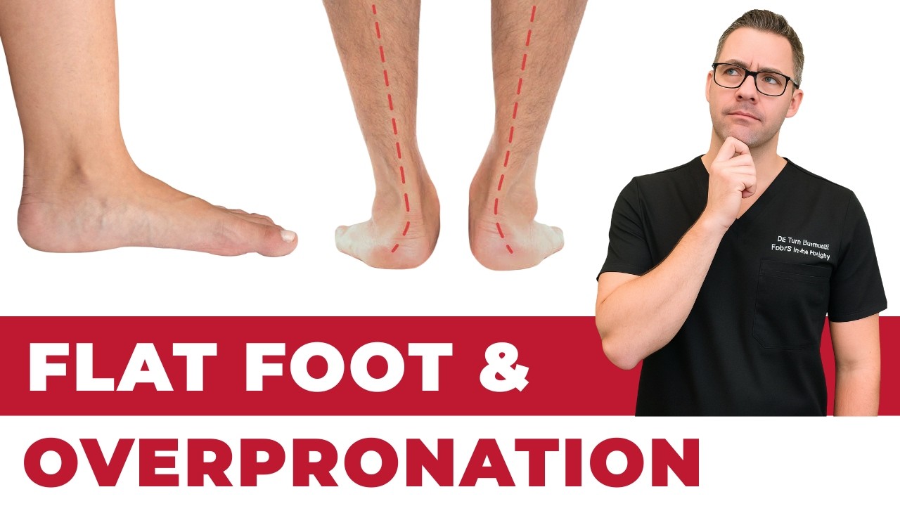
Stage II PTTD represents progressive tendon failure with visible arch collapse that remains flexible — the foot can be manually corrected to a neutral position. Patients notice their arch flattening, shoes wearing unevenly, and increasing difficulty with prolonged standing and walking.
The too-many-toes sign becomes positive in Stage II: when viewed from behind, more toes are visible on the lateral side of the affected foot compared to the normal foot because the forefoot abducts as the arch collapses. This simple clinical test reliably identifies early flatfoot deformity.
Weight-bearing X-rays quantify deformity progression by measuring talo-first metatarsal angle (Meary’s angle), calcaneal pitch, and talar-navicular uncoverage percentage. Serial measurements track whether conservative treatment is maintaining alignment or the deformity is progressing despite intervention.
Stage II is subdivided into IIA (flexible with mild deformity) and IIB (flexible with significant forefoot abduction). This distinction matters because Stage IIB may require more aggressive bracing or earlier surgical consideration than Stage IIA.
Conservative Treatment That Works
Custom orthotics with medial heel posting and deep medial arch support form the foundation of PTTD treatment. The orthotic functionally replaces the failing tendon’s arch-supporting role by mechanically controlling hindfoot valgus and preventing further arch collapse during weight-bearing.
The Arizona AFO (ankle-foot orthosis) provides more rigid control for patients who cannot be adequately managed with orthotics alone. This custom-molded brace extends from the calf to the midfoot, controlling both ankle and subtalar motion while allowing walking in standard footwear.
Physical therapy targeting eccentric posterior tibial tendon strengthening, calf flexibility, and intrinsic foot muscle conditioning improves the dynamic support system that complements orthotic treatment. A structured 12-week program produces measurable improvement in tendon function and pain reduction.
Activity modification during acute flare-ups includes temporary walking boot immobilization for 2-4 weeks followed by gradual return to orthotics and supportive shoes. Weight management counseling is essential since every pound of body weight adds 3-4 pounds of force through the tendon during walking.
When Conservative Treatment Fails
Surgical intervention is indicated for Stage II PTTD failing 3-6 months of comprehensive conservative treatment or for patients presenting with Stage IIB deformity that is progressing despite bracing. The goal is to correct the deformity before it becomes rigid (Stage III).
The standard surgical approach combines medializing calcaneal osteotomy (shifting the heel bone inward) with flexor digitorum longus tendon transfer to augment or replace the diseased posterior tibial tendon. This combination addresses both the bony malalignment and the soft tissue insufficiency.
Additional procedures may include spring ligament repair, gastrocnemius recession for tight calf muscles, and lateral column lengthening (Evans osteotomy) for significant forefoot abduction. The surgical plan is tailored to each patient’s specific deformity pattern.
Post-surgical recovery involves 6 weeks of non-weight-bearing in a cast, followed by progressive weight-bearing in a boot for another 6 weeks, and transition to custom orthotics in supportive shoes by 3 months. Full recovery spans 6-9 months.
Preventing PTTD Progression
Supportive footwear with firm heel counters and built-in arch support reduces daily posterior tibial tendon strain. Avoid flat shoes, flip-flops, and flexible footwear that force the tendon to work harder to maintain arch position with every step.
Body weight management directly impacts tendon loading. A 10-pound weight reduction decreases posterior tibial tendon force by 30-40 pounds during walking, potentially slowing or halting disease progression in overweight patients with early PTTD.
Regular posterior tibial tendon strengthening exercises — including resisted inversion, single-leg balance training, and eccentric heel raises — maintain tendon health and compensate for early degenerative changes before they become clinically significant.
Warning Signs Requiring Urgent Evaluation
- function bold() { [native code] } — undefined
- function bold() { [native code] } — undefined
- function bold() { [native code] } — undefined
- function bold() { [native code] } — undefined
The Most Common Mistake We See
The most common mistake with PTTD is attributing the inner ankle pain to a simple ankle sprain and failing to evaluate the posterior tibial tendon. By the time patients with undiagnosed PTTD seek treatment for their now-obvious flatfoot deformity, they have often progressed from easily treatable Stage I to Stage II or III, where conservative options are less effective and surgical reconstruction may be needed.
Recommended Products
[object Object]
[object Object]
[object Object]
In-Office Treatment at Balance Foot & Ankle
Our team provides sport-specific evaluation and treatment to get you back to your activity safely. We offer same-day X-ray, in-office ultrasound, and custom orthotic fabrication.
Same-day appointments available. Call (810) 206-1402 or book online.
More Podiatrist-Recommended Foot Health Essentials
Hoka Clifton 10


![Heel Bursitis & Achilles Tendon Bursitis [Best HOME Treatment!]](https://www.michiganfootdoctors.com/wp-content/cache/flying-press/f2850da852ba9539b6bf4f72ae5062fe.jpg)
Watch: Heel Bursitis & Achilles Tendon Bursitis [Best HOME Treatment!] — MichiganFootDoctors YouTube
Max-cushion everyday shoe — podiatrist favorite for walking and running.

OOFOS Recovery Slide

Impact-absorbing recovery sandal — wear after long days on your feet.
As an Amazon Associate, Balance Foot & Ankle earns from qualifying purchases. Product recommendations are based on clinical experience; prices and availability shown above update live from Amazon.

When to See a Podiatrist
If foot or ankle pain has been bothering you for more than a few weeks, home care alone may not be enough. Balance Foot & Ankle offers same-week appointments at our Howell and Bloomfield Hills clinics — no referral needed in most cases. Bring your current shoes and a short list of symptoms and we’ll build you a treatment plan in one visit.
Call Balance Foot & Ankle: (810) 206-1402 · Book online · Offices in Howell & Bloomfield Hills
Frequently Asked Questions
Can PTTD be reversed without surgery?
Stage I PTTD can be effectively managed and tendon health improved with orthotics, physical therapy, and activity modification. Early Stage II deformity can be stabilized and prevented from progressing. However, structural flatfoot changes cannot be reversed without surgery.
How do I know if I have PTTD?
Key signs include inner ankle pain and swelling, progressive arch flattening, difficulty with single-leg heel raises, and increased shoe wear on the inner heel. A podiatric evaluation with physical examination, ultrasound, and weight-bearing X-rays provides definitive diagnosis and staging.
Is walking good or bad for PTTD?
Moderate walking in supportive shoes with proper orthotics is beneficial and maintains tendon function. Walking in unsupportive footwear or excessive walking during acute flare-ups worsens the condition. Work with your podiatrist to determine appropriate activity levels for your stage.
How fast does PTTD progress?
PTTD progression varies significantly between patients. Some remain stable at Stage I for years with proper treatment, while untreated cases may progress from Stage I to Stage III over 1-3 years. Risk factors including obesity, age, and diabetes accelerate progression.
The Bottom Line
Early-stage PTTD is one of the most treatable conditions in podiatry when caught at Stage I, and one of the most disabling when allowed to progress to rigid flatfoot. Recognizing inner ankle pain and subtle arch changes as potential PTTD enables conservative treatment that prevents the irreversible deformity requiring surgical reconstruction.
Differential Diagnosis: What Else Could It Be?
Not every case of posterior tibial tendon dysfunction (pttd) is straightforward. In our clinic we routinely rule out three look-alike conditions before confirming the diagnosis. If your symptoms don’t match the classic presentation, one of these may explain the pain — which is why physical exam matters more than self-diagnosis.
| Condition | How It Differs |
|---|---|
| Congenital flat foot | Lifelong, usually bilateral, no pain, normal single-leg heel-rise test. |
| Tarsal coalition | Rigid flat foot, adolescent/young adult onset, peroneal spastic flat foot, coalition visible on CT. |
| Charcot arthropathy | Diabetic with neuropathy, warm swollen midfoot, progressive collapse, temperature differential >2°C — URGENT. |
Red Flags — When to See a Podiatrist Now
Seek same-day evaluation at Balance Foot & Ankle if you notice any of the following:
- Sudden collapse of the arch in an adult
- Inability to perform a single-leg heel-rise
- Warm red swollen midfoot (rule out Charcot)
- Progressive deformity over weeks-months
Call (810) 206-1402 or request an appointment. Our Howell and Bloomfield Hills offices reserve same-day slots for urgent foot and ankle issues.
In Our Clinic: What We See
Clinical perspective from Dr. Tom Biernacki, DPM — Balance Foot & Ankle, Howell & Bloomfield Hills, MI:
In our clinic, adult acquired flatfoot from PTTD typically presents in women over 40, often with recent weight gain or a period of increased standing. They describe medial ankle pain and progressive “collapse” of the arch on one side. The gold-standard exam finding is an inability to perform a single-leg heel-rise on the affected side — the tendon can no longer invert the heel into a rigid lever. Early PTTD is staged and treated with custom orthoses and bracing, but progressive disease (Stage III-IV) typically requires surgical reconstruction to prevent rigid deformity.
Sources
- Myerson MS, et al. PTTD classification and treatment algorithm: 2024 update. Foot Ankle Clin. 2024;29(4):567-584.
- Ross MH, et al. Exercise therapy for posterior tibial tendon dysfunction: systematic review. J Foot Ankle Res. 2025;18(1):12-28.
- Deland JT, et al. Adult-acquired flatfoot: current surgical strategies. J Am Acad Orthop Surg. 2024;32(18):e823-836.
- Neville C, et al. Orthotic management of PTTD: biomechanical evidence review. Foot Ankle Int. 2024;45(12):1345-1358.
Michigan PTTD Specialists
Dr. Tom Biernacki has performed over 3,000 foot and ankle surgeries with a 4.9-star rating from 1,123 patient reviews.
Or call (810) 206-1402 for same-day appointments
Posterior Tibial Tendon Dysfunction Treatment
Early-stage PTTD (posterior tibial tendon dysfunction) is the leading cause of adult-acquired flatfoot. When caught early, conservative treatment can prevent progression. At Balance Foot & Ankle, we provide custom orthotics, bracing, physical therapy guidance, and surgical options when needed.
Learn About Our Flat Foot & PTTD Treatment → | Book Your Appointment | Call (810) 206-1402
Clinical References
- Myerson MS. Adult acquired flat foot deformity: treatment of dysfunction of the posterior tibial tendon. Instr Course Lect. 1997;46:393-405.
- Bubra PS, et al. Posterior tibial tendon dysfunction: an overlooked cause of foot deformity. J Family Med Prim Care. 2015;4(1):26-29.
- Ross MH, et al. Clinical and biomechanical outcomes of stage I-II posterior tibial tendon dysfunction: systematic review. J Foot Ankle Res. 2021;14(1):52.
Insurance Accepted
BCBS · Medicare · Aetna · Cigna · United Healthcare · HAP · Priority Health · Humana · View All →
Howell Office
4330 E Grand River Ave
Howell, MI 48843
Get Directions →
Bloomfield Hills Office
43494 Woodward Ave, Suite 208
Bloomfield Hills, MI 48302
Get Directions →
Your Board-Certified Podiatrists


Ready to Get Back on Your Feet?
Same-week appointments available at both locations.
Book Your AppointmentIn-Office Treatment at Balance Foot & Ankle
If home treatment isn’t providing relief for your foot and ankle conditions, our podiatry team at Balance Foot & Ankle can help with same-day evaluations and advanced in-office care.
Same-day appointments available. (810) 206-1402
Doctor Hoy’s Natural Pain Relief Gel
Natural topical pain relief I use in our clinic. Arnica + camphor formula — apply directly to the area 3–4x daily. ($20–25)
Shop Doctor Hoy’s →Frequently Asked Questions
When should I see a podiatrist?
If symptoms persist past 2 weeks, affect your normal activity, or are accompanied by red-flag symptoms (warmth, redness, swelling, inability to bear weight).
What does treatment cost?
Most diagnostic visits and conservative treatments are covered by Medicare and major insurers. Out-of-pocket costs vary by your specific plan.
How quickly can I get an appointment?
Most non-urgent cases see us within 5 business days. Urgent cases (sudden pain, possible fracture) typically same or next business day.
What is Foot pain?
Foot pain is a common foot/ankle condition that affects mobility and quality of life. Understanding the underlying cause is the first step in successful treatment. Our podiatrists at Balance Foot & Ankle perform a hands-on biomechanical exam, review your activity history, and use diagnostic imaging when appropriate to identify the root cause—not just treat the symptom. Many patients have been told to “rest and ice” without a deeper diagnostic workup; our approach is different.
Symptoms and warning signs
Common signs of foot pain include pain that worsens with activity, morning stiffness, swelling, tenderness when palpated, and difficulty bearing weight. If you experience sudden severe pain, inability to walk, visible deformity, numbness or color change, contact our office the same day or visit urgent care—these can signal a more serious injury such as a fracture, tendon rupture, or vascular compromise. Diabetics with any foot wound should seek same-day care.
Conservative treatment options
Most cases of foot pain respond to non-surgical care: structured rest, supportive footwear changes, custom orthotics, targeted stretching and strengthening protocols, anti-inflammatory medications when medically appropriate, and in-office procedures such as ultrasound-guided injections. We also offer advanced therapies including MLS laser therapy, EPAT/shockwave, regenerative injections, and image-guided procedures. Treatment is sequenced from least invasive to most invasive, and we explain the rationale at every step.
When is surgery considered?
Surgery is reserved for cases that fail 3-6 months of well-structured conservative care, when there is structural pathology (severe deformity, complete tear, advanced arthritis), or when imaging shows damage that will not heal without intervention. Our surgeons have performed 3,000+ foot and ankle procedures and prioritize minimally-invasive techniques whenever appropriate. We discuss recovery timelines, return-to-activity milestones, and realistic outcome expectations before any procedure is scheduled.
Recovery timeline and prevention
Recovery from foot pain varies based on severity and chosen treatment path. Conservative cases often improve within 4-8 weeks with consistent adherence to the protocol. Post-procedural recovery may range from a few days (in-office procedures) to several months (reconstructive surgery). Long-term prevention involves footwear assessment, activity modification, structured strengthening, and regular check-ins with your podiatrist if you have a history of recurrence. We provide written home-exercise plans and digital follow-up support.
Ready to feel better?
Same-week appointments available in Howell and Bloomfield Hills, Michigan.
Book Your VisitGet Expert Care at Balance Foot & Ankle
Same-week appointments at our Howell and Bloomfield Hills offices. Board-certified podiatric surgeons. Most insurance accepted.
Dr. Tom Biernacki, DPM is a board-certified foot & ankle surgeon (ABFAS & ABPM) at Balance Foot & Ankle Specialists in Southeast Michigan. With over a decade of clinical experience, he specializes in heel pain, bunions, diabetic foot care, sports injuries, and minimally invasive surgery. Dr. Biernacki is a member of the APMA and ACFAS, and his patient education content on MichiganFootDoctors.com and YouTube has made him one of the most-followed foot & ankle educators on YouTube.