
Medically reviewed by Dr. Tom Biernacki, DPM
Board-certified podiatric surgeon | Balance Foot & Ankle, Howell & Bloomfield Township, MI
Last reviewed: May 2026

| Tendon | Function | Rupture Mechanism | Key Clinical Sign | Treatment |
|---|---|---|---|---|
| Achilles tendon | Plantarflexion (push-off) | Sudden eccentric load; jumping; sprinting | Positive Thompson test; palpable gap | Functional brace OR surgical repair; equivalent outcomes in trials |
| Posterior tibial tendon | Arch support; foot inversion | Chronic overload in flatfoot; acute trauma | Progressive flatfoot; “too many toes” sign; single heel raise failure | Surgical reconstruction for complete rupture (flatfoot repair) |
| Peroneal tendons | Foot eversion; lateral ankle stability | Ankle inversion sprain; chronic tendinopathy | Lateral ankle snapping; instability; MRI confirms | Conservative for partial; surgical repair for complete or retracted |
| Flexor hallucis longus (FHL) | Big toe plantarflexion; push-off power | Forceful plantarflexion; repetitive ballet en pointe | Weakness of big-toe flexion; possible nodule | Conservative usually adequate; surgery for complete rupture in athletes |
| Extensor digitorum longus | Toe dorsiflexion; foot clearance | Laceration; direct trauma; forced plantarflexion | Dropped toes; inability to dorsiflex digits | Surgical repair for complete tear if functional deficit significant |
| Achilles Rupture: Surgery vs. Conservative | Surgical Repair | Functional Brace Protocol |
|---|---|---|
| Re-rupture rate | 2–5% | 3–8% (slightly higher in some trials) |
| Return to sport timeline | 6–9 months for recreational; 4–6 months for elite | 8–12 months typical |
| Complication risk | Wound infection 2–4%; nerve injury 1–2% | DVT risk without prophylaxis; re-rupture risk if non-adherent |
| Best candidate | Young elite athlete; large gap on MRI; active lifestyle | Older patient; sedentary; medical comorbidities; patient preference |
| Evidence basis | Multiple RCTs show equivalent functional outcomes to conservative | Multiple RCTs — equivalent outcomes when proper protocol followed |
| Cost | Higher (surgical + anesthesia + OR fees) | Lower (boot + physical therapy) |
Medically Reviewed | Dr. Tom Biernacki, DPM | Board-Certified Podiatrist | Balance Foot & Ankle, Michigan
Quick Answer
Foot and ankle tendon ruptures are serious injuries that require prompt diagnosis to avoid permanent functional loss. The four most clinically significant are: Achilles tendon (Thompson test confirms rupture; surgical vs. functional bracing debate), posterior tibial tendon (too-many-toes sign plus single-heel-rise failure plus Stage I-IV classification), peroneal tendons (lateral ankle pain plus groove pathology plus MRI), and extensor hallucis longus (dropped great toe plus laceration history). Each has a distinct mechanism, clinical test, and treatment pathway. Time from injury to treatment directly affects outcome — these injuries should not be managed as simple sprains.
Overview of Foot Tendon Ruptures
Tendon ruptures in the foot and ankle represent some of the most functionally disabling injuries we manage at Balance Foot & Ankle — and some of the most commonly missed on initial evaluation. In our clinic, we see posterior tibial tendon dysfunction diagnosed as ankle sprain months after onset, Achilles ruptures initially treated as calf strains, and peroneal tendon tears attributed to chronic ankle weakness for years before the correct diagnosis is made. The delay matters: every tendon has a window during which primary repair or functional reconstruction is most successful. After that window, scar formation, muscle atrophy, and tendon retraction make repair significantly more difficult and outcomes less predictable. This article covers the four major foot tendon ruptures — their mechanisms, clinical tests, and the treatment decisions that define outcomes.
Achilles Tendon Rupture
Mechanism and Risk Factors
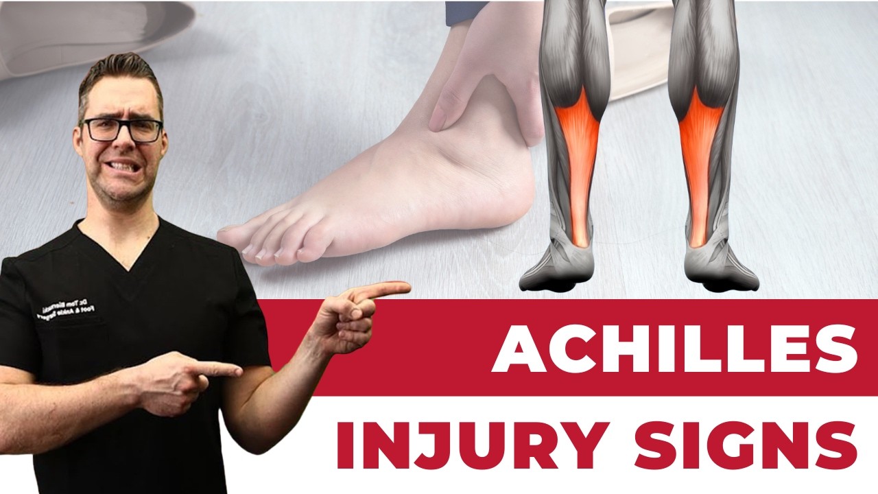
The Achilles tendon is the strongest tendon in the body — it transmits forces up to 12 times body weight during running — and yet it ruptures more frequently than any other tendon in the lower extremity. The classic mechanism is an eccentric overload: a sudden push-off or acceleration with the foot planted and the knee extending, as in the first step of a sprint or pushing off a court surface. The typical patient is a recreational male athlete between 30 and 50 years old. Fluoroquinolone antibiotics (ciprofloxacin, levofloxacin) increase Achilles rupture risk by 3-6x by causing collagen degradation; we ask about recent antibiotic use in every Achilles injury. Prior corticosteroid injection into the tendon itself (not the paratenon) is also a significant risk factor. Most ruptures occur in the watershed zone 2-6 cm proximal to the calcaneal insertion — the area of relatively poor blood supply.
Thompson Test — The Definitive Clinical Test
The Thompson test (Simmonds-Thompson test) is the most important bedside test for Achilles rupture and has sensitivity and specificity above 96%. Perform it with the patient prone on the examination table, knees bent to 90 degrees, feet hanging off the edge. Squeeze the calf muscle belly firmly. A normal result: the foot plantarflexes (the intact tendon transmits the force). A positive result (rupture): the foot does not move, or moves minimally, because the mechanical link between the calf muscle and the heel is disrupted. A false negative can occur when other plantarflexors (flexor hallucis longus, peroneus longus) are intact and produce a small amount of passive plantarflexion — this is why we combine the Thompson test with palpation of the tendon gap (a palpable defect in the tendon substance 2-6 cm above the heel) and observation of the resting foot angle (the ruptured side often rests in more dorsiflexion due to loss of resting tension). Together, these three findings make the diagnosis essentially certain without imaging.
Surgical vs Functional Bracing — The Evidence
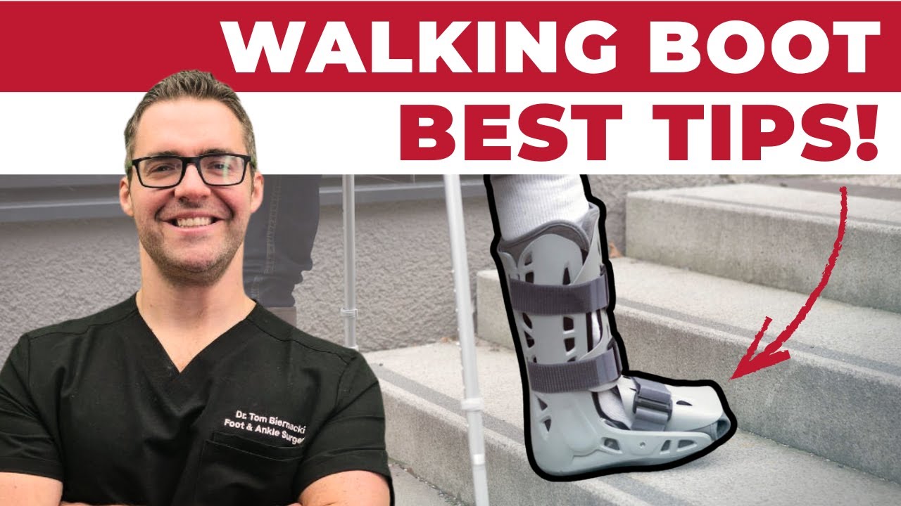
The treatment of acute Achilles tendon rupture has undergone significant evolution. Historical management was surgical repair vs. long-leg casting — and surgery clearly outperformed casting in re-rupture rates. Modern management compares surgical repair to functional bracing (early controlled motion in a boot or hinged brace starting within days of injury), and the distinction is less clear-cut than previously believed. The UKSTAR trial (2020) and multiple meta-analyses show that in healthy patients under 60 with acute complete ruptures, early functional bracing achieves re-rupture rates of 3-4% and functional outcomes equivalent to surgical repair. Surgical repair has traditionally had a re-rupture rate of 1-2% but carries wound complication and infection risks of 3-10%. In our practice, we individualize the decision: young, active, high-demand patients (athletes, laborers) who want to minimize re-rupture risk and can tolerate surgical recovery are offered repair; patients who are older, less active, have diabetes, or have contraindications to surgery are excellent candidates for functional bracing with the same expected functional outcomes. The most important variable is timing — both pathways require initiation within 2 weeks of rupture for optimal results.
Posterior Tibial Tendon Dysfunction (PTTD)
The Too-Many-Toes Sign
Posterior tibial tendon dysfunction (PTTD) — also called adult-acquired flatfoot deformity — is the most common cause of progressive flatfoot in adults and one of the most under-diagnosed tendon conditions in our practice. The posterior tibial tendon (PTT) is the primary dynamic stabilizer of the medial arch; when it fails, the arch collapses, the hindfoot everts, and the forefoot abducts. The too-many-toes sign is the landmark clinical test: stand behind the patient and observe both feet simultaneously. Normally, looking from behind, one to one-and-a-half toes are visible lateral to the heel on each side. In PTTD with forefoot abduction, three to five toes are visible on the affected side — the forefoot has drifted outward relative to the hindfoot. Combined with medial ankle tenderness along the PTT course (from behind the medial malleolus to the navicular insertion) and inability to perform a single-heel rise on the affected side (the PTT inverts the heel at the end of heel rise — a flat, non-inverting heel in single stance is pathognomonic), this clinical triad confirms the diagnosis without imaging.
Stage Classification and Treatment
PTTD is classified in four stages that directly determine treatment. Stage I: tendon is intact but inflamed (tenosynovitis); the deformity is flexible; single-heel rise is painful but possible. Treatment: custom ankle-foot orthosis (AFO) or UCBL orthosis to offload the tendon, combined with physical therapy. Stage II: tendon is elongated or partially torn; flexible flatfoot deformity present; single-heel rise fails. Treatment: rigid custom AFO for non-operative management; surgical reconstruction (FDL tendon transfer plus medial displacement calcaneal osteotomy plus spring ligament repair) for active patients or those who fail orthotic management. Stage III: rigid flatfoot deformity — the hindfoot has become fixed in valgus and the subtalar joint is arthritic; cannot correct passively. Treatment: triple arthrodesis or subtalar fusion. Stage IV: tibiotalar involvement with valgus ankle tilt. Treatment: extensive reconstruction or tibiotalocalcaneal fusion. The critical clinical point: Stage I and II PTTD are treated very differently, and the difference is whether the deformity is flexible (corrects on manual examination) or rigid. Delaying treatment from Stage II to Stage III is a common outcome of missed or dismissed diagnosis — the patient who waited for the pain to become bad enough to see a doctor.
Peroneal Tendon Tears and Rupture
The peroneus brevis and longus tendons run in a fibro-osseous groove behind the lateral malleolus, stabilized by the superior peroneal retinaculum. Peroneal tendon pathology is the most commonly overlooked cause of chronic lateral ankle pain — it is frequently attributed to recurrent ankle sprain when the actual problem is a longitudinal peroneus brevis split tear caused by the retinaculum shearing the tendon against a sharp groove edge with each inversion injury. The peroneus brevis split tear is the most common peroneal pathology: the tendon develops a longitudinal slit (not a transverse rupture) that causes chronic pain, weakness in eversion, and occasional subluxation of the tendon out of its groove. Clinical findings: tenderness directly over the peroneal groove posterior to the lateral malleolus, pain with resisted eversion and plantarflexion combined, and occasionally a snapping or clicking sensation as the tendons sublux over the malleolus. MRI is the definitive imaging study: a split peroneus brevis appears as a C-shape or boomerang shape around the peroneus longus in the groove. Treatment: acute tears in the acute phase can heal with immobilization; chronic symptomatic tears require surgical debridement and repair via tubularization of the tendon. Peroneus longus rupture at the cuboid tunnel causes lateral midfoot pain and requires MRI for diagnosis and fixation or reconstruction for treatment.
Extensor Hallucis Longus Rupture
Extensor hallucis longus (EHL) rupture is far less common than the other tendon injuries discussed here but has a distinctive presentation that makes it immediately recognizable: the patient cannot lift the great toe off the ground (extensor lag), creating a drop toe deformity where the great toe drags on the floor or catches on carpet edges. Most EHL ruptures are caused by lacerations over the dorsum of the foot — a machinery injury, glass cut, or surgical incision — but spontaneous rupture occurs in patients with inflammatory arthritis or after repeated corticosteroid injection. Clinical diagnosis is immediate: ask the patient to extend the great toe against resistance; no active extension confirms the rupture. The tendon stump may be palpable as a tender nodule proximal to the injury. Treatment requires surgical repair or reconstruction; unrepaired EHL rupture leaves a permanent functional deficit. Because many EHL injuries involve lacerations with wound contamination risk, these are often managed as surgical emergencies — laceration repair plus tendon repair in one procedure.
Red Flags — Treat These as Urgent or Emergent:
- Sudden pop or snap in the back of the ankle during activity plus inability to push off = Achilles rupture until proven otherwise — do not walk on it; seek evaluation same day
- Inability to lift the great toe after a foot laceration or injury = EHL rupture requiring urgent surgical evaluation
- Progressive flatfoot deformity — arch collapse that is getting worse over months — is never normal and indicates posterior tibial tendon failure; early treatment preserves reconstructive options
- Chronic lateral ankle pain after repeated sprains that is not improving = peroneal tendon tear until proven otherwise; MRI is diagnostic
- Any foot or ankle tendon injury in a patient who has taken fluoroquinolone antibiotics in the past 6 months — the risk of bilateral or multiple tendon ruptures is elevated
Most Common Mistake We See:
Achilles ruptures treated as calf strains. The presentation is almost identical at first — sudden posterior leg and heel pain during activity — and both conditions cause immediate pain and difficulty walking. The critical difference: a calf strain has a positive Thompson test only if the muscle itself is so torn that it does not transmit force (very rare), while an Achilles rupture consistently fails the Thompson test. We see patients 6-8 weeks post-injury who were told to rest and ice a calf strain — by that point, primary repair is no longer possible and the options are limited to functional reconstruction or chronic disability. Every calf strain in an adult over 30 after a push-off mechanism deserves a Thompson test before the patient walks out of the clinic.

Not ideal for: Acute rupture phase — do not weight-bear until evaluated. PowerStep Pinnacle insoles are appropriate for post-recovery management of posterior tibial tendon dysfunction, providing the arch support and hindfoot control needed to protect the reconstructed or rehabilitated tendon during the return-to-activity phase.

Not ideal for: Open wounds or the acute injury phase. Doctor Hoy’s natural arnica gel is appropriate for the soft tissue soreness and bruising of the peritendinous area during subacute recovery once wounds and surgical incisions are fully closed.
Suspected Tendon Injury? Don’t Wait.
Appointments · Howell & Bloomfield Township, MI
Book Online (810) 206-1402Frequently Asked Questions
Can a ruptured Achilles tendon heal without surgery
Yes — with modern functional bracing protocols, acute complete Achilles ruptures heal without surgery in most patients with outcomes equivalent to surgical repair. The key is early initiation: functional bracing must start within 1-2 weeks of rupture for the tendon ends to heal in proper alignment. The UKSTAR trial demonstrated that a structured accelerated rehabilitation protocol (progressive weight-bearing in a boot with heel wedges, starting within days of injury) achieves re-rupture rates of 3-4% — comparable to surgical repair in low-to-moderate activity patients. Surgery remains preferred for high-demand athletes or patients with chronic or neglected ruptures where the tendon ends have retracted and cannot be opposed by bracing alone.
How is posterior tibial tendon dysfunction different from a flat foot
Most people with flat feet are born with them and are asymptomatic — flexible congenital flatfoot does not require treatment. Posterior tibial tendon dysfunction is an acquired progressive condition in adults where a previously normal arch collapses because the tendon that supports it fails. The distinction matters because PTTD causes pain (medial ankle, arch, and sometimes lateral ankle from subfibular impingement), is progressive without treatment, and has a treatment ladder that can halt or reverse the deformity. A 50-year-old with a progressively flattening arch and medial ankle pain does not have flat feet — they have PTTD until proven otherwise. Congenital flatfoot in adults that has never been symptomatic requires observation only.
How long does recovery take after Achilles tendon repair
Whether treated surgically or with functional bracing, Achilles rupture recovery follows a similar timeline: non-weight-bearing or protected weight-bearing for 2-6 weeks depending on protocol, progressive weight-bearing in a boot from weeks 2-8, transition to regular footwear at 8-12 weeks, return to sport at 6-12 months. Running is typically not recommended before 4-6 months; jumping and cutting sports require 9-12 months. Patients who rush return to sport before the tendon has remodeled (which takes 12-18 months to complete at the cellular level) have higher re-rupture and re-injury rates. Completing formal physical therapy focused on eccentric calf strengthening is the single best predictor of functional outcome at 1 year.
The Bottom Line
Foot and ankle tendon ruptures are time-sensitive injuries where early correct diagnosis determines whether the patient regains full function. The Thompson test diagnoses Achilles rupture with 96%+ accuracy at the bedside; the too-many-toes sign and single-heel-rise failure diagnose posterior tibial tendon dysfunction before irreversible arch collapse; peroneal tendon tears explain the chronic lateral ankle pain that never resolved after sprains. The common thread in all of these: they are initially misattributed to simpler diagnoses — calf strain, ankle sprain, flat feet — and the delay costs the patient their treatment window. If you have persistent ankle or foot pain after an injury that has not fully resolved in 4-6 weeks, or if you notice any change in your foot arch or toe position, a podiatric evaluation to rule out tendon pathology is essential.
Sources
- Lantto I, et al. “Epidemiology of Achilles tendon ruptures.” Foot Ankle Int. 2015.
- Carmont MR, et al. “UKSTAR trial: functional bracing vs surgical repair for Achilles tendon rupture.” BMJ Open. 2020.
- Johnson KA, Strom DE. “Tibialis posterior tendon dysfunction.” Clin Orthop Relat Res. 1989.
- Demetracopoulos CA, Deland JT. “Adult-acquired flatfoot deformity.” J Am Acad Orthop Surg. 2020.
- Dombek MF, et al. “Peroneal tendon tears: a retrospective review.” J Foot Ankle Surg. 2003.
Frequently Asked Questions
🏥 Recommended by Dr. Biernacki — Foundation Wellness Products
These are the same products Dr. Biernacki recommends to his patients at Balance Foot & Ankle in Michigan. Available through our trusted partners.
In-Office Treatment at Balance Foot & Ankle
If home treatment isn’t providing relief for your Achilles tendonitis, our podiatry team at Balance Foot & Ankle can help with expert evaluations and advanced in-office care.
Ready to Get Relief?
Book online 24/7 in Howell & Bloomfield Township, MI
4.9★ | 1,200+ Reviews | 3,000+ Surgeries
Or call: (810) 206-1402

Dr. Tom Biernacki, DPM is a board-certified foot & ankle surgeon (ABFAS & ABPM) at Balance Foot & Ankle Specialists in Southeast Michigan. With over a decade of clinical experience, he specializes in heel pain, bunions, diabetic foot care, sports injuries, and minimally invasive surgery. Dr. Biernacki is a member of the APMA and ACFAS, and his patient education content on MichiganFootDoctors.com and YouTube has made him one of the most-followed foot & ankle educators on YouTube.