
Medically reviewed by Dr. Tom Biernacki, DPM
Board-certified podiatric surgeon | Balance Foot & Ankle, Howell & Bloomfield Township, MI
Last reviewed: May 2026

| Stage | Deformity | PTT Function | Subtalar / Ankle | Treatment |
|---|---|---|---|---|
| Stage I | No deformity; tenosynovitis only | Weak but intact; painful single-leg heel rise | Normal; flexible | Boot immobilization 6-8 weeks; PT; UCBL orthotic |
| Stage II | Flexible flatfoot; hindfoot valgus; forefoot abduction | Incompetent; single-leg heel rise impossible | Flexible subtalar; ankle intact | Custom orthotic or UCBL; PT; surgical reconstruction if failed (FDL transfer + calcaneal osteotomy) |
| Stage IIb | Moderate-severe flexible flatfoot; greater than 40% talonavicular uncoverage | Severely incompetent | Flexible; more severe abduction | Surgical: FDL transfer + lateral column lengthening + medial cuneiform osteotomy |
| Stage III | Rigid flatfoot; fixed hindfoot valgus | Failed | Rigid subtalar; peritalar subluxation | Triple arthrodesis (subtalar + talonavicular + calcaneocuboid) |
| Stage IV | Stage III + valgus ankle deformity | Failed | Rigid subtalar + deltoid incompetence; ankle valgus | Triple arthrodesis + deltoid reconstruction or total ankle replacement |
| Surgical Procedure | Stage | Mechanism | Success Rate | Recovery |
|---|---|---|---|---|
| FDL Tendon Transfer | Stage II; PTT non-reconstructable | FDL rerouted to navicular to replace PTT function | 75-85% good outcomes; maintains hindfoot correction | 6-8 weeks NWB; 4-6 months full activity |
| Medial Displacement Calcaneal Osteotomy | Stage II (combined with FDL transfer) | Shifts calcaneus medially to correct hindfoot valgus | Essential adjunct; reduces recurrence | Combined with FDL transfer recovery |
| Lateral Column Lengthening (Evans osteotomy) | Stage IIb; forefoot abduction greater than 40% | Opens calcaneal osteotomy corrects talonavicular coverage | Corrects abduction component effectively | 6-10 weeks NWB |
| Triple Arthrodesis | Stage III-IV | Fuses subtalar + TN + CC joints in corrected position | 75-85% pain relief; permanent correction | 8-12 weeks NWB; 6-12 months full activity |
| Total Ankle Replacement (TAR) | Stage IV with ankle arthritis | Motion-preserving ankle replacement with hindfoot fusion | 80-85% survivorship at 10 years | 6-12 months full recovery |
Quick answer: Treatment for posterior tibial tendon dysfunction flatfoot stages treatment follows a stepwise approach: 1) conservative care first (rest, ice, supportive footwear, OTC anti-inflammatories), 2) physical therapy and targeted exercises, 3) in-office treatments (injections, custom orthotics) if conservative fails at 4-6 weeks, 4) surgery for refractory cases. Most patients resolve at step 1 or 2. Call (810) 206-1402.
Medically Reviewed | Dr. Tom Biernacki, DPM | Board-Certified Podiatric Surgeon | Balance Foot & Ankle, Michigan

![How to Fix Flat Feet? [Collapsing Arch Pain & Flat Foot Correction!]](https://www.michiganfootdoctors.com/wp-content/cache/flying-press/7d7817e5895b520e184d5a23725fccc6.jpg)
Watch: How to Fix Flat Feet? [Collapsing Arch Pain & Flat Foot Correction!] — MichiganFootDoctors YouTube
What Is Posterior Tibial Tendon Dysfunction?
Posterior tibial tendon dysfunction (PTTD) is the most common cause of adult-acquired flatfoot deformity. The posterior tibial tendon — which runs behind the inner ankle and inserts along the arch — is the primary dynamic support structure that maintains the medial longitudinal arch during weight-bearing. When this tendon becomes inflamed, degenerates, or ruptures, the arch progressively collapses, producing the characteristic “too many toes” sign, a flattened arch, and difficulty with activities requiring push-off.
At Balance Foot & Ankle PLLC, Dr. Tom Biernacki is highly experienced in the staged evaluation and treatment of PTTD — from early-stage tendinopathy managed conservatively to advanced rigid flatfoot deformity requiring complex surgical reconstruction.
The Stages of PTTD
Stage I: The tendon is inflamed and painful but structurally intact. The arch appears normal and the deformity is flexible. A single-leg heel rise is painful but possible. This stage responds well to immobilization, orthotics, and physical therapy.
Stage II: The tendon has partially torn or elongated. The arch has partially collapsed, producing an obvious flatfoot deformity on standing, but the deformity remains flexible — it corrects when the foot is not weight-bearing. The “too many toes” sign is positive. Single-leg heel rise is difficult or impossible. Custom orthotics and bracing are the mainstay of conservative care; tendon reconstruction is the surgical option.
Stage III: The flatfoot deformity has become rigid — it does not correct off weight-bearing. Peritalar subluxation has occurred. Surgery at this stage typically involves hindfoot fusion procedures.
Stage IV: The ankle joint itself has been affected by the deformity, with valgus (outward) tilt of the talus in the ankle mortise. This stage may require combined hindfoot and ankle joint surgery.
Causes and Risk Factors
PTTD most commonly affects women over 40, particularly those with pre-existing flatfoot, obesity, hypertension, diabetes, or prior corticosteroid injection near the tendon. A history of repetitive overuse, trauma to the inner ankle, or seronegative inflammatory arthritis can also initiate or accelerate tendon degeneration.
Diagnosis
Dr. Biernacki’s examination evaluates arch height on weight-bearing, the “too many toes” sign, single-leg heel rise capacity, and foot flexibility. Diagnostic ultrasound provides real-time assessment of posterior tibial tendon integrity. MRI is the gold standard for characterizing tendon degeneration, partial tears, and peritendinous inflammation. Standing X-rays assess the degree of flatfoot deformity and any bony changes.
Conservative Treatment
Custom ankle-foot orthosis (AFO): For Stage I and II PTTD, an AFO or Arizona brace controls hindfoot valgus and supports the arch, dramatically reducing posterior tibial tendon stress. The Arizona brace — a leather AFO that fits inside a regular shoe — is particularly well tolerated for prolonged daily use.
Custom foot orthotics with medial heel post: UCBL-style orthotics that post the medial heel reduce pronation and arch collapse, providing meaningful support in early PTTD.
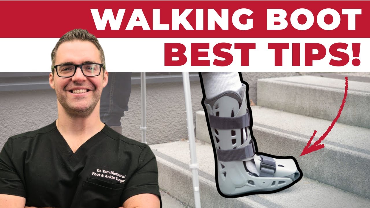
Immobilization: A walking boot for 4–6 weeks allows acute posterior tibial tendon inflammation to resolve, followed by progressive transition to orthotics and bracing.
Physical therapy: Eccentric strengthening of the posterior tibial muscle, intrinsic foot muscle strengthening, and ankle flexibility exercises slow degeneration in early disease.
Surgical Treatment
For Stage II PTTD that fails conservative management, surgical reconstruction typically involves a flexor digitorum longus (FDL) tendon transfer to augment the failed posterior tibial tendon, combined with a calcaneal osteotomy (medializing the heel bone) to correct hindfoot alignment. Isolated medial column procedures may be added for forefoot abduction.
For Stage III PTTD, subtalar or triple arthrodesis (fusion of the hindfoot joints) corrects the rigid deformity and provides stable, pain-free weight-bearing. Stage IV may require additional ankle intervention.
Dr. Tom's Product Recommendations

Ossur Rebound Sport Ankle Brace
⭐ Highly Rated
Hinged ankle brace that controls hindfoot valgus motion — reducing posterior tibial tendon strain during daily activities. Fits inside most athletic shoes.
Stage I PTTD or mild flatfoot with inner ankle pain
Advanced Stage II–IV PTTD requiring custom AFO
Disclosure: We earn a commission at no extra cost to you.

PowerStep Pinnacle BLUE Premium Insoles
⭐ Highly Rated
Semi-rigid biomechanical arch support that reduces overpronation and provides medial arch reinforcement — an accessible option for patients with early PTTD awaiting custom orthotic fitting.
Early flatfoot / mild PTTD with mild to moderate pronation
Moderate-to-severe PTTD requiring custom UCBL orthotic or AFO
Disclosure: We earn a commission at no extra cost to you.
✅ Pros / Benefits
- Conservative bracing highly effective in Stage I and early Stage II PTTD
- Surgical reconstruction achieves excellent outcomes in correctly staged patients
- Early treatment prevents progression to rigid deformity requiring fusion
- Custom AFO allows continued activity during treatment
❌ Cons / Risks
- Bracing is lifelong management — not a cure for degenerated tendon
- Advanced stages require major reconstructive surgery with extended recovery
- PTTD is progressive — early intervention is critical
Dr. Tom Biernacki’s Recommendation
PTTD is one of those conditions where timing is everything. In Stage I, we can often stop the progression with the right orthotic and brace and preserve the tendon. By Stage III, the options become much larger surgeries. I always tell patients: if you notice your arch collapsing, pain on the inner ankle, or inability to do a single-leg heel rise, come see us right away. Early intervention makes a dramatic difference in outcomes.
— Dr. Tom Biernacki, DPM | Board-Certified Podiatric Surgeon | Balance Foot & Ankle
Frequently Asked Questions
What is the first sign of posterior tibial tendon dysfunction?
The earliest sign is typically pain and swelling along the inner ankle and lower leg — along the course of the posterior tibial tendon. The pain is worst with activity and often worsens with single-leg heel rise. Some patients notice their arch beginning to flatten before significant pain develops.
Can PTTD be treated without surgery?
Yes — Stage I and many Stage II cases respond well to conservative treatment with custom orthotics, Arizona bracing, and physical therapy. Stage III and IV deformities with rigid flatfoot typically require surgical correction.
What does PTTD reconstruction surgery involve?
Stage II reconstruction typically combines a flexor digitorum longus tendon transfer (replacing the failed posterior tibial tendon function) with a calcaneal osteotomy (realigning the heel bone). Recovery involves a period of non-weight-bearing followed by gradual return to full activity over 4–6 months.
How is PTTD different from flat feet?
Flat feet (pes planus) is a structural shape of the foot that may be present from childhood. PTTD is an acquired deformity caused by a failing posterior tibial tendon — it develops in adulthood and progresses over time. All PTTD produces flatfoot, but not all flat feet are caused by PTTD.
Michigan Foot Pain? See Dr. Biernacki In Person
4.9★ rated | 1,200+ Reviews | 3,000+ Surgeries
Book online 24/7 · Howell & Bloomfield Township
📞 (810) 206-1402 Book Online →⚕ Doctor Recommended
VitaMedica Arnica GelFragrance-free, non-greasy relief for foot & ankle soreness
View Product →In-Office Treatment at Balance Foot & Ankle
If home treatment isn’t providing relief for your posterior tibial tendon dysfunction flatfoot stages treatment, our podiatry team at Balance Foot & Ankle can help with expert evaluations and advanced in-office care.
OrthoInfo – AAOS: Posterior Tibial Tendon Dysfunction
Ready to Get Relief?
Book online 24/7 in Howell & Bloomfield Township, MI
4.9★ | 1,200+ Reviews | 3,000+ Surgeries
Or call: (810) 206-1402

Dr. Tom Biernacki, DPM is a board-certified foot & ankle surgeon (ABFAS & ABPM) at Balance Foot & Ankle Specialists in Southeast Michigan. With over a decade of clinical experience, he specializes in heel pain, bunions, diabetic foot care, sports injuries, and minimally invasive surgery. Dr. Biernacki is a member of the APMA and ACFAS, and his patient education content on MichiganFootDoctors.com and YouTube has made him one of the most-followed foot & ankle educators on YouTube.