The most important clinical decision with Posterior Tibial Tendon Dysfunction Stage 1 Conservative isn’t which treatment to start with — it’s identifying the correct subtype. That changes everything. Call (810) 206-1402.
Quick Answer
PTTD Stage I: Conservative Management of Early Posterior Tib relates to tendon injury — typically caused by overuse or sudden strain. Most patients improve in 6-12 weeks with conservative care. Same-week appointments in Howell + Bloomfield Hills: (810) 206-1402.
Medically reviewed by Dr. Tom Biernacki, DPM — Board-certified foot & ankle surgeon, 3,000+ surgeries performed. Updated April 2026 with current clinical evidence. This article reflects real practice experience from Balance Foot & Ankle Specialists in Howell and Bloomfield Hills, Michigan.
Quick Answer
Most foot and ankle problems respond to conservative care — proper footwear, supportive inserts, activity modification, and targeted stretching — within 4-8 weeks. Persistent pain beyond that window, or any symptom that prevents walking, warrants a podiatric evaluation to rule out fracture, tendon tear, or systemic cause.
Watch: Dr. Tom Biernacki, DPM
Medically reviewed by Dr. Tom Biernacki, DPM — Board-Certified Podiatric Surgeon — Balance Foot & Ankle, Howell & Bloomfield Hills, MI. Last updated April 2026.
▶ Watch
Medically Reviewed by Dr. Tom Biernacki, DPM — Board-Certified Podiatrist, Balance Foot & Ankle Specialists, Michigan. Last updated April 2026.
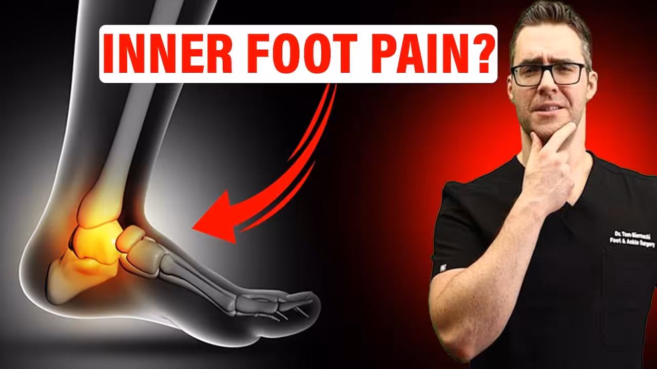
Posterior tibial tendon dysfunction (PTTD) — the progressive failure of the tibialis posterior tendon that supports the medial longitudinal arch — is the most common cause of adult acquired flatfoot deformity. Stage I PTTD (tendinopathy without deformity — the tendon is painful and swollen but the arch and hindfoot alignment remain normal) represents the critical early window where aggressive conservative management can halt progression and prevent the advanced flatfoot deformity that requires complex surgical reconstruction. Recognizing Stage I PTTD and treating it appropriately is the most important podiatric intervention for preventing acquired flatfoot.
Anatomy and Stage I Presentation
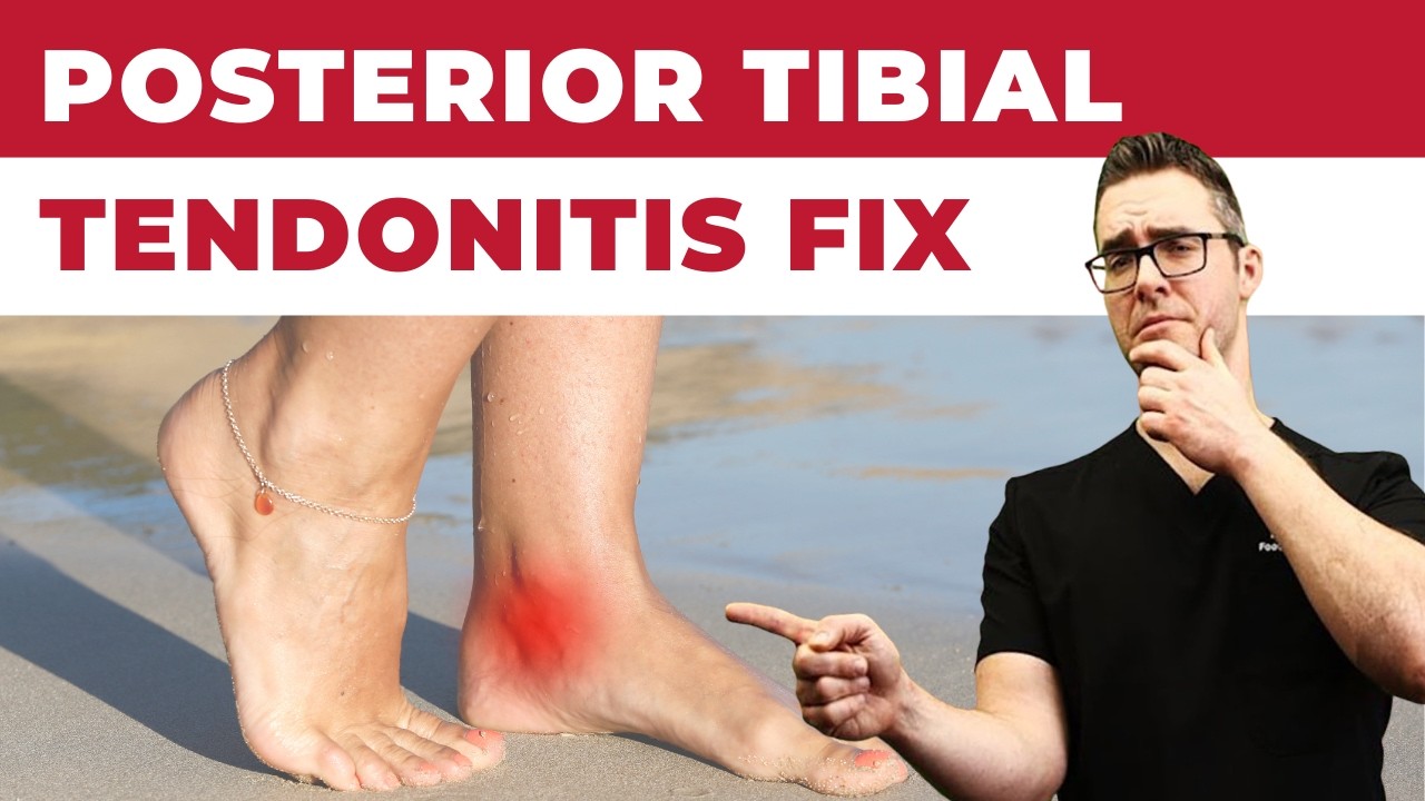
The posterior tibial tendon courses from the tibialis posterior muscle in the deep posterior leg compartment, passes behind the medial malleolus in a fibro-osseous tunnel, and inserts primarily into the navicular tuberosity with secondary insertions to the cuneiforms, cuboid, and metatarsal bases. As the primary dynamic stabilizer and supinator of the subtalar joint, the tibialis posterior tendon is the critical structure preventing hindfoot eversion and arch collapse during weight-bearing. Stage I presentation: medial ankle pain and swelling along the posterior tibial tendon from the medial malleolus to the navicular insertion; tendon is intact but dysfunctional with inflammation and micro-tears; arch height and hindfoot valgus are NORMAL or minimally changed at this stage — the deformity has not yet developed; the single-leg heel rise test is positive (pain with heel rise but the heel still inverts, indicating some tendon function remains; in Stage II the heel does not invert due to tendon insufficiency). MRI confirms tendinopathy (tendon thickening, intrasubstance signal change, peritenon inflammation) and distinguishes Stage I from longitudinal tears (Stage II).
Conservative Management Protocol
Immobilization (acute Phase 1, 4–6 weeks): removable cam boot in neutral or slight supination; strict activity modification to reduce tendon load; NSAIDs for inflammatory component; avoid cortisone injection (risk of tendon rupture). Orthotic management (Phase 2, after acute inflammation subsides): custom UCBL orthotic with medial hindfoot posting and forefoot valgus post controls hindfoot valgus and reduces tibialis posterior tendon demand; Arizona brace for more significant involvement. Physical therapy: eccentric tibialis posterior strengthening protocol (progressive loading under control); gastrocnemius stretching (equinus is a major contributing factor); proprioception rehabilitation. Duration and monitoring: Stage I patients require 3–6 months of consistent conservative management; failure to improve or progression to arch collapse despite compliance warrants surgical consultation. Avoiding progression: Stage I → Stage II PTTD occurs when the medial soft tissue constraints (spring ligament, plantar fascia, plantar calcaneonavicular ligament) stretch under the uncompensated hindfoot valgus — maintaining tendon function prevents this cascade. Dr. Biernacki at Balance Foot & Ankle diagnoses PTTD at Stage I with tendon ultrasound and implements aggressive conservative management to halt progression to flatfoot deformity. Call (810) 206-1402 at our Bloomfield Hills or Howell office.
📧 Get Dr. Tom’s Free Lab Test Guide
Discover the 5 lab tests every person over 35 should ask their doctor about — explained in plain English by a board-certified physician.
class=”mfd-patient-scenario” id=”in-our-clinic”>In Our Clinic: What We See
Clinical perspective from Dr. Tom Biernacki, DPM — Balance Foot & Ankle, Howell & Bloomfield Hills, MI:
In our clinic, adult acquired flatfoot from PTTD typically presents in women over 40, often with recent weight gain or a period of increased standing. They describe medial ankle pain and progressive “collapse” of the arch on one side. The gold-standard exam finding is an inability to perform a single-leg heel-rise on the affected side — the tendon can no longer invert the heel into a rigid lever. Early PTTD is staged and treated with custom orthoses and bracing, but progressive disease (Stage III-IV) typically requires surgical reconstruction to prevent rigid deformity.
class=”mfd-differential” id=”differential-diagnosis”>Differential Diagnosis: What Else Could It Be?
Not every case of posterior tibial tendon dysfunction (pttd) is straightforward. In our clinic we routinely rule out three look-alike conditions before confirming the diagnosis. If your symptoms don’t match the classic presentation, one of these may explain the pain — which is why physical exam matters more than self-diagnosis.
Condition
How It Differs
Congenital flat foot
Lifelong, usually bilateral, no pain, normal single-leg heel-rise test.
class=”wp-block-heading mfd-treatment-bridge” id=”in-office-treatment”>In-Office Treatment at Balance Foot & Ankle
If home care isn’t resolving your your foot or ankle concern, a visit with a board-certified podiatrist is the fastest path to accurate diagnosis and a personalized plan. At Balance Foot & Ankle Specialists, Dr. Tom Biernacki, Dr. Carl Jay, and Dr. Daria Gutkin offer same-day and next-day appointments at both our Howell and Bloomfield Hills offices. We perform on-site diagnostic ultrasound, digital X-ray, conservative care, advanced regenerative treatments, and minimally invasive surgery when indicated.
Impact-absorbing recovery sandal — wear after long days on your feet.
As an Amazon Associate, Balance Foot & Ankle earns from qualifying purchases. Product recommendations are based on clinical experience; prices and availability shown above update live from Amazon.
When to See a Podiatrist
If foot or ankle pain has been bothering you for more than a few weeks, home care alone may not be enough. Balance Foot & Ankle offers same-week appointments at our Howell and Bloomfield Hills clinics — no referral needed in most cases. Bring your current shoes and a short list of symptoms and we’ll build you a treatment plan in one visit.
No — many people with flat feet have no pain at all. Pain develops when flat feet lead to excessive pronation that stresses tendons, ligaments, and joints. Risk factors for developing pain include obesity, prolonged standing, high-impact activities, and aging.
Can flat feet be corrected?
In children, arch development can sometimes be supported. In adults, the arch cannot be rebuilt without surgery, but custom orthotics, physical therapy, and appropriate footwear can effectively control symptoms and prevent progression.
What is the treatment for adult flatfoot deformity?
Early stages respond well to custom orthotics, physical therapy, and supportive footwear. Advanced cases with Achilles tightness may benefit from stretching and bracing. Severe cases — especially stage III-IV posterior tibial tendon dysfunction — may require reconstruction surgery.
Need Treatment at Balance Foot & Ankle?
Dr. Tom Biernacki, Dr. Carl Jay, and Dr. Daria Gutkin see patients at our Howell and Bloomfield Hills offices.
The most common mistake we see is: Waiting too long before seeking care. Fix: any foot pain lasting more than 4 weeks, or any sudden severe symptom, deserves a professional evaluation rather than more rest.
Warning Signs That Need Same-Day Care
Seek immediate evaluation at Balance Foot & Ankle if you experience any of the following:
Dr. Tom Biernacki, DPM · Board-Certified Foot & Ankle Surgeon. Specializes in conservative-first care, minimally invasive bunion surgery, and complex reconstruction.
Dr. Carl Jay, DPM · Accepting new patients. Specializes in sports medicine, athletic injuries, and routine podiatric care.
Dr. Daria Gutkin, DPM, AACFAS · Accepting new patients. Specializes in surgical reconstruction and pediatric podiatry.
Locations: 4330 E Grand River Ave, Howell, MI 48843 · 43494 Woodward Ave Suite 208, Bloomfield Hills, MI 48302
If symptoms persist past 2 weeks, affect your normal activity, or are accompanied by red-flag symptoms (warmth, redness, swelling, inability to bear weight).
What does treatment cost?
Most diagnostic visits and conservative treatments are covered by Medicare and major insurers. Out-of-pocket costs vary by your specific plan.
How quickly can I get an appointment?
Most non-urgent cases see us within 5 business days. Urgent cases (sudden pain, possible fracture) typically same or next business day.
Foot pain is a common foot/ankle condition that affects mobility and quality of life. Understanding the underlying cause is the first step in successful treatment. Our podiatrists at Balance Foot & Ankle perform a hands-on biomechanical exam, review your activity history, and use diagnostic imaging when appropriate to identify the root cause—not just treat the symptom. Many patients have been told to “rest and ice” without a deeper diagnostic workup; our approach is different.
Symptoms and warning signs
Common signs of foot pain include pain that worsens with activity, morning stiffness, swelling, tenderness when palpated, and difficulty bearing weight. If you experience sudden severe pain, inability to walk, visible deformity, numbness or color change, contact our office the same day or visit urgent care—these can signal a more serious injury such as a fracture, tendon rupture, or vascular compromise. Diabetics with any foot wound should seek same-day care.
Conservative treatment options
Most cases of foot pain respond to non-surgical care: structured rest, supportive footwear changes, custom orthotics, targeted stretching and strengthening protocols, anti-inflammatory medications when medically appropriate, and in-office procedures such as ultrasound-guided injections. We also offer advanced therapies including MLS laser therapy, EPAT/shockwave, regenerative injections, and image-guided procedures. Treatment is sequenced from least invasive to most invasive, and we explain the rationale at every step.
When is surgery considered?
Surgery is reserved for cases that fail 3-6 months of well-structured conservative care, when there is structural pathology (severe deformity, complete tear, advanced arthritis), or when imaging shows damage that will not heal without intervention. Our surgeons have performed 3,000+ foot and ankle procedures and prioritize minimally-invasive techniques whenever appropriate. We discuss recovery timelines, return-to-activity milestones, and realistic outcome expectations before any procedure is scheduled.
Recovery timeline and prevention
Recovery from foot pain varies based on severity and chosen treatment path. Conservative cases often improve within 4-8 weeks with consistent adherence to the protocol. Post-procedural recovery may range from a few days (in-office procedures) to several months (reconstructive surgery). Long-term prevention involves footwear assessment, activity modification, structured strengthening, and regular check-ins with your podiatrist if you have a history of recurrence. We provide written home-exercise plans and digital follow-up support.
Reviewed by Dr. Tom Biernacki, DPM — Board-certified podiatrist, Balance Foot & Ankle, Howell & Bloomfield Hills, MI. 4.9-star rating across 1,123+ patient reviews. Schedule an evaluation | (810) 206-1402
Ready to feel better?
Same-week appointments available in Howell and Bloomfield Hills, Michigan.
Dr. Tom Biernacki, DPM is a board-certified foot & ankle surgeon (ABFAS & ABPM) at Balance Foot & Ankle Specialists in Southeast Michigan. With over a decade of clinical experience, he specializes in heel pain, bunions, diabetic foot care, sports injuries, and minimally invasive surgery. Dr. Biernacki is a member of the APMA and ACFAS, and his patient education content on MichiganFootDoctors.com and YouTube has made him one of the most-followed foot & ankle educators on YouTube.
Balance Foot & Ankle surgeons are affiliated with Trinity Health Michigan, Corewell Health, and Henry Ford Health — three of Michigan’s largest health systems.


![Heel Bursitis & Achilles Tendon Bursitis [Best HOME Treatment!]](https://www.michiganfootdoctors.com/wp-content/cache/flying-press/f2850da852ba9539b6bf4f72ae5062fe.jpg)