
Medically reviewed by Dr. Tom Biernacki, DPM
Board-certified podiatric surgeon | Balance Foot & Ankle, Howell & Bloomfield Township, MI
Last reviewed: May 2026

| Grade | Structures Involved | Stability | Treatment | Return to Sport |
|---|---|---|---|---|
| Grade 1 | AITFL partial tear; no interosseous membrane involvement | Stable — no mortise widening | Protected weight bearing; boot 2–3 weeks; physical therapy | 3–6 weeks |
| Grade 2 | AITFL complete + partial interosseous membrane tear | Stable under stress; no widening at rest | Boot 4–6 weeks; progressive weight bearing; PT for 8–12 weeks | 6–12 weeks |
| Grade 3 | AITFL + complete interosseous membrane + PITFL tear | Unstable — stress test positive; mortise may widen | Surgical stabilization (screw or suture button); 6 weeks NWB | 4–6 months |
| Grade 3 + fracture (Maisonneuve) | All ligaments + proximal fibula fracture | Severely unstable | Surgical fixation; NWB cast 6 weeks; screw removal at 3–4 months | 5–7 months |
| Test | How Performed | Positive Finding | Sensitivity |
|---|---|---|---|
| Squeeze test | Compress fibula toward tibia at mid-calf level | Pain at ankle (not at squeeze point) | Moderate; 30–40% sensitivity for Grade 1 but high for Grade 3 |
| External rotation stress test | Knee at 90°; stabilize tibia; externally rotate foot | Pain at anterior ankle / syndesmosis | Good for Grade 2–3; combined with squeeze test improves accuracy |
| Cotton test (manual) | Stabilize tibia; translate fibula laterally with thumb | Abnormal lateral fibular translation vs contralateral | Good for Grade 3; performed under anesthesia or fluoroscopy |
| Stress X-ray (mortise + ER stress) | Mortise view + externally rotated stress view | Medial clear space >4mm or >1mm asymmetry vs contralateral | Best objective test for instability; guides surgical decision |
| MRI | Imaging of ligament complex | AITFL/PITFL tear; interosseous membrane edema/tear | High for ligament tears; does not assess dynamic stability |

Quick answer: Syndesmotic Injury Ankle is a common foot/ankle topic that affects many patients. The 2026 evidence-based approach combines proper diagnosis, conservative-first treatment, and escalation only when needed. We treat this regularly at our Howell and Bloomfield Township practices. Call (810) 206-1402.
Medically Reviewed | Dr. Tom Biernacki, DPM | Board-Certified Podiatrist | Balance Foot & Ankle, Michigan | 5,000+ patients/year
The most important clinical decision with Syndesmotic Injury Ankle isn’t which treatment to start with — it’s identifying the correct subtype. That changes everything. Call (810) 206-1402.
The most important clinical decision with Syndesmotic Injury Ankle isn’t which treatment to start with — it’s identifying the correct subtype. That changes everything. Call (810) 206-1402.
The Syndesmosis: What Gets Injured
The ankle syndesmosis is the fibrous joint between the distal tibia and fibula, stabilized by the anterior inferior tibiofibular ligament (AITFL), posterior inferior tibiofibular ligament (PITFL), interosseous membrane, and interosseous ligament. Injury disrupts the ankle mortise — the precise bony socket that grips the talus — allowing the mortise to widen and causing instability and cartilage damage.
Grading Syndesmotic Injuries
Grade I: sprain of AITFL without widening — stable, treat conservatively. Grade II: partial tear with potential widening — borderline, may need fixation. Grade III: complete tear with widening — requires surgical stabilization. Pronation-external rotation fracture patterns (bimalleolar, Maisonneuve) almost always involve syndesmotic disruption.
Diagnosis
Squeeze test (compress tibia and fibula at mid-calf reproduces syndesmotic pain) and external rotation stress test are clinical indicators. Stress X-rays and weight-bearing films assess widening. CT scan is best for detecting subtle syndesmotic diastasis. MRI characterizes ligament tears.
Treatment
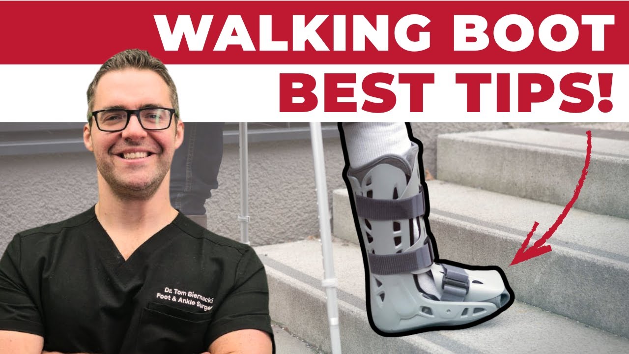
Stable (Grade I): boot immobilization 4–6 weeks, prolonged rehabilitation. Unstable (Grade II–III): surgical stabilization — trans-syndesmotic screws (rigid, require removal) or suture-button devices (flexible, allowing normal syndesmotic micromotion, do not require routine removal). Recovery post-fixation: 6–12 weeks non-weight-bearing, then progressive loading, return to sport 3–6 months.
FAQs
Why does a high ankle sprain take so much longer to heal than a regular sprain? The syndesmosis is a weight-bearing joint under repeated rotational stress with every step. It is under constant load during rehabilitation, making the ligament take significantly longer to heal than the non-weight-bearing lateral ligaments.
💊 Dr. Tom’s Recovery Support Picks
Between appointments and after procedures, these are the products I recommend for at-home recovery support.
My go-to topical for post-procedure soreness. Arnica + menthol — apply 3-4x daily. Plant-based, FSA-eligible, no greasy residue.
View on Amazon →
Graduated compression helps reduce post-op swelling and supports recovery. True graduated design — not just tight socks.
View on Amazon →
FTC Disclosure: As an Amazon Associate and Foundation Wellness affiliate, we earn from qualifying purchases. This never affects our clinical recommendations.
Michigan Foot & Ankle? See Dr. Biernacki In Person
Same-week appointments at our Howell and Bloomfield Township offices.
📞 (810) 206-1402 Book Online →In-Office Treatment at Balance Foot & Ankle
If home treatment isn’t providing relief for your foot and ankle conditions, our podiatry team at Balance Foot & Ankle can help with same-day evaluations and advanced in-office care.
AAOS OrthoInfo: Syndesmotic Ankle Injury
Ready to Get Relief?
Same-day appointments available in Howell & Bloomfield Township, MI
4.9★ | 1,123 Reviews | 3,000+ Surgeries
Or call: (810) 206-1402

Dr. Tom Biernacki, DPM is a board-certified foot & ankle surgeon (ABFAS & ABPM) at Balance Foot & Ankle Specialists in Southeast Michigan. With over a decade of clinical experience, he specializes in heel pain, bunions, diabetic foot care, sports injuries, and minimally invasive surgery. Dr. Biernacki is a member of the APMA and ACFAS, and his patient education content on MichiganFootDoctors.com and YouTube has made him one of the most-followed foot & ankle educators on YouTube.