
Quick answer: Treatment for adult residual clubfoot deformity treatment options follows a stepwise approach: 1) conservative care first (rest, ice, supportive footwear, OTC anti-inflammatories), 2) physical therapy and targeted exercises, 3) in-office treatments (injections, custom orthotics) if conservative fails at 4-6 weeks, 4) surgery for refractory cases. Most patients resolve at step 1 or 2. Call (810) 206-1402.
Medically reviewed by Dr. Tom Biernacki, DPM · Board-Certified Podiatric Surgeon · Last reviewed: April 2026 · Editorial Policy
Medically reviewed by Dr. Tom Biernacki, DPM — Board-Certified Podiatric Surgeon — Balance Foot & Ankle, Howell & Bloomfield Township, MI. Last updated April 2026.
▶ Watch
Medically Reviewed by Dr. Tom Biernacki, DPM — Board-Certified Podiatrist, Balance Foot & Ankle Specialists, Michigan. Last updated April 2026.
Congenital talipes equinovarus (clubfoot) affects approximately 1 in 1,000 live births. The Ponseti method — serial casting followed by percutaneous Achilles tenotomy and a foot abduction orthosis — has become the worldwide standard of care, achieving excellent correction in the vast majority of patients. Nevertheless, a significant proportion of adult patients present with residual or recurrent deformity that requires ongoing management. Understanding the spectrum of residual adult clubfoot and the available interventions is essential for podiatric surgeons treating this population.
Pathoanatomy of Residual Deformity
Adult residual clubfoot deformity encompasses several patterns: residual hindfoot varus (the most common residual), forefoot adductus, dynamic supination (from overactive tibialis anterior with inadequate peroneal function), and equinus at the ankle or midfoot. The underlying pathology involves persistent underdevelopment of lateral column structures, hypoplastic peroneal muscles, abnormal tarsal bone morphology with persistent navicular subluxation on the talar head, and adaptive shortening of posterior and medial soft tissues. The talus remains externally rotated in the ankle mortise in severe cases, altering biomechanics throughout the kinetic chain.
Clinical Assessment and Imaging
Assessment begins with standing alignment photographs and gait analysis. The Coleman block test distinguishes flexible hindfoot varus driven by forefoot-driven supination from fixed structural hindfoot deformity — a critical distinction that determines the surgical approach. Weight-bearing foot and ankle radiographs quantify talar-first metatarsal angle, calcaneal pitch, and talo-calcaneal divergence. CT is essential for surgical planning when multiple tarsal joints require assessment or osteotomies are contemplated. MRI identifies chondral degeneration in the ankle and subtalar joints, which significantly influences prognosis and surgical decision-making.
Non-Surgical Management
Mild residual deformity with preserved joint function benefits from custom foot orthotics with medial hindfoot posting, lateral forefoot wedging to accommodate forefoot adductus, and ankle-foot orthoses when dynamic instability is significant. Intensive physical therapy addressing tibialis anterior/peroneal strength imbalance can reduce dynamic supination in flexible deformities. Serial stretching and night splinting address residual equinus contracture in compliant patients. Properly fitted footwear — often requiring extra-depth or custom-molded shoes — reduces pressure-related complications from deformity-driven plantar pressure concentration.
Surgical Options for Structural Correction
Surgical correction is stratified by deformity severity and joint preservation potential. For flexible hindfoot varus with forefoot-driven supination, tibialis anterior tendon transfer to the lateral cuneiform (Garceau procedure) corrects the dynamic deforming force and is highly effective in skeletally mature patients with flexible hindfoot. Calcaneal osteotomy — lateral closing wedge (Dwyer) or lateral displacement (sliding) — directly corrects rigid hindfoot varus when the subtalar joint remains functional. Triple arthrodesis (talocalcaneal, talonavicular, calcaneocuboid fusions) remains the definitive salvage for rigid, painful multi-joint deformity with advanced subtalar and midtarsal arthritis; it reliably corrects multi-plane deformity at the cost of hindfoot motion. Supramalleolar osteotomy is added when tibial torsion contributes to overall limb alignment. Achilles lengthening or posterior capsulotomy addresses residual equinus. Modern techniques emphasize maximal joint preservation, reserving arthrodesis for salvage situations. Dr. Biernacki at Balance Foot & Ankle evaluates adult residual clubfoot with comprehensive biomechanical and imaging assessment. Call (810) 206-1402.
📍 Located in Michigan?
Our board-certified podiatrists treat this condition at two convenient locations. Book online 24/7 at either office.
Insurance Accepted
BCBS · Medicare · Aetna · Cigna · United Healthcare · HAP · Priority Health · Humana · View All →
Howell Office
4330 E Grand River Ave
Howell, MI 48843
Get Directions →
Bloomfield Township Office
43494 Woodward Ave, #208
Bloomfield Township, MI 48302
Get Directions →
More Podiatrist-Recommended Foot Health Essentials
Hoka Clifton 10

Max-cushion everyday shoe — podiatrist favorite for walking and running.

OOFOS Recovery Slide

Impact-absorbing recovery sandal — wear after long days on your feet.
As an Amazon Associate, Balance Foot & Ankle earns from qualifying purchases. Product recommendations are based on clinical experience; prices and availability shown above update live from Amazon.
When to See a Podiatrist
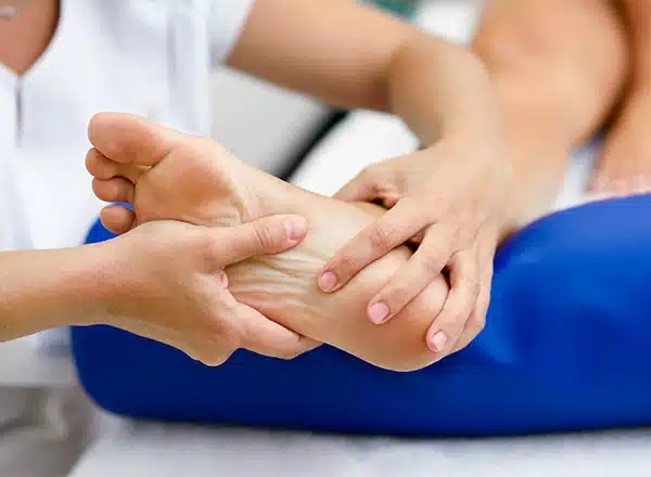
If foot or ankle pain has been bothering you for more than a few weeks, home care alone may not be enough. Balance Foot & Ankle offers online booking 24/7 at our Howell and Bloomfield Township clinics — no referral needed in most cases. Bring your current shoes and a short list of symptoms and we’ll build you a treatment plan in one visit.
Call Balance Foot & Ankle: (810) 206-1402 · Book online · Offices in Howell & Bloomfield Township
Pros & Cons of Conservative Care for foot care
Advantages
- ✓ Conservative care first
- ✓ Book online 24/7
- ✓ Multiple insurance accepted
Considerations
- ✗ Self-treatment can mask issues
- ✗ See a podiatrist if pain >2 weeks
Dr. Tom’s Recommended Products for foot care
Affiliate disclosure: As an Amazon Associate, Balance Foot & Ankle earns from qualifying purchases. We only recommend products we use with patients.
Footnanny Heel Cream Dr. Tom’s Pick
Best for: Daily moisturizer for cracked heels
Ready to Get Back on Your Feet?
Appointments in Howell + Bloomfield Township. Most insurance accepted. Dr. Tom Biernacki, DPM & team.
Call Now: (810) 206-1402
Visit Balance Foot & Ankle — Online Booking 24/7
Our podiatry team serves patients throughout Michigan including Howell, Brighton, and Bloomfield Township. If you’re dealing with heel pain, ingrown toenails, or a foot injury, you can book online 24/7.
Book online 24/7. (810) 206-1402
VitaMedica Arnica Gel
Arnica gel plus 15 plant compounds for sore feet, plantar fasciitis flares and post-activity soreness. Apply a thin layer to the sore area.
Shop VitaMedica Arnica Gel →Ready for Expert Care?
Appointments in Howell & Bloomfield Township, MI.
4.9★ | 1,200+ Reviews | 3,000+ Surgeries
Or call: (810) 206-1402

Dr. Tom Biernacki, DPM is a board-certified foot & ankle surgeon (ABFAS & ABPM) at Balance Foot & Ankle Specialists in Southeast Michigan. With over a decade of clinical experience, he specializes in heel pain, bunions, diabetic foot care, sports injuries, and minimally invasive surgery. Dr. Biernacki is a member of the APMA and ACFAS, and his patient education content on MichiganFootDoctors.com and YouTube has made him one of the most-followed foot & ankle educators on YouTube.