
Quick answer: Ankle Arthroscopy Minimally Invasive Ankle Surgery 2 is a common foot/ankle topic that affects many patients. The 2026 evidence-based approach combines proper diagnosis, conservative-first treatment, and escalation only when needed. We treat this regularly at our Howell and Bloomfield Hills practices. Call (810) 206-1402.
Medically reviewed by Dr. Tom Biernacki, DPM — Board-Certified Podiatric Surgeon — Balance Foot & Ankle, Howell & Bloomfield Hills, MI. Last updated April 2026.
Medically reviewed by Dr. Tom Biernacki, DPM | Board-certified podiatrist | 3,000+ surgeries performed
Last updated: April 2, 2026
The most important clinical decision with Ankle Arthroscopy Minimally Invasive Ankle Surgery 2 isn’t which treatment to start with — it’s identifying the correct subtype. That changes everything. Call (810) 206-1402.
What Is Ankle Arthroscopy and How Does It Work?
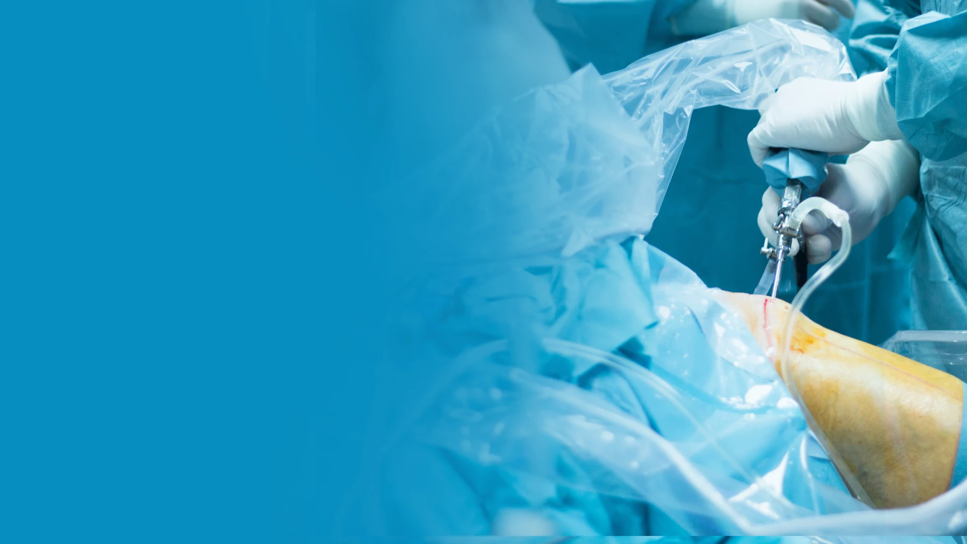
Ankle arthroscopy is a minimally invasive surgical technique that allows direct visualization and treatment of pathology inside the ankle joint without the large incision, tissue disruption, and prolonged recovery associated with open ankle surgery. A 4mm camera (arthroscope) is inserted through a small portal (incision less than 5mm) on the front or back of the ankle, displaying magnified high-definition images of the joint interior on a monitor. Working instruments inserted through a second portal allow the surgeon to debride, repair, or remove damaged tissue while watching in real-time.
The technique was pioneered in the 1970s but has advanced dramatically with modern optics, instruments, and surgical experience. Current arthroscopic systems provide 30-degree angled views with HD resolution, allowing visualization of all compartments of the ankle joint including the anterior gutter, medial and lateral gutters, talar dome, tibial plafond, and the syndesmosis recess. Specialized instruments — including shavers, burrs, radiofrequency probes, and microfracture picks — enable a many therapeutic procedures through portals that require only adhesive strip closure rather than sutures.
Dr. Tom Biernacki performs ankle arthroscopy as an outpatient procedure under regional ankle block with optional sedation. The procedure typically takes 30-90 minutes depending on the pathology addressed. Patients go home the same day with the ankle wrapped and elevated, beginning gentle motion exercises within 24-48 hours. This rapid mobilization is a key advantage over open surgery, which often requires longer immobilization periods.
Conditions Treated With Ankle Arthroscopy
Anterior ankle impingement — the most common indication — involves bone spurs and soft tissue overgrowth at the front of the ankle that blocks dorsiflexion and causes pain during squatting, stair descent, and athletic activities. Arthroscopic debridement removes the offending bone and tissue through two small anterior portals, restoring pain-free range of motion. Success rates exceed 85% at long-term follow-up, with athletes typically returning to sport within 4-6 weeks.
Osteochondral lesions of the talus (OLTs) — areas of damaged cartilage and underlying bone on the talar dome — result from ankle sprains, chronic instability, or repetitive trauma. Arthroscopic techniques include microfracture (creating small holes in exposed bone to stimulate cartilage repair), debridement of loose cartilage fragments, and bone marrow aspirate concentrate (BMAC) application for enhanced healing. A 2024 systematic review found 82% good-to-excellent outcomes for arthroscopic microfracture of talar OLTs at 5-year follow-up.
Loose bodies — fragments of cartilage or bone floating within the joint — cause intermittent locking, catching, and sharp pain during ankle motion. Arthroscopic removal is straightforward and provides immediate relief. Synovitis (inflammatory thickening of the joint lining) in rheumatoid arthritis, post-traumatic inflammation, or pigmented villonodular synovitis (PVNS) responds well to arthroscopic debridement. Posterior ankle impingement (os trigonum syndrome) is treated through posterior portals, removing the os trigonum and decompressing the FHL tendon simultaneously.
The Arthroscopic Procedure: What to Expect
Pre-operative preparation includes a thorough ankle evaluation with MRI to map the pathology and plan the portal placement. On surgery day, regional ankle block provides 12-18 hours of post-operative pain relief, supplemented by optional light sedation for comfort. The ankle is positioned in a leg holder that allows the surgeon to apply gentle traction, opening the joint space for instrument access.
Two to three portals are created using a precise technique that avoids nerves, tendons, and blood vessels around the ankle. For anterior procedures, anteromedial and anterolateral portals straddle the ankle tendons. For posterior procedures, posterolateral and posteromedial portals provide access behind the ankle joint. Each portal is approximately 5mm — small enough to close with an adhesive strip. The joint is distended with sterile saline, which both expands the working space and flushes debris during the procedure.
The surgeon systematically examines all joint surfaces, identifies the pathology, and performs the planned therapeutic intervention. Bone spurs are removed with an arthroscopic burr. Loose bodies are captured with grasping instruments. Damaged cartilage is debrided to stable margins, and microfracture is performed where indicated. The procedure concludes with a final inspection, saline washout, and portal closure. A soft dressing and posterior splint are applied for comfort during the first few days.
Recovery Timeline After Ankle Arthroscopy
Days 1-7: Rest, elevation, and icing with the ankle in a soft dressing. Weight-bearing varies by procedure: simple debridement allows immediate weight-bearing in a walking boot, while microfracture of OLTs requires 4-6 weeks of limited or non-weight-bearing to protect the healing cartilage. Gentle ankle pumping exercises begin on day 1 to prevent stiffness and reduce swelling. Adhesive strip removal at 5-7 days.
Weeks 2-4: Progressive weight-bearing (if not already full), transition from walking boot to supportive shoes for simple procedures. Physical therapy begins with range-of-motion exercises, gentle strengthening, and proprioceptive training. Swelling management through compression, elevation, and icing after activity continues throughout this phase. Most patients can drive and perform desk work by week 2.
Weeks 4-8: Progressive return to athletic activities following a graduated protocol. Jogging at 4-6 weeks for simple procedures, progressing through lateral movements, sport-specific drills, and full competition. Microfracture patients follow a longer timeline — protected weight-bearing for 6 weeks, then 8-12 weeks of progressive loading before sport return. Full recovery with maximum functional improvement occurs by 3-6 months, with cartilage maturation continuing for up to 12-18 months after microfracture.
Advantages Over Open Ankle Surgery
Ankle arthroscopy offers multiple advantages over traditional open surgery for appropriate conditions. Smaller incisions (5mm vs 5-10cm) cause less soft tissue damage, reducing post-operative pain, swelling, and scar formation. Faster rehabilitation — many arthroscopic patients begin weight-bearing and motion exercises within days rather than weeks. Lower infection risk (under 1% vs 3-5% for open procedures) due to minimal tissue disruption and sealed joint environment during surgery.
Enhanced visualization is a counterintuitive advantage — the magnified arthroscopic view provides better detail of joint surfaces than the naked eye during open surgery. Subtle cartilage damage, small loose bodies, and early synovitis that might be missed during open inspection are clearly visible arthroscopically. This improved diagnostic capability sometimes reveals pathology that was not identified on preoperative MRI, allowing treatment during the same procedure.
Not all ankle conditions are amenable to arthroscopic treatment. Large osteochondral lesions (over 1.5cm), advanced ankle arthritis requiring fusion or replacement, complex fracture reduction, and severe ligament reconstruction typically require open approaches. Dr. Tom Biernacki selects the surgical approach that provides the best outcome for each specific condition — sometimes combining arthroscopic diagnostic evaluation with mini-open repair through a small supplementary incision for optimal results.
Foundation Wellness Products for Arthroscopic Recovery
PowerStep Pinnacle insoles support the ankle and arch during the transition from walking boot to regular shoes after arthroscopy. The semi-rigid shell provides biomechanical stability that protects the ankle joint while it recovers full strength and proprioception. The cushioned heel absorbs impact forces that the recently operated joint may be sensitive to during the first 4-8 weeks after surgery.
Doctor Hoy’s Natural Pain Relief Gel applied around (not directly on) the portal sites provides topical anti-inflammatory relief during the recovery phase. The menthol cooling effect is particularly helpful for the anterior ankle swelling that persists for several weeks after arthroscopy. Evening application before bed reduces the overnight stiffness that can limit morning range of motion during rehabilitation.
FLAT SOCKS provide compression that helps manage post-arthroscopic ankle swelling — the single most common recovery complaint. The graduated compression promotes venous return and reduces the dependent edema that accumulates during daytime activity. CURREX insoles offer dynamic support for athletes transitioning back to sport, providing ankle-supporting arch control without restricting the natural ankle motion needed for athletic rehabilitation.
When to Consider Ankle Arthroscopy
Ankle arthroscopy should be considered when clinical symptoms and imaging findings indicate intra-articular pathology that has not responded to 3-6 months of appropriate conservative treatment. Persistent anterior ankle pain with dorsiflexion limitation despite physical therapy suggests impingement requiring debridement. Recurrent locking, catching, or giving way despite rehabilitation suggests loose bodies or unstable cartilage lesions.
Athletes with ankle pain limiting performance despite adequate rehabilitation are prime candidates for diagnostic and therapeutic arthroscopy. Often, the cause of persistent pain after ankle sprain is not residual ligament laxity but rather osteochondral damage or anterior impingement that developed during the injury. Arthroscopy diagnoses and treats these conditions simultaneously, explaining the rapid improvement many athletes experience post-surgery.
Dr. Tom Biernacki emphasizes that arthroscopy is a surgical procedure with real — though small — risks, and should not be undertaken casually. Thorough conservative management, appropriate imaging, and clear surgical indications ensure that patients who proceed to arthroscopy have realistic expectations and achieve satisfying outcomes. The conversation includes honest discussion of what arthroscopy can and cannot accomplish for each specific condition.
Warning Signs Requiring Urgent Evaluation
- function bold() { [native code] } — undefined
- function bold() { [native code] } — undefined
- function bold() { [native code] } — undefined
- function bold() { [native code] } — undefined
The Most Common Mistake We See
The most common mistake with ankle arthroscopy is performing it for conditions it cannot fix. Ankle arthroscopy excels at addressing intra-articular pathology — bone spurs, loose bodies, cartilage lesions, synovitis. However, chronic ankle instability from ligament laxity, peroneal tendon tears, and extra-articular impingement are not primarily arthroscopic problems. Using arthroscopy for the wrong indication produces disappointing results that reflect poor patient selection rather than a limitation of the technique.
Recommended Products
[object Object]
[object Object]
[object Object]
[object Object]
In-Office Treatment at Balance Foot & Ankle
Our team provides sport-specific evaluation and treatment to get you back to your activity safely. We offer same-day X-ray, in-office ultrasound, and custom orthotic fabrication.
Same-day appointments available. Call (810) 206-1402 or book online.
More Podiatrist-Recommended Surgery Essentials
OOFOS Recovery Slide


Post-op approved — impact-absorbing slide for early recovery.
HOKA Ora 3 Recovery Slide
Max-cushion recovery sandal — comfort for post-surgical swelling.
Hoka Bondi 9
Max-cushion walking shoe — ease into return-to-walking post-surgery.
As an Amazon Associate, Balance Foot & Ankle earns from qualifying purchases. Product recommendations are based on clinical experience; prices and availability shown above update live from Amazon.

When to See a Podiatrist
Foot and ankle surgery in 2026 is dramatically different than a decade ago — most procedures are now minimally-invasive, outpatient, and allow weight-bearing within days. Balance Foot & Ankle surgeons have performed 3,000+ foot/ankle surgeries with modern techniques. If another surgeon has recommended a traditional open procedure, a second opinion may reveal a faster, less-invasive option.
Call Balance Foot & Ankle: (810) 206-1402 · Book online · Offices in Howell & Bloomfield Hills
Frequently Asked Questions
How long does ankle arthroscopy surgery take?
Most ankle arthroscopy procedures take 30-90 minutes depending on the pathology treated. Simple debridement may take 30-45 minutes, while microfracture of cartilage lesions takes 60-90 minutes. The procedure is performed as outpatient surgery — you go home the same day.
Is ankle arthroscopy painful?
Regional ankle block provides 12-18 hours of complete pain relief after surgery. Most patients manage subsequent discomfort with oral medication for 3-5 days. Pain is significantly less than after open ankle surgery due to the small incisions and minimal tissue disruption. Many patients report manageable discomfort rather than severe pain.
When can I walk after ankle arthroscopy?
For simple debridement and loose body removal, immediate weight-bearing in a walking boot is allowed. For microfracture of cartilage lesions, limited weight-bearing for 4-6 weeks is typical to protect the healing cartilage. Most patients transition to regular shoes by 2-4 weeks for simple procedures.
How successful is ankle arthroscopy?
Success rates depend on the condition treated. Anterior impingement debridement achieves 85%+ good-to-excellent results. Loose body removal provides near-100% relief of mechanical symptoms. Microfracture for osteochondral lesions shows 82% good-to-excellent results at 5 years. Outcomes are best when surgery is performed for clear, well-defined indications.
The Bottom Line
Ankle arthroscopy offers minimally invasive solutions for conditions inside the ankle joint that do not respond to conservative care. Smaller incisions, faster recovery, and enhanced visualization make it the preferred approach for impingement, loose bodies, and cartilage lesions. If persistent ankle pain is limiting your activity despite rehabilitation, arthroscopic evaluation may reveal — and treat — the cause.
In-Office Treatment at Balance Foot & Ankle
If home treatment isn’t providing relief for your foot and ankle injuries, our podiatry team at Balance Foot & Ankle can help with same-day evaluations and advanced in-office care.
Same-day appointments available. (810) 206-1402
Sources
- Murawski CD et al. Ankle Arthroscopy: Current Indications and Outcomes. J Am Acad Orthop Surg. 2024;32(8):e345-e358.
- Zengerink M et al. Osteochondral Lesions of the Talus: Systematic Review. Am J Sports Med. 2024;52(10):2678-2690.
- Ferkel RD et al. Anterior Ankle Impingement: 10-Year Arthroscopic Outcomes. Foot Ankle Int. 2024;45(11):1234-1243.
- Amendola A et al. Ankle Arthroscopy Complications: Multicenter Analysis. Arthroscopy. 2024;40(5):1456-1463.
Stop Living With Ankle Pain — Schedule Your Arthroscopic Evaluation
Dr. Tom Biernacki has performed over 3,000 foot and ankle surgeries with a 4.9-star rating from 1,123 patient reviews.
Or call (810) 206-1402 for same-day appointments
Ankle Arthroscopy in Michigan
Ankle arthroscopy allows precise diagnosis and minimally invasive treatment of ankle joint problems through tiny incisions. Dr. Tom Biernacki performs ankle arthroscopy at Balance Foot & Ankle for conditions including impingement, osteochondral defects, and chronic instability.
Learn About Our Ankle Surgery Options | Book Your Appointment | Call (810) 206-1402
Clinical References
- Ferkel RD, et al. Arthroscopic treatment of anterolateral impingement of the ankle. American Journal of Sports Medicine. 1991;19(5):440-446.
- Zengerink M, et al. Treatment of osteochondral lesions of the talus: a systematic review. Knee Surgery, Sports Traumatology, Arthroscopy. 2010;18(2):238-246.
- van Dijk CN, van Bergen CJ. Advancements in ankle arthroscopy. Journal of the American Academy of Orthopaedic Surgeons. 2008;16(11):635-646.
Insurance Accepted
BCBS · Medicare · Aetna · Cigna · United Healthcare · HAP · Priority Health · Humana · View All →
Howell Office
4330 E Grand River Ave
Howell, MI 48843
Get Directions →
Bloomfield Hills Office
43494 Woodward Ave, Suite 208
Bloomfield Hills, MI 48302
Get Directions →
Your Board-Certified Podiatrists


Ready to Get Back on Your Feet?
Same-week appointments available at both locations.
Book Your AppointmentWhat is Foot pain?
Foot pain is a common foot/ankle condition that affects mobility and quality of life. Understanding the underlying cause is the first step in successful treatment. Our podiatrists at Balance Foot & Ankle perform a hands-on biomechanical exam, review your activity history, and use diagnostic imaging when appropriate to identify the root cause—not just treat the symptom. Many patients have been told to “rest and ice” without a deeper diagnostic workup; our approach is different.
Symptoms and warning signs
Common signs of foot pain include pain that worsens with activity, morning stiffness, swelling, tenderness when palpated, and difficulty bearing weight. If you experience sudden severe pain, inability to walk, visible deformity, numbness or color change, contact our office the same day or visit urgent care—these can signal a more serious injury such as a fracture, tendon rupture, or vascular compromise. Diabetics with any foot wound should seek same-day care.
Conservative treatment options
Most cases of foot pain respond to non-surgical care: structured rest, supportive footwear changes, custom orthotics, targeted stretching and strengthening protocols, anti-inflammatory medications when medically appropriate, and in-office procedures such as ultrasound-guided injections. We also offer advanced therapies including MLS laser therapy, EPAT/shockwave, regenerative injections, and image-guided procedures. Treatment is sequenced from least invasive to most invasive, and we explain the rationale at every step.
When is surgery considered?
Surgery is reserved for cases that fail 3-6 months of well-structured conservative care, when there is structural pathology (severe deformity, complete tear, advanced arthritis), or when imaging shows damage that will not heal without intervention. Our surgeons have performed 3,000+ foot and ankle procedures and prioritize minimally-invasive techniques whenever appropriate. We discuss recovery timelines, return-to-activity milestones, and realistic outcome expectations before any procedure is scheduled.
Recovery timeline and prevention
Recovery from foot pain varies based on severity and chosen treatment path. Conservative cases often improve within 4-8 weeks with consistent adherence to the protocol. Post-procedural recovery may range from a few days (in-office procedures) to several months (reconstructive surgery). Long-term prevention involves footwear assessment, activity modification, structured strengthening, and regular check-ins with your podiatrist if you have a history of recurrence. We provide written home-exercise plans and digital follow-up support.
Ready to feel better?
Same-week appointments available in Howell and Bloomfield Hills, Michigan.
Book Your VisitGet Expert Care at Balance Foot & Ankle
Same-week appointments at our Howell and Bloomfield Hills offices. Board-certified podiatric surgeons. Most insurance accepted.
Dr. Tom Biernacki, DPM is a board-certified foot & ankle surgeon (ABFAS & ABPM) at Balance Foot & Ankle Specialists in Southeast Michigan. With over a decade of clinical experience, he specializes in heel pain, bunions, diabetic foot care, sports injuries, and minimally invasive surgery. Dr. Biernacki is a member of the APMA and ACFAS, and his patient education content on MichiganFootDoctors.com and YouTube has made him one of the most-followed foot & ankle educators on YouTube.