
Medically reviewed by Dr. Tom Biernacki, DPM
Board-certified podiatric surgeon | Balance Foot & Ankle, Howell & Bloomfield Township, MI
Last reviewed: May 2026
Balance Foot & Ankle offers same-day appointments for urgent foot and ankle conditions across Southeast Michigan — but the most important factor in outcomes isn’t getting seen quickly. Our podiatrists explain what to do in the first 24-48 hours before your appointment that most patients skip entirely. Call (810) 206-1402 — expert podiatric care across Michigan.

| Classification | Fibula Level | Syndesmosis | Deltoid / Medial | Stability | Treatment |
|---|---|---|---|---|---|
| Weber A (Danis-Weber) | Below tibial plafond (infrasyndesmotic) | Intact | Intact | Stable | Short leg cast or functional brace 4–6 weeks; no surgery in most |
| Weber B | At tibial plafond level (transsyndesmotic) | Partially torn or intact | Variable — key to stability determination | Stable if medial intact; unstable if medial torn or >2 mm talar shift | Conservative if stable; ORIF if unstable (talar shift, positive stress X-ray) |
| Weber C | Above tibial plafond (suprasyndesmotic) | Torn — syndesmosis disrupted | Usually torn (bimalleolar or equivalent) | Unstable — always | ORIF required; syndesmotic fixation (screw or tightrope) + lateral plate |
| Bimalleolar Fracture | Both malleoli fractured | Variable | Medial malleolus fracture | Unstable | ORIF both malleoli; plate lateral + screw or tension band medial |
| Trimalleolar Fracture | Both malleoli + posterior tibia | Disrupted | Medial torn | Highly unstable; talar subluxation risk | ORIF all three; posterior malleolus fixed if >25% articular surface or unstable |
| Treatment | Indication | Fixation Detail | Weight-Bearing Protocol | Return to Activity |
|---|---|---|---|---|
| Functional Bracing / Walking Boot | Stable Weber A; stable isolated fibula fracture; no talar shift | No fixation; CAM boot or functional brace | WB as tolerated in boot; X-ray at 2 weeks to confirm stability | 6–10 weeks; standard shoe at 8–10 weeks |
| NWB Short Leg Cast | Stable Weber B with good alignment; patient compliance concern | No fixation; cast maintains reduction | Strict NWB 6 weeks; serial X-rays | 10–12 weeks |
| ORIF — Lateral Plate (Fibula) | Unstable Weber B; Weber C; all displaced fibula fractures | 3.5 mm LCP anatomic fibula plate; or 1/3 tubular plate; lag screw for long oblique | NWB 2 weeks → PWB 4–6 weeks → FWB 8–10 weeks | 4–5 months to sport |
| ORIF — Medial Malleolus Screw/TBW | Bimalleolar fracture; medial malleolus fragment >1 cm | Two 4.0 mm cancellous screws; or tension band wire for small fragments | Combined with lateral fixation protocol | 4–5 months |
| Syndesmotic Fixation | Weber C; positive external rotation stress test; widened mortise | 3.5 mm quadricortical screw (removed 8–12 weeks) or suture-button tightrope (permanent) | NWB 4–6 weeks post-ORIF | 6 months to sport if screw removed; 5 months with tightrope |
Foot pain isn't resolving?
Same-week appointments at Howell & Bloomfield Township
Medically Reviewed | Dr. Tom Biernacki, DPM | Board-Certified Podiatric Surgeon | Balance Foot & Ankle, Michigan
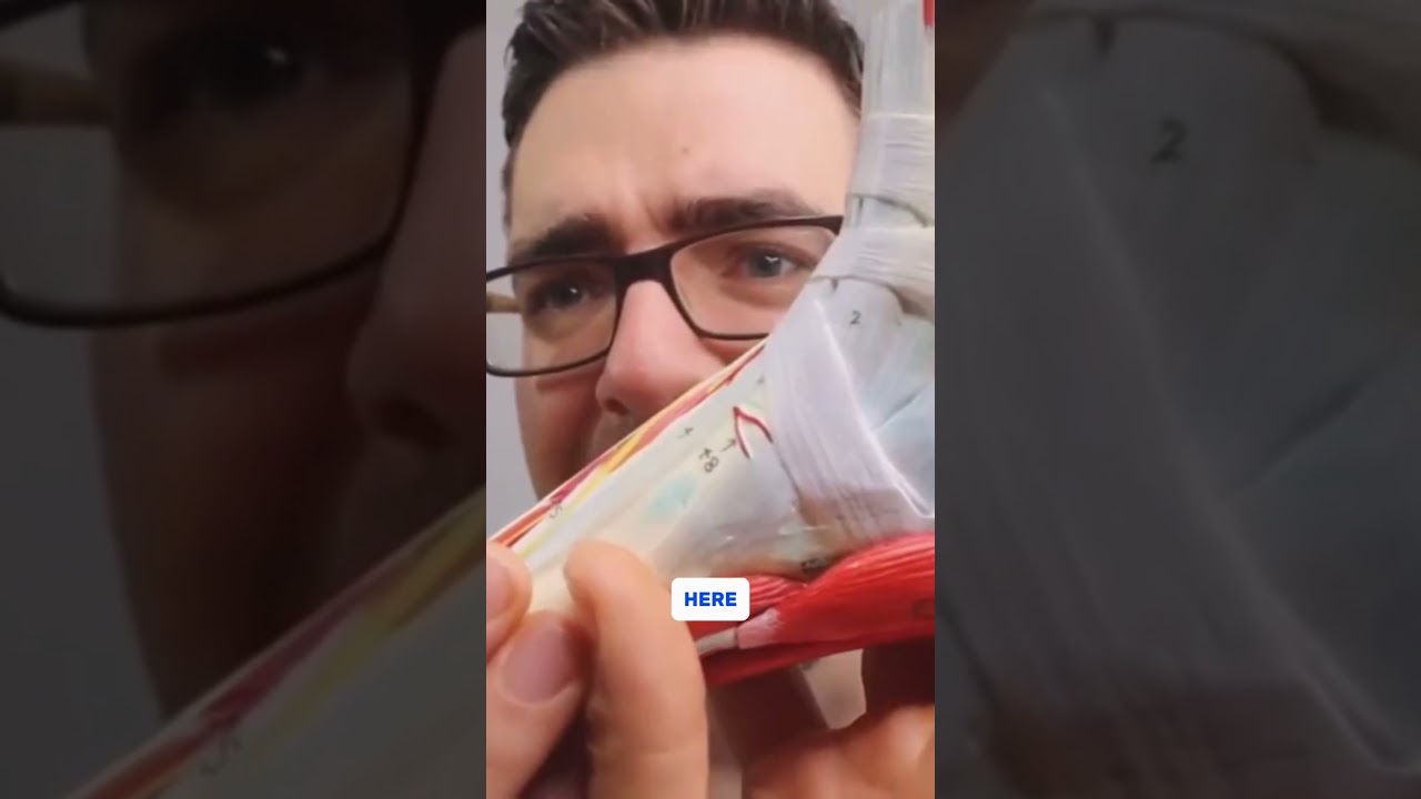

Ankle fractures are among the most common skeletal injuries, accounting for approximately 10% of all fractures. At Balance Foot & Ankle, Dr. Biernacki provides comprehensive ankle fracture care — from acute evaluation and splinting through weight-bearing casting, surgical fixation when indicated, and complete post-operative rehabilitation. The critical initial decision — operative vs. non-operative management — depends on fracture pattern, stability, alignment, and patient factors including bone quality, activity level, and medical comorbidities.
Ankle Fracture Classification
Dr. Biernacki uses both the Weber (Danis-Weber) classification for fibular fractures and the Lauge-Hansen classification for injury mechanism to guide treatment. Weber A (below the level of the ankle joint, below the syndesmosis): typically stable, managed non-operatively. Weber B (at the level of the joint, involving the syndesmosis): stability depends on medial-sided integrity — stress views or MRI assess syndesmotic stability; unstable patterns require fixation. Weber C (above the syndesmosis, with obligate syndesmotic injury): typically unstable, requires ORIF with syndesmotic fixation. Special patterns: bimalleolar (fibula + medial malleolus) — unstable, requires ORIF; trimalleolar (fibula + medial malleolus + posterior malleolus) — high instability, typically requires ORIF including posterior malleolus fixation if fragment >25% of articular surface; Maisonneuve fracture (proximal fibula fracture with deltoid ligament injury) — proximal fibula fracture with ankle instability, requires syndesmotic fixation.
Non-Operative Management
Stable ankle fractures — isolated Weber A injuries and stable isolated fibular fractures without medial-sided involvement — are managed non-operatively. Initial management: posterior short-leg splint with elevation and ice for 48–72 hours until swelling subsides, followed by transition to a removable pneumatic walking boot. Weight-bearing as tolerated is permitted for most stable fractures. Serial radiographs at 1 week and 3 weeks confirm maintained alignment. Transition to supportive footwear with physical therapy begins at 6 weeks with confirmed healing on X-ray. Return to full unrestricted activity: typically 3–4 months from injury for stable fractures.
Surgical Fixation (ORIF)
Unstable ankle fractures are treated with open reduction internal fixation (ORIF) to restore the anatomic ankle mortise and allow early functional rehabilitation. The fibula is fixed with an intramedullary nail or lateral plate and screws; the medial malleolus with partially threaded cancellous screws or tension band construct; the posterior malleolus with posterior-to-anterior lag screws for large fragments. Syndesmotic disruption is addressed with suture-button devices or temporary syndesmotic screws. Dr. Biernacki performs ankle ORIF at accredited surgical facilities; patients are splinted post-operatively with transition to non-weight-bearing cast/boot for 6 weeks, then progressive weight-bearing with physical therapy. Most patients return to full activity 4–6 months post-operatively.
Special Considerations: Diabetic Ankle Fractures
Ankle fractures in diabetic patients — particularly those with peripheral neuropathy — carry significantly higher complication rates: delayed union, non-union, hardware failure, infection, and Charcot neuroarthropathy development. Non-operative management in neuropathic patients requires extended immobilization (12+ weeks) and strict non-weight-bearing. Surgical fixation uses augmented constructs with additional fixation points and extended weight-bearing restrictions. Dr. Biernacki applies specialized diabetic fracture protocols to this high-risk population, with frequent follow-up and aggressive monitoring for complications.
Dr. Tom's Product Recommendations
Vive Pneumatic Walking Boot — Short Leg Cam Walker
⭐ Highly Rated
Pneumatic cam walker boot for stable ankle fracture management. Adjustable air cells provide customized compression and support during weight-bearing rehabilitation phase after cast removal.
Dr. Tom says: “”After my fibula fracture, my podiatrist transitioned me to this boot at 3 weeks. Much better than the cast for showering and sleeping.””
Stable ankle fracture patients transitioning from initial splint to weight-bearing rehabilitation (as directed by physician)
Unstable or surgically treated fractures — require strict non-weight-bearing with surgical boot or cast
Disclosure: We earn a commission at no extra cost to you.
Aircast AirSport Ankle Brace — Return to Sport
⭐ Highly Rated
Semi-rigid ankle brace for return-to-sport protection after ankle fracture healing. Provides lateral stability and proprioceptive feedback during the transition back to athletic activity.
Dr. Tom says: “”My podiatrist had me wear this Aircast brace when returning to basketball after my ankle fracture. Great lateral support.””
Athletes returning to sport after ankle fracture healing — lateral stability and proprioceptive support
Acute fracture phase — rigid immobilization in boot or cast required until fracture heals
Disclosure: We earn a commission at no extra cost to you.
✅ Pros / Benefits
- Comprehensive ankle fracture care from acute evaluation through surgical fixation and rehabilitation
- Weber/Lauge-Hansen classification allows evidence-based operative vs. non-operative decision-making
- Specialized diabetic fracture protocols address the unique high-risk complication profile
❌ Cons / Risks
- Surgical ORIF requires 4–6 weeks non-weight-bearing followed by 2–4 months rehabilitation
- Diabetic and osteoporotic patients have significantly higher complication rates regardless of treatment approach
- Trimalleolar and high-energy fracture patterns require complex multi-fragment surgical reconstruction
Dr. Tom Biernacki’s Recommendation
Ankle fractures are one of those injuries where the initial evaluation really matters. Missing a syndesmotic injury on a Weber B fracture because you didn’t do stress views — and then treating it non-operatively — results in a chronic unstable ankle mortise and early arthritis. My practice is to classify every ankle fracture completely, get appropriate stress imaging when stability is uncertain, and make a definitive operative vs. non-operative decision at the first visit. Patients deserve a clear plan from day one, not weeks of ‘wait and see.’
— Dr. Tom Biernacki, DPM | Board-Certified Podiatric Surgeon | Balance Foot & Ankle
Frequently Asked Questions
How do I know if my ankle is broken or just sprained?
A high-impact injury with significant immediate swelling, inability to bear weight, and point tenderness directly over the bone (not just the ligaments) warrants X-ray evaluation. The Ottawa Ankle Rules are a validated clinical tool — tenderness over the medial or lateral malleolus, or inability to bear weight, has high sensitivity for fracture. When in doubt, get X-rays.
Can a broken ankle heal without surgery?
Yes — many stable isolated fibular fractures heal excellently with boot or cast immobilization. Operative treatment is reserved for unstable patterns where non-operative management carries high risk of malunion, chronic instability, or arthritis. The fracture classification determines which treatment is appropriate.
How long does a broken ankle take to heal?
Stable fractures managed non-operatively: 4–6 weeks to fracture union, 3–4 months to full activity. Surgical ORIF cases: 6 weeks non-weight-bearing, then 4–6 months total to full unrestricted activity. Diabetic patients and elderly patients with osteoporosis typically require extended healing times.
When can I go back to sports after an ankle fracture?
Return-to-sport criteria include: radiographic evidence of fracture union, full or near-full range of motion, symmetric strength compared to the uninjured side, and functional testing (single-leg balance, jumping, cutting). For most ankle fractures, return to sport occurs 3–5 months after injury with appropriate rehabilitation.
Michigan Foot Pain? See Dr. Biernacki In Person
4.9★ rated | 1,123 Reviews | 3,000+ Surgeries
Same-week appointments · Howell & Bloomfield Township
📞 (810) 206-1402 Book Online →Frequently Asked Questions
How long does treatment take to work?
Most patients see improvement in 4-8 weeks with consistent conservative care. Persistent symptoms after 8 weeks need imaging and escalation.
When is surgery needed?
Surgery is reserved for cases that fail 3-6 months of conservative care, structural deformities, or fractures requiring stabilization.
Is this covered by insurance?
Most diagnostic visits and conservative treatments are covered by Medicare and major insurers. Custom orthotics often require diabetic or post-surgical justification.
Visit Balance Foot & Ankle — Same-Day Appointments Available
Our podiatry team serves patients throughout Michigan including Howell, Brighton, and Bloomfield Township. If you’re dealing with heel pain, ingrown toenails, or a foot injury, we have same-day appointment availability.
AAOS: Ankle Fractures (Broken Ankle)
Ready to Get Relief?
Same-day appointments available in Howell & Bloomfield Township, MI
4.9★ | 1,123 Reviews | 3,000+ Surgeries
Or call: (810) 206-1402

Dr. Tom Biernacki, DPM is a board-certified foot & ankle surgeon (ABFAS & ABPM) at Balance Foot & Ankle Specialists in Southeast Michigan. With over a decade of clinical experience, he specializes in heel pain, bunions, diabetic foot care, sports injuries, and minimally invasive surgery. Dr. Biernacki is a member of the APMA and ACFAS, and his patient education content on MichiganFootDoctors.com and YouTube has made him one of the most-followed foot & ankle educators on YouTube.
More questions patients ask
What types of ankle fractures are there and how are they classified?
Ankle fractures involve one or more of the three bones that form the ankle mortise -- the tibia (medial malleolus and plafond), the fibula (lateral malleolus), and occasionally the posterior tibia (posterior malleolus); classification guides treatment decisions. The ankle mortise: the ankle is a mortise-and-tenon joint in which the dome of the talus sits within the mortise created by the medial malleolus (tibia), the tibial plafond (ceiling), and the lateral malleolus (fibula); the stability of the ankle depends on maintaining the exact dimensional integrity of this mortise -- any widening of the mortise allows the talus to shift laterally, causing abnormal loading of the cartilage and accelerated arthritis; Fracture classification: lateral malleolus fractures (most common): isolated fibula fractures at or below, at, or above the syndesmosis (the tibiofibular ligament); below the syndesmosis (infrasyndesmotic): typically stable; above the syndesmosis (transsyndesmotic or suprasyndesmotic): more likely to be unstable; bimalleolar fractures: fractures of both the lateral and medial malleoli; inherently unstable (loss of both sides of the mortise); trimalleolar fractures: all three malleoli are fractured; the posterior malleolus fracture affects the posterior tibial articular surface; inherently unstable; syndesmotic injuries: the tibiofibular syndesmotic ligament complex tears, causing widening of the mortise even without bony fracture; requires specific assessment (gravity stress test or external rotation stress view); Stability assessment: the most critical determination in ankle fracture management is whether the mortise is stable (the talus remains correctly centered within the ankle joint); stress X-rays (gravity stress or manual stress views) are used to determine stability when the initial films are inconclusive.
When does an ankle fracture need surgery?
The fundamental surgical indication for ankle fracture is an unstable mortise -- when the talus shifts out of its normal centered position within the ankle joint, the articular cartilage bears asymmetric loading that leads to post-traumatic arthritis if not corrected. Surgical indications: unstable fracture patterns on stress X-ray: any ankle fracture where gravity stress or external rotation stress imaging shows more than 1mm of lateral talar shift (medial clear space widening greater than 4mm) requires surgical fixation; bimalleolar and trimalleolar fractures: both malleoli are fractured, eliminating bilateral bony restraint for the talus; these are virtually always unstable and require surgery; diastasis (syndesmotic disruption): separation of the tibia and fibula at the syndesmosis widens the mortise and requires fixation; significantly displaced fractures: even an isolated lateral malleolus fracture that is significantly displaced may require surgery for proper alignment and bone healing; posterior malleolus fractures: fractures involving more than 25-33% of the tibial articular surface require fixation; Non-surgical management (stable fractures): isolated lateral malleolus fractures below or at the level of the syndesmosis that are non-displaced and show no mortise widening on stress views; a short leg walking cast or walking boot for 4-6 weeks; X-ray monitoring to confirm maintained alignment; What surgery involves: lateral malleolus fixation: a plate and screws (lateral plate fixation) or an intramedullary nail holds the fibula in length and alignment; medial malleolus fixation: two partially threaded cancellous screws or a tension band wire fixes the medial fragment; syndesmotic fixation: a screw or suture-button device (TightRope) holds the tibia and fibula in correct alignment while the ligaments heal; posterior malleolus: if large enough, fixed with screws from anterior to posterior or with a posterior plate.
What is the recovery from ankle fracture surgery?
Ankle fracture surgery recovery is determined by fracture healing time (6-8 weeks for bony consolidation) and the complexity of the injury -- more complex fractures with multiple fixation constructs require longer protection. General recovery timeline: Week 0-2: splint immobilization; strict non-weight-bearing; elevation of the leg above heart level is critical for the first 2 weeks; significant swelling is expected; wound care and suture removal at 10-14 days; Week 2-6: transition from splint to a removable walking boot; continued non-weight-bearing; range of motion exercises for the ankle begin (pumping exercises within the boot); the goal is to prevent the ankle from becoming stiff while protecting the healing bone; X-ray at 6 weeks: confirming fracture healing; Week 6-8: if X-ray shows bridging callus and healing, protected weight-bearing in the boot begins; progressive from toe-touch to full weight-bearing over 2-3 weeks; Week 8-12: transition from boot to regular footwear; physical therapy begins formally: ankle strengthening (peroneal, calf), proprioception training, gait retraining; the ankle swelling after fracture surgery persists for 6-12 months; compression garments and elevation are important throughout; Month 3-6: return to low-impact activity; the ankle regains most of its functional range and strength; Month 6-12: higher-impact activity and sport; full return to activity; return to work: desk work: 4-6 weeks (with boot); standing work: 8-12 weeks; physical labor: 4-6 months; driving: right ankle -- minimum 6 weeks; left ankle -- 2-4 weeks with an automatic transmission; hardware removal: syndesmotic screws are removed at 3-4 months to allow the fibula to resume its normal slight motion; other hardware (plates, lateral screws) typically remains permanently unless symptomatic.
What are the long-term consequences of ankle fractures?
Even well-treated ankle fractures carry a risk of post-traumatic arthritis -- the long-term outlook depends on the initial cartilage injury, the accuracy of reduction, and patient factors. Post-traumatic ankle arthritis: the most significant long-term complication; occurs when the initial impact damages the articular cartilage of the tibial plafond and talar dome, or when imperfect reduction allows the talus to bear asymmetrically within the mortise; timeline of arthritis development: X-ray changes typically appear within 3-7 years of injury; clinical symptoms may appear earlier or later; risk factors for worse outcomes: high-energy injuries (car accidents, falls from height) -- more cartilage damage at impact; trimalleolar fractures; syndesmotic injuries; older patient age; obesity; diabetes; rates of post-traumatic arthritis: after bimalleolar fractures with anatomic reduction: 15-30% develop significant arthritis at 10-20 years; after trimalleolar or complex fractures: higher rates; Stiffness after ankle fracture: the ankle commonly loses some range of motion after fracture; the lost range is typically dorsiflexion (the ability to bend the foot upward); this stiffness contributes to altered gait mechanics and may cause calf tightness; physical therapy and time improve stiffness; a gastrocnemius recession may be performed if significant equinus contracture persists; Complex regional pain syndrome (CRPS): an uncommon but serious complication producing disproportionate pain, swelling, skin changes, and limited mobility; treated with physical therapy, nerve blocks, and medication; Malunion: if the fracture heals in a non-anatomic position (fibula short, laterally shifted, or rotated), the mortise is not restored to normal geometry, and accelerated arthritis is predictable; fibular malunion osteotomy can correct this if identified early; Long-term management: patients with post-traumatic ankle arthritis are managed initially with orthotics and activity modification; when arthritis becomes disabling, total ankle replacement or ankle fusion are the surgical options.
Still have a question about coverage or cost? Book online and we will check your benefits before your visit: Book in Howell · Book in Bloomfield Township. Prefer to talk it through first? Call (810) 206-1402.