

This page covers the clinical evaluation, evidence-based treatment options, and recovery timeline for foot arch pain at Balance Foot & Ankle in Michigan. For same-week appointments at our Howell or Bloomfield Hills offices, call (810) 206-1402.
| Arch Pain Condition | Pain Location | Classic Symptom Pattern | Age Group | Diagnosis Tool | First Treatment |
|---|---|---|---|---|---|
| Plantar Fasciitis | Medial heel / arch origin | Worst first steps AM; improves then worsens with activity | 30–60; runners | Clinical; ultrasound if uncertain | Stretching, orthotic, night splint |
| Posterior Tibial Tendon Dysfunction | Medial arch / inner ankle | Arch collapse over months; single-leg heel raise fails | 40–70; overweight women | MRI for tendon tear staging | UCBL orthotic, PT, brace; surgery Stage II–IV |
| Navicular Stress Fracture | Dorsal midfoot / arch | Aching during run; N-spot focal tenderness | Athletes 15–35 | MRI (X-ray often negative) | NWB boot 6–8 weeks; strict protocol |
| Spring Ligament Injury | Plantar medial midfoot | Arch collapse after ankle sprain; chronic flatfoot | Active adults post-trauma | MRI; weight-bearing X-ray | Orthotic; surgical reconstruction if complete tear |
| Tarsal Coalition | Midfoot / subtalar region | Progressive flatfoot in adolescent; rigid subtalar joint | Adolescents 8–16 | CT scan (calcaneonavicular bar) | Boot; resection for symptomatic coalition |
| Plantar Fibromatosis | Central arch | Firm nodule(s) in arch; aching with standing | 40–70 | Ultrasound or MRI (nodule confirmed) | Orthotic offloading; intralesional collagenase; surgery last resort |
| Arch Type | Description | Associated Conditions | Footwear Recommendation | Orthotic Type |
|---|---|---|---|---|
| High Arch (Cavus) | Elevated medial arch; rigid foot | Lateral ankle instability, metatarsalgia, stress fractures | Cushioned neutral shoe; avoid motion control | Accommodative with lateral posting |
| Normal Arch | Moderate arch height; flexible | Plantar fasciitis (common), general fatigue | Stability shoe with moderate arch support | Semi-rigid functional orthotic |
| Low / Flat Arch (Pes Planus) | Minimal to absent medial arch | PTTD, plantar fasciitis, knee/hip pain | Motion-control or stability shoe; medial post | Rigid functional orthotic with deep heel cup |
| Acquired Flatfoot (Adult) | Progressive arch collapse in adult | PTTD, spring ligament tear | Stability shoe + UCBL insert or AFO brace | UCBL or custom AFO depending on PTTD stage |
![How to Cure Plantar Fasciitis in One Week? [FAST Heel Pain Relief!]](https://www.michiganfootdoctors.com/wp-content/cache/flying-press/b5600c3bfb9845a6095da8266d8b5363.jpg)
Watch: How to Cure Plantar Fasciitis in One Week? [FAST Heel Pain Relief!] — MichiganFootDoctors YouTube
Foot pain isn't resolving?
Same-week appointments at Howell & Bloomfield Hills
Table of Contents
Arch pain is one of the most frustrating foot complaints we see at Balance Foot & Ankle — not because it’s mysterious, but because it gets mislabeled as plantar fasciitis almost universally when the actual cause could be any of eight distinct conditions, each requiring different treatment. In our clinic, Dr. Tom Biernacki sees dozens of patients per week who have been stretching for months for “plantar fasciitis” when they actually have posterior tibial tendon dysfunction or a navicular stress fracture. Getting the diagnosis right is the difference between six weeks of recovery and six months of worsening pain.
1. Plantar Fasciitis: The Most Common Arch Pain Cause
Plantar fasciitis is the leading cause of arch and heel pain, affecting roughly 2 million Americans per year. The plantar fascia is a thick band of connective tissue running from the heel bone (calcaneus) to the base of the toes, and when it’s repeatedly strained beyond its elastic capacity, micro-tears develop near the heel attachment. The resulting inflammation causes the characteristic sharp stabbing pain with first steps in the morning or after prolonged sitting — the “first-step pain” that is pathognomonic for this condition. Pain is typically worst at the medial heel and arch junction.
Who gets it: Runners logging sudden mileage increases, people who work on hard floors in non-supportive footwear, individuals with both flat feet and high arches (tight fascia in both cases), and those with tight calf muscles (gastrocnemius-soleus complex restriction is the single most modifiable risk factor). In our practice, we estimate 40–50% of new arch pain presentations are plantar fasciitis.
Key distinguishing feature: Pain is sharpest in the morning with first 10–15 steps, then improves with walking, then returns after prolonged standing. Pain is located at the medial heel and proximal arch — not mid-arch or forefoot. Palpation of the calcaneal insertion reproduces the pain precisely.
Treatment: 85–90% of cases resolve conservatively within 6–12 months. First-line: calf stretching (gastrocnemius + soleus stretches, 3×/day minimum), plantar fascia-specific stretch, supportive footwear with cushioned heel and arch support, and orthotic insoles. We recommend PowerStep Pinnacle Maxx insoles — they provide the firm medial arch support and deep heel cup that clinical trials show reduce plantar fascia load by 14–18% versus flat insoles. Night splints for patients with severe morning pain. If conservative care fails at 3 months: corticosteroid injection, platelet-rich plasma (PRP), or extracorporeal shockwave therapy (ESWT).
Not ideal for: patients needing custom orthotics for rigid deformities, or those with very high arches requiring custom support.
2. Posterior Tibial Tendon Dysfunction (PTTD): The Flatfoot Crisis
Posterior tibial tendon dysfunction (PTTD) is the most under-diagnosed cause of progressive arch pain and is the primary driver of adult-acquired flatfoot deformity. The posterior tibial tendon runs from the calf muscle, behind the medial ankle, and inserts into the navicular bone and midfoot — it’s the primary dynamic stabilizer of the arch. When this tendon degenerates or ruptures (often insidiously over months), the arch progressively collapses, producing pain along the inner ankle and midfoot that is often mistaken for ankle sprains or simple arch strain.
Who gets it: Women over 40 with flat feet, individuals with hypertension, diabetes, or obesity (all increase tendon degeneration risk), and athletes involved in impact sports. PTTD is dramatically more common than most patients realize — it’s often Stage I or II before any visible flatfoot develops, meaning the tendon is painful and swollen but the arch hasn’t fully collapsed yet.
Key distinguishing feature: Pain is located along the inner ankle and extending into the arch, not at the heel. The single-leg heel rise test is the gold-standard bedside test: inability to perform 10 single-leg heel raises on the affected side strongly suggests PTTD. You may also see an asymmetric flatfoot deformity and a positive “too many toes” sign (excessive hindfoot valgus causing the lateral toes to be visible when viewed from behind).
Treatment: Stage I (inflammation, no deformity): aggressive conservative care — medial arch orthotic, UCBL device, physical therapy, NSAIDs, and occasionally a short leg walking cast for 6 weeks to rest the tendon. Stage II (flexible flatfoot): custom UCBL orthotics, Arizona brace, or surgical reconstruction (medializing calcaneal osteotomy + flexor digitorum longus transfer). Stage III–IV: surgical reconstruction or fusion. Early diagnosis is critical — Stage I treated aggressively can arrest progression; Stage IV requires complex reconstruction. Doctor Hoy’s Natural Pain Relief Gel (arnica + camphor) is excellent for topical management of tendon inflammation in Stage I.
3. Navicular Stress Fracture: The Overlooked Athlete’s Injury
The navicular stress fracture is one of the most commonly missed diagnoses in athletes presenting with midfoot and arch pain. The navicular bone sits at the apex of the medial longitudinal arch, receiving enormous compressive and shear forces with every step. Because it has a relatively avascular central zone (the “watershed zone”), stress fractures here heal poorly and are notorious for delayed presentation on plain X-rays — up to 80% of navicular stress fractures are X-ray negative at initial presentation, which is why so many are dismissed and progress to complete fractures.
Who gets it: Runners, basketball players, soccer players, and other court/field athletes — typically young athletes in their teens to 30s who have ramped training too quickly. The “N-spot test” (direct palpation of the dorsal navicular) is highly sensitive: focal pain at the navicular with direct pressure in an athlete with midfoot pain should prompt MRI regardless of X-ray findings.
Key distinguishing feature: Diffuse dorsal midfoot pain that worsens with activity and improves with rest. No first-step pain (distinguishes from plantar fasciitis). The N-spot test is positive. Pain is often described as a deep ache in the foot arch during or after running. Plain X-rays are often negative — MRI or CT scan is required for diagnosis.
Treatment: Non-weight-bearing cast for 6–8 weeks is the standard of care. Attempting to run through a navicular stress fracture is one of the most common catastrophic errors we see — it converts a stress fracture to a complete fracture requiring surgery (screw fixation). After casting: graduated return to activity with biomechanical assessment. CURREX RunPro insoles are excellent during return-to-sport to optimize midfoot load distribution.
4. Tarsal Tunnel Syndrome: The Arch’s Nerve Problem
Tarsal tunnel syndrome is the foot’s equivalent of carpal tunnel syndrome — compression of the posterior tibial nerve as it passes through the tarsal tunnel (a fibro-osseous canal behind the medial malleolus). The nerve branches into the medial and lateral plantar nerves within the tunnel, and compression at this level produces burning, tingling, and numbness in the arch, heel, and toes. It’s frequently misdiagnosed as plantar fasciitis because both cause arch and heel pain — but the character and distribution of pain are distinctly different.
Who gets it: Patients with flat feet (increased traction on the posterior tibial nerve), individuals with space-occupying lesions in the tarsal tunnel (ganglion cysts, varicosities, lipomas), post-traumatic cases (ankle fractures, severe sprains with scar tissue formation), and patients with systemic conditions like hypothyroidism, diabetes, or rheumatoid arthritis that predispose to nerve compression.
Key distinguishing feature: Burning, tingling, or electric shock-type pain radiating from the inner ankle into the arch and toes — this is not typical of plantar fasciitis. Tinel’s sign (tapping posterior to the medial malleolus reproduces the radiating pain/tingling) is the key physical exam finding. Pain is often worse at night and with prolonged standing. Nerve conduction studies (EMG/NCS) confirm the diagnosis and localize the level of compression.
Treatment: Conservative: orthotics to reduce nerve traction, NSAIDs, corticosteroid injection into the tarsal tunnel, activity modification. Surgical: tarsal tunnel release (decompression of the flexor retinaculum) with release of medial and lateral plantar nerve branches — success rates of 75–85% in appropriate surgical candidates. Identifying and removing any space-occupying lesion dramatically improves outcomes.
5. Cavus Foot (High Arch): When the Arch Is Too High
While most arch pain discussions focus on flat feet, high-arched feet (pes cavus) are an equally significant but less appreciated cause of arch pain and foot pathology. A cavus foot has an excessively elevated medial longitudinal arch, which concentrates ground reaction forces at the heel and metatarsal heads rather than distributing them across the full foot. Over time, this leads to lateral ankle instability, metatarsal stress fractures, plantar fascia overload, peroneal tendon tears, and arch pain from fascial and intrinsic muscle strain.
Who gets it: A neurological cause must always be evaluated when pes cavus is identified — up to 60% of progressive cavus foot deformities have an underlying neurological etiology (Charcot-Marie-Tooth disease being most common, followed by Friedreich’s ataxia, spinal cord tumors, and other neuromuscular conditions). Hereditary cavus without neurological cause is also common. In our practice, any new cavus foot diagnosis prompts a neurology referral to rule out CMT.
Key distinguishing feature: High-arched appearance that doesn’t flatten with weight-bearing. Calluses under the first and fifth metatarsal heads (lateral loading pattern). Frequent lateral ankle sprains. A Coleman Block test helps differentiate flexible (forefoot-driven) from rigid (hindfoot-driven) deformity and guides surgical planning. Foot pain is more diffuse — arch, ball of foot, and lateral border.
Treatment: Conservative: accommodative orthotics with lateral posting to redistribute load, lateral ankle bracing for instability, CURREX performance insoles for lower-demand activities. Surgical (rigid deformity with functional impairment): combination of soft tissue procedures (plantar fascia release, peroneus longus to brevis transfer) and osteotomies (calcaneal, first metatarsal) — highly individualized to the deformity pattern.
6. Spring Ligament Tear: The Hidden Arch Destabilizer
The spring ligament (calcaneonavicular ligament complex) is the primary static stabilizer of the medial longitudinal arch, forming a sling that supports the talar head. When it tears — typically in the setting of PTTD, severe ankle sprains, or acute trauma — the arch loses a critical structural support and progressively collapses even faster than with isolated PTTD. Spring ligament tears are frequently overlooked on standard MRI reads and require dedicated foot MRI protocol with musculoskeletal radiology review.
Who gets it: Often co-occurs with PTTD (the two conditions are biomechanically linked — as the posterior tibial tendon weakens, the spring ligament compensates until it also fails). Also seen in severe ankle sprains, in patients with generalized ligamentous laxity, and occasionally as an isolated athletic injury. The combination of PTTD + spring ligament tear produces a much more severe and rapidly progressive flatfoot than either condition alone.
Key distinguishing feature: Medial midfoot pain and progressive flatfoot deformity, similar to PTTD but often with more rapid progression. Instability and a sense of the foot “giving way” medially. Definitive diagnosis requires MRI — specifically, look for superomedial calcaneonavicular ligament disruption. Examination alone cannot reliably distinguish PTTD-only from PTTD + spring ligament tear.
Treatment: Conservative management similar to PTTD (aggressive bracing, custom orthotics) but with lower success rates when both structures are compromised. Surgical repair or reconstruction of the spring ligament (using tibialis anterior tendon allograft or FHL autograft) is often necessary and should be performed simultaneously with PTTD reconstruction to achieve durable arch realignment.
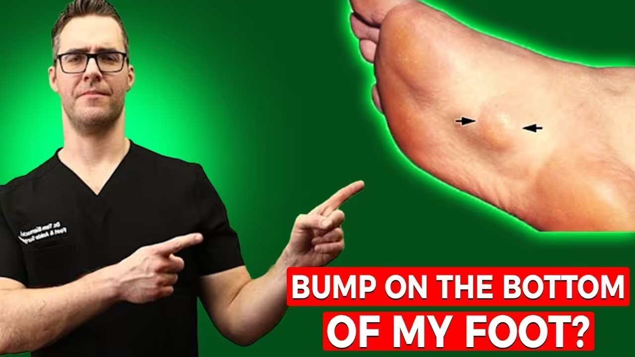
7. Plantar Fibromatosis (Ledderhose Disease): The Arch Nodule
Plantar fibromatosis (Ledderhose disease) is a benign but potentially progressive proliferative condition of the plantar fascia in which fibroblasts abnormally proliferate to form firm, fixed nodules within the fascia. Unlike plantar fasciitis, which causes diffuse inflammation, Ledderhose produces discrete, palpable nodules — typically in the central or medial arch — that can cause significant pain with weight-bearing as they enlarge. It’s part of the spectrum of fibromatoses (related to Dupuytren’s contracture in the hand and Peyronie’s disease).
Who gets it: Middle-aged to older adults, with a higher incidence in men; associated with diabetes mellitus, chronic liver disease, epilepsy medications (especially phenytoin), and family history of fibromatosis. Bilateral involvement in 25–50% of cases. The condition is benign but can progress to nodule coalescence and toe contracture in severe cases.
Key distinguishing feature: Palpable, firm, non-tender-to-lateral-pressure nodules in the arch that ARE tender with direct axial (standing) pressure. Unlike a ganglion cyst, they do not transilluminate. Unlike plantar fasciitis, there is no first-step pain — pain is present throughout the day with any weight-bearing on the nodule. MRI distinguishes Ledderhose from other soft tissue masses.
Treatment: Conservative (first line for small, stable nodules): custom orthotics with cutout accommodation to offload the nodule, corticosteroid injection (reduces inflammation, doesn’t eliminate nodule), topical verapamil. Intralesional collagenase injection (Xiaflex) — emerging evidence. Radiation therapy for progressive disease. Surgical excision: reserved for refractory cases with significant disability; high recurrence rate (up to 60%) if plantar fascia is not excised completely.
How to Tell Them Apart: Diagnostic Comparison Table
Because so many arch pain conditions overlap in their presentation, this table gives you the key clinical features that differentiate each cause. Bring this to your appointment and note which column best describes your symptoms.
| Condition | Pain Location | First-Step Pain | Tingling/Numbness | Palpable Mass | Best Imaging |
|---|---|---|---|---|---|
| Plantar Fasciitis | Medial heel + arch | Yes (hallmark) | No | No | US or MRI |
| PTTD | Inner ankle + arch | No | No | Tendon swelling | MRI |
| Navicular Stress Fx | Dorsal midfoot | No | No | N-spot tenderness | MRI or CT |
| Tarsal Tunnel | Inner ankle → arch → toes | No | Yes | Possible | EMG/NCS + MRI |
| Cavus Foot | Diffuse + lateral | No | Sometimes | No | Weight-bearing X-ray |
| Spring Lig. Tear | Medial midfoot | No | No | No | Dedicated MRI |
| Plantar Fibromatosis | Central/medial arch | No | No | Yes | MRI |
Red Flags: When Arch Pain Requires Urgent Evaluation
⚠ Seek Prompt Podiatric Evaluation For:
- Progressive flatfoot deformity — arch visibly collapsing over weeks to months (PTTD/spring ligament emergency)
- Inability to perform single-leg heel raise — strong indicator of posterior tibial tendon dysfunction
- Tingling, burning, or numbness radiating into toes from the arch area (tarsal tunnel, neuropathy)
- Palpable arch nodule that is growing or becoming more painful (plantar fibromatosis vs. soft tissue tumor)
- Midfoot pain in an athlete that doesn’t improve with 2 weeks of rest (navicular stress fracture until proven otherwise)
- Arch pain associated with trauma — Lisfranc injury can mimic arch strain and requires urgent imaging
- Night pain that wakes you from sleep — can indicate infection, tumor, or inflammatory arthritis
Treatment Options by Severity
Appropriate treatment depends entirely on accurate diagnosis. The following framework applies broadly, but remember that conditions like navicular stress fracture, spring ligament tear, and Stage III–IV PTTD require condition-specific protocols rather than generic arch pain management.
Sale

PowerStep Pinnacle High Arch Orthotic Insoles, Plantar Fasciitis Relief, Supination Heel Pain, Arch Support, PowerStep Insoles for Women and Men, Made in USA (Men’s 10-10.5, Women’s 12)
- High Arch Support: PowerStep supination insoles deliver firm, flexible high arch support plus a deep heel cradle for comfort, stability & motion control, helping align feet, reduce pain, and protect against ball & heel pressure.
- All Day Comfort & Support: PowerStep Pinnacle High shoe inserts for women and men use premium dual layer cushioning to deliver heel to toe comfort and responsive bounce back with every step, without going flat.
- Relieves & Helps Prevent Pain: PowerStep Pinnacle High insoles for supination can help alleviate common foot conditions often linked to supination, including plantar fasciitis, Achilles tendonitis, fat pad atrophy, and Morton’s neuroma.
- No Trimming: PowerStep insoles move easily from shoe to shoe. Inserts are sized by shoe size for footwear with removable factory insoles. Designed for walking, running, work & casual dress shoes; pairs well with best walking shoes for women and men.
- Made in the USA: We stand behind our PowerStep Insoles for women and men. Proudly made in the USA & backed by a 30-day money-back guarantee. HSA & FSA Eligible
| Severity | Intervention | Timeline |
|---|---|---|
| Mild (0–6 weeks) | Supportive footwear, OTC arch support (PowerStep Pinnacle Maxx), activity modification, calf stretching, Doctor Hoy’s topical gel | Start immediately |
| Moderate (6–12 weeks) | Podiatry evaluation, custom orthotics, physical therapy, diagnostic imaging (US or MRI), DASS compression if edema present | After 6 weeks no improvement |
| Severe / Non-responsive | Corticosteroid injection, PRP, ESWT, walking boot, or surgical consultation depending on diagnosis | After 12 weeks conservative care |
View at our shop
Not ideal for: open wounds, broken skin, or patients with camphor sensitivity.
Get an Accurate Arch Pain Diagnosis
Dr. Tom Biernacki, DPM has performed 3,000+ procedures and sees arch pain patients daily at both locations. Same-day appointments available.
Howell: 4330 E Grand River Ave | Bloomfield Hills: 43494 Woodward Ave #208
Frequently Asked Questions About Arch Pain
What is the most common cause of arch pain?
Plantar fasciitis is by far the most common cause of arch pain, accounting for roughly 40–50% of cases in clinical practice. It produces characteristic sharp heel and arch pain with first steps in the morning, caused by inflammation and micro-tearing at the calcaneal attachment of the plantar fascia. However, because plantar fasciitis is so well-known, other conditions are frequently missed — particularly posterior tibial tendon dysfunction and navicular stress fractures in athletic patients.
Can flat feet cause arch pain even without a specific injury?
Yes — flat feet (pes planus) chronically overload the plantar fascia, posterior tibial tendon, and spring ligament, leading to gradual-onset arch pain without any single traumatic event. The progressive overload model explains why flat-footed individuals are at higher risk for plantar fasciitis, PTTD, and spring ligament degeneration. Supportive orthotics with medial arch support (such as PowerStep Pinnacle Maxx) can significantly reduce this load and prevent progression.
When should I see a podiatrist for arch pain?
See a podiatrist if arch pain persists beyond 2–3 weeks despite rest and supportive footwear, if you notice progressive flatfoot deformity, if pain is associated with tingling or numbness, if you feel a palpable lump in your arch, or if you’re an athlete with midfoot pain that doesn’t improve with 2 weeks of rest (navicular stress fracture must be ruled out). Early evaluation prevents conditions like PTTD and navicular stress fractures from progressing to severe stages requiring surgery.
Does insurance cover arch pain treatment and custom orthotics?
Most insurance plans cover podiatric evaluation and diagnostic imaging for arch pain as a medically necessary service. Custom orthotics are covered by many plans when documented medically necessary conditions are present (PTTD, plantar fasciitis unresponsive to conservative care, cavus foot). OTC insoles like PowerStep are not covered but are significantly less expensive ($40–$60) and often effective for milder cases. Our front desk team verifies benefits before your appointment — call (810) 206-1402 for details.
Is it safe to keep exercising with arch pain?
It depends entirely on the cause. Mild plantar fasciitis often tolerates low-impact activity (cycling, swimming) well. But navicular stress fractures require strict non-weight-bearing — continuing to run converts a stress fracture to a complete fracture. PTTD worsens with impact loading. The safest approach: stop impact activity until you have a diagnosis. Two weeks off running to get an accurate diagnosis is far better than six months off recovering from a complete navicular fracture or ruptured posterior tibial tendon.
Sources
- Buchbinder R. Plantar fasciitis. N Engl J Med. 2004;350(21):2159-2166.
- Deland JT. Adult-acquired flatfoot deformity. J Am Acad Orthop Surg. 2008;16(7):399-406.
- Torg JS, et al. The natural history of the posterior tibial tendon — clinical and histological evaluation. Foot Ankle Int. 2005;26(10):833-838.
- Khan KM, et al. Navicular stress fracture in athletes. Sports Med. 1994;17(1):65-76.
- Gondring WH, et al. Tarsal tunnel syndrome. Foot Ankle Int. 2003;24(7):545-548.
- Zgonis T, et al. Plantar fibromatosis and surgical management. Foot Ankle Spec. 2021;14(5):437-444.
In-Office Treatment at Balance Foot & Ankle
If home treatment isn’t providing relief for your arch pain, our podiatry team at Balance Foot & Ankle can help with same-day evaluations and advanced in-office care.
Same-day appointments available. (810) 206-1402
Learn about our plantar fascia & arch treatment → | Book online →
Get Expert Care at Balance Foot & Ankle
Same-week appointments at our Howell and Bloomfield Hills offices. Board-certified podiatric surgeons. Most insurance accepted.
Same-Week Appointments in Howell & Bloomfield Hills
Three board-certified podiatric surgeons. 1,123+ five-star reviews. Most insurance accepted.

Dr. Tom Biernacki, DPM is a board-certified foot & ankle surgeon (ABFAS & ABPM) at Balance Foot & Ankle Specialists in Southeast Michigan. With over a decade of clinical experience, he specializes in heel pain, bunions, diabetic foot care, sports injuries, and minimally invasive surgery. Dr. Biernacki is a member of the APMA and ACFAS, and his patient education content on MichiganFootDoctors.com and YouTube has made him one of the most-followed foot & ankle educators on YouTube.