
| Cause | Prevalence | Associated Features | Diagnostic Clue | Treatment Direction |
|---|---|---|---|---|
| Onychomycosis (fungal) | Most common (50%) | Yellow/brown discoloration, thickening | Positive KOH or fungal culture | Oral terbinafine or efinaconazole topical |
| Trauma (repetitive) | Common — athletes, long nails | Distal separation; no discoloration | History of pressure/impact; correct nail length | Trim nail; protective footwear; time |
| Psoriasis | ~50% of psoriasis patients | “Oil spot,” pitting, subungual hyperkeratosis | Skin or scalp psoriasis present | Topical calcipotriol; intralesional steroid |
| Thyroid disease | Uncommon; check if bilateral | Plummer’s nails (hyperthyroid); brittle (hypo) | TSH level | Treat underlying thyroid disorder |
| Reaction to nail products | Increasingly common | History of gel/acrylic nails | Temporal link to nail application | Remove products; avoid acrylates |
| Systemic disease (iron deficiency, syphilis) | Rare | Multiple nails; systemic symptoms | CBC, ferritin, RPR | Treat underlying condition |
| Treatment Step | Action | Rationale |
|---|---|---|
| 1. Clip separated nail | Trim the detached portion as far back as painlessly possible | Removes the “tent” that harbors moisture and microbes |
| 2. Keep nail dry | Avoid prolonged water exposure; wear gloves for wet work | Moisture under the nail perpetuates separation |
| 3. Treat infection | Antifungal topical (efinaconazole 10%) if fungal confirmed | Clears the underlying pathogen promoting separation |
| 4. Footwear modification | Extra-depth shoe; avoid high heels | Eliminates ongoing nail trauma during healing |
| 5. Monitor regrowth | Toenail regrows 9–18 months; fingernail 4–6 months | Patience required — the nail must grow out completely |
| 6. Treat root cause | Psoriasis, thyroid, iron — address systemic driver | Without treating the cause, separation recurs |
Quick Answer
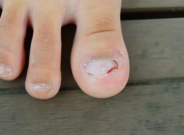
Onycholysis is the separation of the nail plate from the nail bed, beginning at the free edge and progressing proximally. In the foot, the most common causes are repetitive trauma from tight shoes, onychomycosis (fungal nail infection), psoriasis, and systemic conditions such as thyroid disease. Treatment depends on the cause: trim the separated portion, keep the nail dry, treat any underlying infection or systemic disease, and correct the mechanical factors. The separated nail does not reattach — the nail must regrow from the matrix.
What Is Onycholysis
Onycholysis describes the detachment of the nail plate from the nail bed. The separation typically begins at the distal (free) edge of the nail and advances proximally — toward the cuticle — creating a white, yellow, or greenish discoloration where air or debris fills the space between plate and bed. The color change is not the nail itself changing; it is the optical effect of the air gap and any secondary colonization beneath. The nail plate itself remains intact; it has simply lost its adherence to the underlying nail bed epithelium.
In our clinic, onycholysis in toenails is an extremely common finding — and frequently misdiagnosed. Patients often assume the discoloration means fungal infection, when in fact trauma from shoe pressure is the underlying driver in the majority of cases we see. The distinction matters because the treatment differs entirely. Trimming the separated portion and widening the toe box resolves trauma-induced onycholysis; oral antifungals are not indicated unless fungal infection is confirmed by culture or KOH preparation. Treatment directed at the wrong cause produces no improvement and delays resolution.
Causes of Onycholysis in the Foot
- Repetitive trauma — the most common cause in toenails; the nail plate impacts the shoe’s toe box repeatedly during ambulation, progressively lifting the free edge from the nail bed; runners, hikers, and anyone in tight footwear are high-risk; the hallux and second toenail are most commonly affected
- Onychomycosis (fungal nail infection) — dermatophytes (most commonly Trichophyton rubrum) invade the subungual space, producing hyperkeratosis and separation; the nail thickens, discolors, and lifts from the nail bed; confirmed by culture or PCR before treating with systemic antifungals
- Psoriasis — nail psoriasis occurs in 50-80% of psoriasis patients; produces the “oil drop” sign (salmon-colored patch beneath the nail plate), pitting, and onycholysis; often bilateral and symmetric; associated with psoriatic arthritis
- Thyroid disease — both hyperthyroidism and hypothyroidism can cause onycholysis; in hyperthyroidism, the Plummer nail (onycholysis beginning on the ring finger, but also affecting toenails) is a classic sign; thyroid function testing is warranted in unexplained or bilateral cases
- Contact irritants and chemical exposure — nail polish solvents, prolonged moisture exposure, cleaning chemicals; these dissolve the nail bed attachment directly
- Iron deficiency anemia — can produce koilonychia (spoon nails) and onycholysis simultaneously; iron studies indicated in atypical presentation
- Drug-induced onycholysis — tetracyclines (photo-onycholysis after sun exposure), fluoroquinolones, taxane chemotherapy agents, and retinoids are notable causes; medication history is essential
- Subungual exostosis or tumor — bony outgrowth beneath the nail plate lifts it from below; presents with focal onycholysis and nail deformity; confirmed on X-ray
Symptoms and Diagnosis
The classic presentation is a white, yellow, or pale discoloration of part of the nail plate beginning at the free edge — the “white crescent” of air beneath the plate. There is typically no pain unless secondary bacterial or fungal colonization has occurred in the subungual space. Pressing on the lifted nail plate may produce discomfort. In long-standing cases, debris (keratin, dirt, and secondary organisms) accumulates under the plate, producing a foul odor and increasing the risk of secondary infection.
Diagnosis is clinical in most cases. When fungal infection is suspected as a contributing cause, a nail clipping and subungual debris sample should be sent for KOH preparation and culture before initiating antifungal therapy — oral antifungals have significant drug interactions and a treatment duration of 3-6 months, making accurate diagnosis important. When onycholysis is bilateral, unexplained, or accompanied by skin changes, systemic workup (thyroid function, iron studies, complete blood count) is warranted. Subungual X-ray is obtained when the nail is elevated by a focal hard mass suggesting exostosis.
Treatment
Trim the Separated Nail
The separated nail plate does not reattach to the nail bed — the bond has been permanently broken. The goal is to remove the separated portion back to the point of adhesion, eliminating the lever arm that allows further separation and removing the dead space that harbors debris and secondary organisms. This is done conservatively in clinic with a nail nipper, trimming the free edge back to the point where the nail is still attached. The nail bed is left exposed but will re-epithelialize; the nail plate regrows from the matrix and, if the underlying cause is corrected, will reattach as it advances distally.
Keep the Nail Bed Dry
Moisture beneath a lifted nail plate promotes secondary infection. After trimming, the nail bed should be kept dry — soaking is contraindicated. Patients are instructed to dry the foot thoroughly after showering, apply a thin layer of topical antifungal cream (ciclopirox or clotrimazole) to the exposed nail bed if fungal colonization is suspected, and avoid prolonged moisture exposure. Occlusive footwear worn for extended periods without breathable socks creates the moist environment that delays healing.
Correct the Mechanical Cause
For trauma-induced onycholysis — by far the most common cause in our clinic — widening the toe box is the single most effective intervention. The nail that impacts the toe box with every step will never fully reattach as long as that impact continues. A proper shoe fitting with at least a thumb’s width between the longest toe and the end of the shoe eliminates the repetitive lifting force. Runners with onycholysis of the hallux or second toe almost universally need a half-size increase and a toe box with more vertical clearance. Taping the toenails before running with athletic tape provides additional protection during the healing phase.
Treat Onychomycosis When Confirmed
When culture or KOH confirms onychomycosis, treatment with oral terbinafine (250 mg daily for 12 weeks for toenails) is the most effective option — topical antifungals have limited penetration into the nail plate and nail bed and achieve cure rates under 10% for established fungal nail disease. Liver function must be screened before oral terbinafine in patients with hepatic risk factors. Laser treatment is available as an adjunct but is not covered by insurance and has variable long-term efficacy data. The separated nail will not reattach during antifungal treatment; the goal is to allow the new nail plate growing from the matrix to emerge healthy and attached.
Address Systemic Causes
Onycholysis from psoriasis, thyroid disease, or anemia does not resolve without treating the underlying condition. Psoriatic nail disease responds to the same systemic agents used for skin psoriasis (biologics, methotrexate) — the podiatrist’s role is to manage the mechanical complications and refer appropriately. Thyroid normalization typically resolves thyroid-related nail changes over several months. Iron repletion for iron-deficiency onycholysis is straightforward and effective.
See a Podiatrist If:
- The lifted nail is painful, warm, or producing discharge — secondary bacterial infection requires treatment
- Onycholysis is bilateral or affecting multiple nails without clear trauma history — systemic cause workup needed
- The nail plate is thickened, crumbly, or yellow-brown with subungual debris — fungal culture before antifungal treatment
- A hard mass is palpable under the nail plate — subungual exostosis or fibroma requires X-ray and possible excision
- Onycholysis recurs despite footwear correction — biomechanical assessment and custom orthotics may be needed to offload the nail
Most Common Mistake We See:
Treating trauma-induced onycholysis with oral antifungals. The nail is lifting because the shoe is hitting it — not because of a fungal infection. We routinely see patients who have completed two full courses of terbinafine for a “fungal nail” that, on careful examination, is traumatic onycholysis with secondary debris accumulation and no fungal growth on culture. Oral antifungals have real risks (drug interactions, hepatotoxicity surveillance, cost) and a 3-month treatment duration. Before prescribing, culture the nail. The separated nail will look exactly the same whether the cause is trauma, fungus, psoriasis, or thyroid disease — the color and appearance do not tell you the cause.

Not ideal for: Active nail infection with open skin. PowerStep Pinnacle insoles with deep heel cups and arch support reduce forefoot pressure and metatarsal loading — an important adjunct for patients whose onycholysis is driven by abnormal gait mechanics or excessive forefoot impact.

Not ideal for: Open subungual infections or broken skin adjacent to the nail. Doctor Hoy’s natural arnica gel provides topical relief for the periungual soreness and nail bed tenderness associated with onycholysis and subungual inflammation.
Nail Separation or Discoloration That Won’t Resolve?
Same-day appointments · Howell & Bloomfield Township, MI
Book Online (810) 206-1402Frequently Asked Questions
Will an onycholysis nail grow back normally
Yes — if the underlying cause is corrected. The nail matrix (the growth center at the base of the nail) is unaffected by onycholysis; it continues producing new nail plate that advances distally. If the cause of separation — tight shoes, fungal infection, psoriasis — is treated, the new nail plate will adhere to the nail bed as it grows. Toenails grow approximately 1.5-2 mm per month, meaning full regrowth from matrix to free edge takes 12-18 months. Patience and consistent cause correction are the formula for a normal result.
How do you treat onycholysis at home
Trim the separated nail back to the point of adhesion (eliminating the dead space), keep the nail bed dry, switch to shoes with adequate toe box width and length, and apply topical antifungal cream (over-the-counter clotrimazole or terbinafine) to the nail bed if fungal infection is suspected while awaiting a culture. Do not attempt to forcibly reattach or glue the nail plate. Do not soak the foot — moisture delays healing. Home management is appropriate for mild, clearly trauma-related cases; see a podiatrist if the nail is painful, infected, or not improving after 2-3 months of conservative care.
Is onycholysis the same as a fungal nail
No — onycholysis (nail separation) is a finding, not a diagnosis. Onychomycosis (fungal nail infection) is one of many causes of onycholysis, but trauma, psoriasis, thyroid disease, and contact irritants also cause identical-appearing nail separation. The appearance of the nail does not reliably distinguish the cause. A nail clipping sent for KOH preparation and fungal culture is the only way to confirm or exclude onychomycosis. This distinction is clinically important because oral antifungals are an unnecessary 3-month treatment with real risks if the cause is mechanical rather than infectious.
The Bottom Line
Onycholysis is one of the most common nail problems we see in a podiatry clinic — and one of the most over-treated. The separated nail will not reattach on its own or with any topical application; the treatment is trimming the detached portion, correcting the cause, and allowing the nail to regrow from the matrix. For most toenail cases, the cause is mechanical: the shoe is too short, too narrow, or too low in the toe box. Fix the footwear, trim the nail, keep it dry, and the nail grows back normally over 12-18 months. For cases with systemic signs, bilateral involvement, or suspected fungal infection, a podiatric evaluation gets you the correct diagnosis and treatment the first time.
How Long Does It Take for the Nail to Reattach?
This is the question patients ask first, and the answer surprises most of them: a separated nail does not reattach. Once the plate has lifted off the bed, that bond is gone. What actually happens is that a new, attached nail grows out from the base and gradually pushes the separated portion forward until it can be trimmed away.
That means the timeline is set entirely by nail growth rate, and fingernails and toenails are very different:
- Fingernails grow roughly 3 mm a month, so full replacement takes about 4 to 6 months.
- Toenails grow roughly 1 mm a month — about a third as fast. Full replacement takes 12 to 18 months, and the big toenail is the slowest of all.
Growth is slower in older adults, in people with reduced circulation, and in diabetes. Cold weather slows it slightly too.
The practical consequence is that you should judge progress by the new growth at the base, not by the separated part. If the nail emerging from the cuticle is attached, pink and normal, the problem is resolved and you are simply waiting out the calendar. If new growth is also separating, the underlying cause is still active and needs addressing.
Gel Nails, Acrylics and Aggressive Cleaning
A large share of onycholysis has nothing to do with disease. It is caused by things done to the nail.
Gel and acrylic manicures are a common culprit, through three mechanisms: the buffing that roughens the plate before application thins it, the UV curing and adhesives can irritate the nail bed, and removal — particularly picking or prying a gel layer off rather than soaking it — can pull the plate away from the bed with it. Repeated cycles compound the damage.
Aggressive cleaning under the nail is the one we see most in the office and the one patients least expect. Digging under a lifted nail with a file, an orange stick or a fingernail to clear the debris feels productive. It is not. Every pass extends the separation a little further back, and the nail responds by lifting more. If your nail is separating, the correct instruction is counterintuitive: stop cleaning under it, keep it trimmed short, and let it grow out.
Other mechanical contributors include nail polish removers used frequently, prolonged wet work, and in the foot specifically, shoes with a toe box that is too shallow — the repeated ceiling contact during walking or running lifts the nail over months without any single memorable injury.
When Nail Separation Needs to Be Looked At
Most onycholysis is benign and resolves once the cause is removed. These features are the exceptions, and they matter:
- A single nail affected with pigmentation — a brown or black band, or pigment spreading onto the surrounding skin. Subungual melanoma is uncommon, but it is most often missed precisely because it is mistaken for a bruise or a fungal nail. One nail behaving differently from all the others deserves an examination rather than a wait.
- Pus, spreading redness, or increasing pain. That is infection, not simple separation, and it needs treatment rather than time.
- Several nails separating at once with no injury to explain it. This pattern points toward a systemic driver — psoriasis, thyroid disease, a medication reaction — and treating the nail alone will not fix it.
- New growth that separates too. The cause is still active. Continuing to trim and wait will not resolve it.
- Diabetes or peripheral neuropathy. Any nail problem in a foot with reduced sensation deserves earlier evaluation, because the usual warning signal — pain — is unreliable.
A diagnosis is usually straightforward: examination, and a nail clipping sent for testing when fungus is suspected. That last step matters more than it sounds, because roughly half of nails that look fungal are not, and treating a non-fungal nail with antifungals wastes months of a 12-to-18-month growth window.
Preventing It From Coming Back
Because the nail takes so long to replace itself, prevention is worth more here than in almost any other nail condition.
- Get the shoe length right. About a thumb’s width between the longest toe and the end of the shoe, measured standing, late in the day. Toe length is not always in the order you expect — a second toe longer than the big toe changes the fit you need.
- Keep nails trimmed straight across and short, but not so short that the corner digs in. A long nail meets the shoe first.
- Keep the area dry. Moisture-wicking socks, changed after exercise; shoes given a day to dry between wears.
- Give the nail breaks from gel and acrylic, and always soak rather than pick when removing.
- Do not clean underneath it. Worth repeating, because it is the most common way patients extend their own separation.
- Treat the underlying condition — confirmed fungal infection, psoriasis, or a thyroid problem — or the nail will keep lifting no matter what you do locally.
If a nail has been separating for months, or one nail is behaving differently from the rest, it is worth having looked at properly rather than waiting out another growth cycle. Balance Foot & Ankle sees patients in Howell (4330 E Grand River Ave, Howell, MI 48843) and Bloomfield (43494 Woodward Ave #208, Bloomfield Township, MI 48302). Call (810) 206-1402.
Sources
- Piraccini BM, Alessandrini A. “Onychomycosis: a review.” J Fungi. 2015.
- Rigopoulos D, et al. “Nail psoriasis: a combined treatment using calcipotriol cream and clobetasol propionate cream.” Acta Derm Venereol. 2002.
- Fawcett RS, et al. “Nail abnormalities: clues to systemic disease.” Am Fam Physician. 2004.
- Tosti A, Piraccini BM. “Onycholysis.” Dermatol Clin. 2006.
- Gupta AK, et al. “Onychomycosis: a review of clinical features, diagnosis, and management.” Int J Dermatol. 2023.

Dr. Tom Biernacki, DPM is a board-certified foot & ankle surgeon (ABFAS & ABPM) at Balance Foot & Ankle Specialists in Southeast Michigan. With over a decade of clinical experience, he specializes in heel pain, bunions, diabetic foot care, sports injuries, and minimally invasive surgery. Dr. Biernacki is a member of the APMA and ACFAS, and his patient education content on MichiganFootDoctors.com and YouTube has made him one of the most-followed foot & ankle educators on YouTube.