
Quick answer: Peroneal tendon subluxation is when the tendons slip out of the groove behind the outer ankle, usually from a stretched or torn superior peroneal retinaculum. When bracing and rest don’t hold them in place, surgery to repair or deepen the retinaculum (often with groove-deepening) restores stability — most patients return to activity in about 3–4 months.

Quick answer: Peroneal Tendon Subluxation Surgery Retinaculum Repair is a common foot/ankle topic that affects many patients. The 2026 evidence-based approach combines proper diagnosis, conservative-first treatment, and escalation only when needed. We treat this regularly at our Howell and Bloomfield Township practices. Call (810) 206-1402.
The most important clinical decision with Peroneal Tendon Subluxation Surgery Retinaculum Repair isn’t which treatment to start with — it’s identifying the correct subtype. That changes everything. Call (810) 206-1402.
What Is Peroneal Tendon Subluxation?
Peroneal tendon subluxation occurs when one or both peroneal tendons (peroneus longus and brevis) slip out of their normal position behind the lateral malleolus, displacing anteriorly (forward). In the most dramatic cases, the tendons can be seen and felt snapping around the malleolus with ankle movement — a phenomenon called peroneal tendon dislocation.
The tendons are normally held in position by the superior peroneal retinaculum (SPR) — a fibrous band that acts as a restraining strap. Subluxation occurs when the SPR tears or becomes stretched, allowing the tendons to escape.
How It Happens
Acute peroneal subluxation most commonly occurs with sudden, forceful ankle dorsiflexion combined with reflexive peroneal muscle contraction — a defensive mechanism during a fall or landing. Skiers, soccer players, basketball players, and gymnasts are commonly affected. The injury is often misdiagnosed as a lateral ankle sprain at initial presentation, delaying appropriate management.
Chronic subluxation can develop gradually without a clear acute event, particularly in patients with a congenitally shallow fibular groove — the bony groove behind the lateral malleolus in which the tendons normally sit — that provides insufficient mechanical restraint.
Symptoms
The hallmark is painful snapping or clicking at the posterolateral ankle with activities requiring ankle dorsiflexion (running, hiking, descending stairs). Patients may be able to voluntarily reproduce the subluxation by dorsiflexing and everting the ankle. The posterolateral ankle may be tender and swollen. If subluxation is not recognized initially, patients often develop peroneal tendon tears from the repeated mechanical abrasion of the tendons against the fibular rim.
Non-Surgical Treatment
Acute peroneal subluxation (within 2–3 weeks of injury with an intact reduction) may be managed with 4–6 weeks of non-weight-bearing in a short leg cast, followed by structured rehabilitation. Conservative success rates are 50–75% for acute presentations. Chronic subluxation and cases where the tendons have already sustained tears have lower conservative success rates and typically require surgery.
Surgical Options
Superior Peroneal Retinaculum Repair
The primary surgical approach is anatomic repair and tightening of the torn or stretched SPR — reattaching it to the fibula with suture anchors and plicating (folding) any redundant tissue to restore appropriate tension. This is the most direct treatment of the underlying pathology and provides reliable results when the fibular groove is of adequate depth.
Fibular Groove Deepening
When the fibular groove is congenitally shallow or has been worn down, groove deepening (fibular groove deepening osteoplasty) is performed simultaneously. A portion of the fibular cortex behind the lateral malleolus is removed and the groove is deepened to provide a more secure mechanical pocket for the tendons. This reduces recurrence in patients with anatomically predisposing groove morphology.
Peroneal Tendon Repair
Concurrent longitudinal tears of the peroneus brevis (which occur from mechanical abrasion against the fibular rim during subluxation) are repaired simultaneously via direct suture repair and tubularization of the tendon.
Recovery
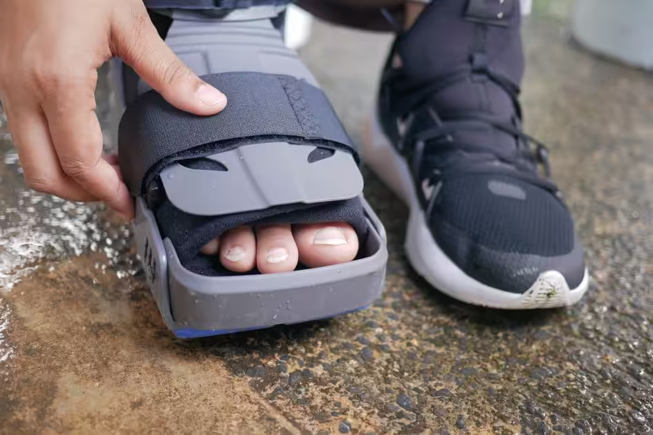
Recovery follows a structured progression: 4–6 weeks non-weight-bearing in a boot or cast, followed by progressive weight-bearing and physical therapy focusing on peroneal strengthening and proprioception. Return to running at 3–4 months and full sport at 4–6 months. Recurrence rates after surgical treatment are low (5–10%) when appropriate fixation is achieved and rehabilitation is completed.
Foot or Ankle Pain? We Can Help.
Balance Foot & Ankle — Howell & Bloomfield Township, MI
📅 Book Online
📞 (810) 206-1402
Insurance Accepted
BCBS · Medicare · Aetna · Cigna · United Healthcare · HAP · Priority Health · Humana · View All →
Howell Office
4330 E Grand River Ave
Howell, MI 48843
Get Directions →
Bloomfield Township Office
43494 Woodward Ave, Suite 208
Bloomfield Township, MI 48302
Get Directions →
Your Board-Certified Podiatrists



Ready to Get Back on Your Feet?
Same-week appointments available at both locations.
Book Your AppointmentDifferential Diagnosis: What Else Could It Be?
Not every case of peroneal tendonitis is straightforward. In our clinic we routinely rule out three look-alike conditions before confirming the diagnosis. If your symptoms don’t match the classic presentation, one of these may explain the pain — which is why physical exam matters more than self-diagnosis.
| Condition | How It Differs |
|---|---|
| Lateral ankle sprain | Acute inversion mechanism, bruising along anterior talofibular ligament, pain with anterior drawer. |
| 5th metatarsal base stress fracture | Point tenderness at 5th metatarsal base, pain with weight-bearing, fracture line on imaging. |
| Sinus tarsi syndrome | Deep ache in the sinus tarsi, pain reproduced with lateral palpation just anterior to the lateral malleolus. |
Red Flags — When to See a Podiatrist Now
Seek same-day evaluation at Balance Foot & Ankle if you notice any of the following:
- Snapping or popping behind the lateral malleolus (subluxation)
- Inability to evert the foot actively
- Persistent lateral ankle swelling >4 weeks
- Sudden pop with inability to continue walking
Call (810) 206-1402 or request an appointment. Our Howell and Bloomfield Township offices reserve same-day slots for urgent foot and ankle issues.
In Our Clinic: What We See
Clinical perspective from Dr. Tom Biernacki, DPM — Balance Foot & Ankle, Howell & Bloomfield Township, MI:
In our clinic, peroneal tendonitis patients usually come in after a recent ankle sprain — the pain started as a “sprain that didn’t fully heal.” They report lateral ankle pain that’s worse with turning the foot outward or walking on uneven surfaces. On exam we palpate specifically along the peroneal tendons behind the fibula and resist eversion. If we feel or see snapping behind the lateral malleolus, that’s peroneal subluxation, which usually needs surgical repair. Isolated peroneal tendonitis responds well to ankle bracing, peroneal eccentric strengthening, and temporary activity modification.
In-Office Treatment at Balance Foot & Ankle
If home treatment isn’t providing relief for your peroneal tendon, our podiatry team at Balance Foot & Ankle can help with same-day evaluations and advanced in-office care.
Same-day appointments available. (810) 206-1402
Peroneal tendon treatment in Michigan → | Learn about our ankle and tendon treatment → | Book online →
Frequently Asked Questions
When should I see a podiatrist?
If symptoms persist past 2 weeks, affect your normal activity, or are accompanied by red-flag symptoms (warmth, redness, swelling, inability to bear weight).
What does treatment cost?
Most diagnostic visits and conservative treatments are covered by Medicare and major insurers. Out-of-pocket costs vary by your specific plan.
How quickly can I get an appointment?
Most non-urgent cases see us within 5 business days. Urgent cases (sudden pain, possible fracture) typically same or next business day.
Get Expert Care at Balance Foot & Ankle
Same-week appointments at our Howell and Bloomfield Township offices. Board-certified podiatric surgeons. Most insurance accepted.

Dr. Tom Biernacki, DPM is a board-certified foot & ankle surgeon (ABFAS & ABPM) at Balance Foot & Ankle Specialists in Southeast Michigan. With over a decade of clinical experience, he specializes in heel pain, bunions, diabetic foot care, sports injuries, and minimally invasive surgery. Dr. Biernacki is a member of the APMA and ACFAS, and his patient education content on MichiganFootDoctors.com and YouTube has made him one of the most-followed foot & ankle educators on YouTube.
More questions patients ask
What is peroneal tendon subluxation and who needs surgery?
Peroneal tendon subluxation occurs when the peroneal tendons slip out of their groove behind the outer ankle bone due to a torn or stretched superior peroneal retinaculum. Surgery is recommended for patients with recurrent subluxation after conservative treatment fails, athletes wanting to return to sport, or cases with significant retinaculum tearing confirmed on MRI.
What does peroneal retinaculum repair surgery involve?
Peroneal retinaculum repair surgery involves tightening or reconstructing the superior peroneal retinaculum — the tissue band that holds the peroneal tendons in place behind the lateral malleolus. The surgeon may also deepen the bony groove (retromalleolar sulcus) if it is too shallow. The procedure is typically performed under general or regional anesthesia as an outpatient.
What is the recovery time for peroneal tendon subluxation surgery?
Recovery from peroneal retinaculum repair takes approximately 3–4 months for return to daily activities and 6–9 months for full athletic return. The first 2–4 weeks involve non-weight-bearing in a cast or boot, followed by progressive weight-bearing and physical therapy. Return to running and cutting sports typically begins around 4–6 months post-operatively.
Still have a question about coverage or cost? Book online and we will check your benefits before your visit: Book in Howell · Book in Bloomfield Township. Prefer to talk it through first? Call (810) 206-1402.