The most important clinical decision with Peroneal Tendon Surgery isn’t which treatment to start with — it’s identifying the correct subtype. That changes everything. Call (810) 206-1402.
Peroneal tendon injury is a problem with the tendons on the outside of the ankle — typically caused by overuse or sudden strain. Most patients improve in 6-12 weeks with conservative care. Same-week appointments in Howell + Bloomfield Hills: (810) 206-1402.
Medically reviewed by Dr. Tom Biernacki, DPM — Board-certified foot & ankle surgeon, 3,000+ surgeries performed. Updated April 2026 with current clinical evidence. This article reflects real practice experience from Balance Foot & Ankle Specialists in Howell and Bloomfield Hills, Michigan.
Quick Answer
Most foot and ankle problems respond to conservative care — proper footwear, supportive inserts, activity modification, and targeted stretching — within 4-8 weeks. Persistent pain beyond that window, or any symptom that prevents walking, warrants a podiatric evaluation to rule out fracture, tendon tear, or systemic cause.
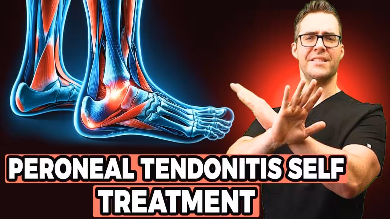
Watch: Dr. Tom Biernacki, DPM
Medically reviewed by Dr. Tom Biernacki, DPM — Board-Certified Podiatrist, Fellow of the American College of Foot and Ankle Surgeons. Updated April 2026.
The peroneal tendons—peroneus longus and peroneus brevis—run along the outer (lateral) side of the ankle, behind the lateral malleolus (the bony bump on the outside of the ankle), and attach to the foot. The peroneus brevis inserts at the base of the fifth metatarsal; the peroneus longus runs under the foot to the medial cuneiform and first metatarsal base. Together, these tendons evert the foot (turn it outward) and stabilize the ankle against inversion sprains. Peroneal tendon injuries are a common but frequently underdiagnosed cause of lateral ankle pain—often misdiagnosed as chronic ankle sprain.
Peroneal tendon problems fall into three main categories: tendinopathy (degenerative thickening and pain), longitudinal split tears (tearing along the length of the tendon, most commonly the peroneus brevis), and subluxation or dislocation (the tendons popping out of their groove behind the fibula due to superior peroneal retinaculum tear). Each requires a different treatment approach, and distinguishing between them requires MRI or ultrasound in addition to clinical examination.
Peroneal Tendon Tears
Longitudinal split tears of the peroneus brevis are the most common peroneal tendon injury. The peroneus brevis is compressed in the fibular groove during ankle inversion, causing it to split lengthwise rather than rupture completely. These tears produce lateral ankle pain behind the fibula, swelling along the tendon, and pain with resisted eversion. They frequently coexist with ankle ligament sprains, which is why they’re often missed initially. MRI or ultrasound demonstrates the split tear and its extent.
Conservative treatment (physical therapy, bracing, activity modification) is attempted for 3–6 months in partial tears. When conservative treatment fails or the tear is complete, surgical repair is indicated. Surgery involves exploring the tendon sheath, debridement of damaged tissue, and tubularization (suturing the split edges closed to restore tendon shape). If more than 50% of the tendon cross-section is involved, tenodesis to the peroneus longus (connecting the damaged tendon to the healthy one) may be performed. Peroneus longus tears are less common but occur at the cuboid tunnel and can cause lateral midfoot pain.
Peroneal Tendon Subluxation and Dislocation
Peroneal tendon subluxation occurs when the superior peroneal retinaculum—the fibrous band that holds the peroneal tendons in their groove behind the fibula—tears during a forceful ankle dorsiflexion-eversion injury. The tendons snap out of their groove (subluxate) and may dislocate anteriorly over the fibula. Patients feel and hear a snapping or popping sensation on the outer ankle with activity. This injury is common in skiers, football players, and basketball players.
Acute subluxation in young athletes is typically treated surgically to prevent recurrent dislocation. The retinaculum is repaired and reinforced, and sometimes the fibular groove is deepened (groove-deepening procedure) to better contain the tendons. Chronic or recurrent subluxation almost always requires surgical stabilization. Success rates with surgical repair are excellent—approximately 90–95% of patients return to sport without recurrent subluxation after appropriate repair.
Recovery After Peroneal Tendon Surgery
Recovery from peroneal tendon surgery depends on the procedure performed. Tendon repair (tubularization or tenodesis) requires 4–6 weeks non-weight-bearing in a boot or cast, followed by progressive weight-bearing through 8–10 weeks. Physical therapy begins with gentle range of motion and progresses to strengthening and proprioceptive training. Return to sport after isolated tendon repair typically occurs at 4–6 months.
Retinaculum repair for subluxation follows a similar timeline: 4–6 weeks non-weight-bearing, boot weaning by 10–12 weeks, and return to sport at 4–6 months. Groove-deepening procedures add complexity and may extend the timeline to 6–9 months. Physical therapy throughout recovery focuses on strengthening the peroneal muscles, restoring ankle proprioception (position sense), and sport-specific functional progression. Ankle taping or bracing is often used during the return-to-sport phase.
class=”mfd-patient-scenario” id=”more-peroneal-tendonitis-guides-from-dr-tom-perone”>In Our Clinic: What We See
Clinical perspective from Dr. Tom Biernacki, DPM — Balance Foot & Ankle, Howell & Bloomfield Hills, MI:
In our clinic, peroneal tendonitis patients usually come in after a recent ankle sprain — the pain started as a “sprain that didn’t fully heal.” They report lateral ankle pain that’s worse with turning the foot outward or walking on uneven surfaces. On exam we palpate specifically along the peroneal tendons behind the fibula and resist eversion. If we feel or see snapping behind the lateral malleolus, that’s peroneal subluxation, which usually needs surgical repair. Isolated peroneal tendonitis responds well to ankle bracing, peroneal eccentric strengthening, and temporary activity modification.
class=”mfd-differential” id=”more-peroneal-tendonitis-guides-from-dr-tom-perone”>Differential Diagnosis: What Else Could It Be?
Not every case of peroneal tendonitis is straightforward. In our clinic we routinely rule out three look-alike conditions before confirming the diagnosis. If your symptoms don’t match the classic presentation, one of these may explain the pain — which is why physical exam matters more than self-diagnosis.
Condition
How It Differs
Lateral ankle sprain
Acute inversion mechanism, bruising along anterior talofibular ligament, pain with anterior drawer.
5th metatarsal base stress fracture
Point tenderness at 5th metatarsal base, pain with weight-bearing, fracture line on imaging.
Sinus tarsi syndrome
Deep ache in the sinus tarsi, pain reproduced with lateral palpation just anterior to the lateral malleolus.
Red Flags — When to See a Podiatrist Now
Seek same-day evaluation at Balance Foot & Ankle if you notice any of the following:
Snapping or popping behind the lateral malleolus (subluxation)
class=”wp-block-heading mfd-treatment-bridge” id=”more-peroneal-tendonitis-guides-from-dr-tom-perone”>In-Office Treatment at Balance Foot & Ankle
If home care isn’t resolving your your foot or ankle concern, a visit with a board-certified podiatrist is the fastest path to accurate diagnosis and a personalized plan. At Balance Foot & Ankle Specialists, Dr. Tom Biernacki, Dr. Carl Jay, and Dr. Daria Gutkin offer same-day and next-day appointments at both our Howell and Bloomfield Hills offices. We perform on-site diagnostic ultrasound, digital X-ray, conservative care, advanced regenerative treatments, and minimally invasive surgery when indicated.
Post-op approved — impact-absorbing slide for early recovery.
HOKA Ora 3 Recovery Slide
Max-cushion recovery sandal — comfort for post-surgical swelling.
Hoka Bondi 9
Max-cushion walking shoe — ease into return-to-walking post-surgery.
As an Amazon Associate, Balance Foot & Ankle earns from qualifying purchases. Product recommendations are based on clinical experience; prices and availability shown above update live from Amazon.
When to See a Podiatrist
Foot and ankle surgery in 2026 is dramatically different than a decade ago — most procedures are now minimally-invasive, outpatient, and allow weight-bearing within days. Balance Foot & Ankle surgeons have performed 3,000+ foot/ankle surgeries with modern techniques. If another surgeon has recommended a traditional open procedure, a second opinion may reveal a faster, less-invasive option.
Peroneal tendon tears typically cause pain and tenderness directly behind the lateral malleolus (the outer ankle bone)—not at the ligament insertion slightly further forward. Pain is worse with activities requiring ankle stability (running, lateral movements, uneven terrain) and with resisted eversion (pushing the foot outward against resistance). Swelling along the tendon sheath behind the fibula is often present. If you’ve had multiple ankle sprains or have persistent lateral ankle pain beyond 6–8 weeks, MRI evaluation can diagnose peroneal tendon pathology. These injuries are commonly missed because the symptoms overlap significantly with lateral ankle ligament sprains.
Can a peroneal tendon tear heal without surgery?
Small partial peroneal tendon tears can sometimes stabilize with conservative treatment: 6–12 weeks of immobilization and physical therapy, followed by activity modification and bracing. Tendinopathy (degenerative thickening without complete tearing) often responds to physical therapy, eccentric strengthening exercises, and orthotics. However, longitudinal split tears—especially complete tears or those involving more than 50% of the tendon—have poor healing capacity without surgery and typically worsen over time without intervention. Peroneal subluxation in active patients almost always requires surgery, as the retinaculum cannot reliably heal with conservative treatment and recurrent subluxation causes progressive tendon damage.
What causes peroneal tendons to snap or pop on the outside of the ankle?
A snapping or popping sensation on the outer ankle during activity—especially with dorsiflexion (pulling the foot up) or eversion—is the hallmark of peroneal tendon subluxation. The tendons are slipping out of their groove behind the fibula as the retinaculum (the restraining band) is torn or loose. Some patients can voluntarily reproduce the snapping. This is distinct from the snapping sometimes felt at the ankle joint itself. Any reproducible tendon snapping on the outer ankle warrants evaluation—it typically indicates mechanical instability that will progress without treatment and commonly requires surgical retinaculum repair.
Dr. Tom Biernacki, DPM is a board-certified podiatric surgeon at Balance Foot & Ankle in Howell and Bloomfield Hills, Michigan. He diagnoses and treats peroneal tendon tears, subluxation, and tendinopathy with both conservative management and surgical repair.
Join 950,000+ Learning About Foot Health
Dr. Tom shares honest medical advice, supplement reviews, and treatment guides you won’t find anywhere else.
The right footwear can make or break your recovery. Dr. Tom’s complete guide to the best shoes for plantar fasciitis, flat feet, neuropathy, bunions & more — with clinical picks for every foot type.
The most common mistake we see is: Waiting too long before seeking care. Fix: any foot pain lasting more than 4 weeks, or any sudden severe symptom, deserves a professional evaluation rather than more rest.
Warning Signs That Need Same-Day Care
Seek immediate evaluation at Balance Foot & Ankle if you experience any of the following:
Dr. Tom Biernacki, DPM is a board-certified foot & ankle surgeon (ABFAS & ABPM) at Balance Foot & Ankle Specialists in Southeast Michigan. With over a decade of clinical experience, he specializes in heel pain, bunions, diabetic foot care, sports injuries, and minimally invasive surgery. Dr. Biernacki is a member of the APMA and ACFAS, and his patient education content on MichiganFootDoctors.com and YouTube has made him one of the most-followed foot & ankle educators on YouTube.
Balance Foot & Ankle surgeons are affiliated with Trinity Health Michigan, Corewell Health, and Henry Ford Health — three of Michigan’s largest health systems.