

| Feature | Primary Raynaud Phenomenon | Secondary Raynaud Phenomenon |
|---|---|---|
| Underlying Cause | No identifiable cause; vasospasm only | Associated with autoimmune disease (scleroderma, lupus, RA, Sjogren); medications; occupational |
| Age of Onset | Teens to 30s; female predominant | Typically >30; often with other systemic symptoms |
| Triphasic Color Change | White (vasospasm) → Blue (deoxygenation) → Red (reperfusion); classic but not always all 3 | Same but more severe; may not fully recover; digital ulcers possible |
| Tissue Damage | No — benign; no ischemic damage | Yes — digital ulcers; pitting; gangrene in severe scleroderma |
| ANA / Autoimmune Testing | Negative | Often positive; anti-Scl-70, anti-centromere in scleroderma |
| Nailfold Capillaroscopy | Normal capillary pattern | Abnormal — giant loops, avascular areas, hemorrhages |
| Treatment Urgency | Conservative; lifestyle modification | Pharmacologic; treat underlying disease; vascular specialist if ulcers |
| Treatment | Indication | Mechanism | Evidence Level | Efficacy |
|---|---|---|---|---|
| Cold Avoidance + Layered Socks / Heated Insoles | All Raynaud — first-line lifestyle | Prevents vasospasm trigger; maintains digit temperature | Level IV (universal recommendation) | Reduces attack frequency 40–60% |
| Nifedipine (Calcium Channel Blocker) | Moderate-severe primary; secondary Raynaud | Vasodilation of peripheral arterioles; reduces vasospasm | Level I | Reduces attack frequency 33–66%; reduces severity |
| Amlodipine | Primary/secondary; better tolerated than nifedipine | Long-acting CCB; smooth muscle relaxation | Level II | Similar to nifedipine; better compliance |
| Sildenafil (Viagra / Revatio) | Severe/refractory secondary Raynaud; digital ulcers | PDE5 inhibitor; increases nitric oxide; vasodilates digital vessels | Level I (scleroderma) | Reduces attack frequency and healing of digital ulcers |
| Topical Nitrates (Nitroglycerine Gel) | Digital ulcers; severe attacks at specific sites | Local vasodilation; applied to affected digit | Level II | 60–70% improvement in digital perfusion |
| Sympathectomy (Digit or Periarterial) | Refractory; limb-threatening ischemia; failed pharmacotherapy | Interrupts sympathetic vasoconstrictive signals to digital vessels | Level III | 60–75% short-term improvement; may recur at 2 years |
Medically reviewed by Dr. Tom Biernacki, DPM
Board-certified podiatric surgeon | Balance Foot & Ankle
Last reviewed: April 2026
If your toes turn white, then blue, then painful red when you step outside in winter — and then the sequence happens again in an air-conditioned grocery store, or even when you’re simply stressed — you’re experiencing one of the most striking vascular phenomena in medicine. Raynaud’s phenomenon affects an estimated 3–5% of the general population, with a significant proportion of sufferers experiencing symptoms primarily in the feet.
At Balance Foot & Ankle, we see Raynaud’s-related foot problems across a spectrum — from patients seeking reassurance that their color-changing toes are benign, to patients with secondary Raynaud’s and autoimmune disease who’ve developed toe ulcers or critical ischemia that requires urgent intervention. The management approach is completely different for these two ends of the spectrum.
The most important clinical decision with Raynauds Phenomenon Feet isn’t which treatment to start with — it’s identifying the correct subtype. That changes everything. Call (810) 206-1402.
What Is Raynaud’s Phenomenon?
Raynaud’s phenomenon is a vasospastic disorder — characterized by episodic, reversible spasm of the small arterioles supplying the fingers and toes (and less commonly the nose, ears, and nipples). During an attack, these small vessels constrict dramatically, reducing or completely halting blood flow to the affected area. The result is a characteristic tricolor change:
- White (pallor) — vasoconstriction halts blood flow; the toes turn white or pale
- Blue (cyanosis) — oxygen-depleted blood pools in the capillaries; the toes turn blue or purple
- Red (rubor) — vasospasm releases and blood rushes back in; the toes turn red and may throb or burn
Not all patients experience all three phases. Some primarily notice pallor and cyanosis; others predominantly experience the painful rewarming phase. Episodes typically last minutes to hours and resolve with rewarming.
Primary Raynaud’s (also called Raynaud’s disease) is idiopathic — no underlying disease, benign course, manageable with lifestyle measures. It typically begins in young women and rarely causes tissue damage.
Secondary Raynaud’s (Raynaud’s syndrome) is associated with an underlying condition — most commonly scleroderma, lupus, Sjögren’s syndrome, mixed connective tissue disease, or occupational vibration exposure. Secondary Raynaud’s is more severe, more likely to cause toe ulcers and digital infarcts, and requires aggressive systemic treatment alongside podiatric management.
![Diabetic Foot Care 101 [Diabetic Foot Pain: Podiatrist TREATMENT]](https://www.michiganfootdoctors.com/wp-content/cache/flying-press/a386d125d030385b9d7b05fcf4b12e37.jpg)
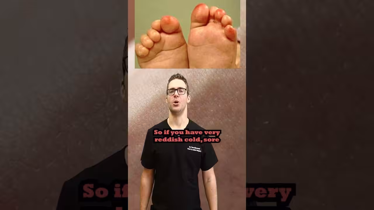
Symptoms of Raynaud’s in the Feet
Raynaud’s foot symptoms during episodes are highly distinctive — few conditions produce the same tricolor pattern. Between episodes, the feet may appear completely normal.
- Episodic color change in the toes — the classic white-to-blue-to-red sequence triggered by cold or emotional stress
- Numbness and tingling during the ischemic phase — when circulation is shut down, toes feel numb, heavy, or “dead”
- Pain and throbbing during the rewarming phase — as blood rushes back in, patients often experience intense pain or burning
- Cold toes disproportionate to ambient temperature — even in mild temperatures, Raynaud’s toes feel dramatically colder than the rest of the body
- Asymmetric involvement — some toes are affected more severely than others; the pattern can vary between attacks
- Skin changes in severe secondary Raynaud’s — chronic ischemia leads to skin thinning, nail changes, and in the most severe cases, toe ulcers or digital gangrene
Key takeaway: The color-change sequence (white → blue → red) triggered by cold or stress is pathognomonic of Raynaud’s. Persistent blue or black discoloration that doesn’t resolve with rewarming suggests a more severe vascular problem requiring urgent evaluation.
Causes and Triggers of Raynaud’s Foot Episodes
Raynaud’s attacks are triggered by vasospasm in response to identifiable stimuli. Understanding your specific triggers is one of the most effective management tools available.
- Cold exposure — the most universal trigger; even minor temperature drops can provoke attacks in susceptible individuals
- Emotional stress — the sympathetic nervous system activation during stress causes the same adrenergic vasoconstriction as cold
- Vibration — power tools, machinery, and even prolonged driving can trigger attacks (particularly in occupational Raynaud’s)
- Caffeine and nicotine — both are vasoconstrictors that lower the threshold for Raynaud’s attacks
- Certain medications — beta-blockers, migraine medications (triptans, ergotamines), decongestants, and some chemotherapy agents can trigger or worsen Raynaud’s
- Underlying autoimmune disease — in secondary Raynaud’s, systemic inflammation directly affects vascular reactivity
Diagnosis and Evaluation
Diagnosing Raynaud’s phenomenon is primarily clinical — the history of episodic tricolor change triggered by cold or stress is usually sufficient to confirm the diagnosis. The more important diagnostic question is whether it’s primary or secondary Raynaud’s, because that determines workup and management intensity.
History and examination: We ask about attack frequency, duration, severity, asymmetry, and triggers. We examine the toes for skin changes suggesting chronic ischemia (atrophy, ulcers, nail dystrophy). We assess the vascular examination — capillary refill time, pulses, ankle-brachial index — to evaluate for coexisting peripheral arterial disease.
Laboratory workup: When secondary Raynaud’s is suspected, we order an ANA (antinuclear antibody) panel, anti-Scl-70, anti-centromere antibodies, anti-dsDNA, ESR, CRP, and CBC. Positive autoimmune serology prompts rheumatology co-management.
Vascular studies: Doppler ultrasound and toe pressure measurements assess the severity of vascular compromise. In severe secondary Raynaud’s with digit-threatening ischemia, angiography may be required before interventional procedures.
Differential diagnosis: Peripheral arterial disease, acrocyanosis (persistent blue discoloration without pallor or rubor, and without pain), pernio/chilblains (inflammatory response to damp cold, producing persistent red-purple skin lesions), blue toe syndrome (cholesterol emboli or thromboembolism), and frostbite.
Treatment of Raynaud’s in the Feet
Treatment is stratified by severity and the primary-vs-secondary distinction. For primary Raynaud’s, the focus is lifestyle measures and trigger avoidance. For secondary Raynaud’s, pharmacological treatment is typically required alongside podiatric wound care.
Lifestyle and Conservative Measures
Cold protection: The cornerstone of primary Raynaud’s management. Thermal socks, insulated footwear, foot warmers, and keeping the entire body warm (vasospasm responds to core temperature, not just local warming) are essential:
Smoking cessation: Nicotine is a powerful vasoconstrictor — cessation consistently reduces Raynaud’s attack frequency and severity. This is non-negotiable in any Raynaud’s patient who smokes.
Caffeine reduction: Minimizing coffee, tea, and energy drinks can meaningfully reduce attack frequency in susceptible patients.
Stress management: Since emotional stress triggers attacks through the same sympathetic pathway as cold, stress reduction techniques (biofeedback, mindfulness, regular aerobic exercise) have documented benefit for Raynaud’s symptom frequency.
Pharmacological Treatment
Calcium channel blockers (nifedipine, amlodipine): The most evidence-based pharmacological treatment for Raynaud’s. These vasodilators reduce attack frequency and severity in both primary and secondary Raynaud’s. A 2023 Cochrane review confirmed nifedipine reduces Raynaud’s attack frequency by approximately 33%.
Phosphodiesterase-5 inhibitors (sildenafil, tadalafil): Used for moderate-to-severe secondary Raynaud’s, particularly in scleroderma patients. Produce sustained vasodilation and have a strong evidence base for reducing attack severity.
Topical nitroglycerin: Applied locally to affected digits to provide direct vasodilation. Useful for acute attacks and as an adjunct to oral therapy.
Prostacyclin infusions (iloprost): For severe secondary Raynaud’s with digital ulcers or critical ischemia — administered intravenously in a clinical setting and provides potent vasodilation lasting weeks to months.
Podiatric Wound Care
In severe secondary Raynaud’s, toe ulcers and digital ischemia require active wound care:
Specialized offloading footwear, debridement of necrotic tissue, biofilm management, and close monitoring for infection are essential. Digital sympathectomy (surgical interruption of the sympathetic nerves supplying the digital arteries) is a last-resort option for refractory digit-threatening ischemia that has failed all medical therapy.
⚠️ When Raynaud’s requires urgent evaluation:
- Toe color that doesn’t return to normal within 30–60 minutes of rewarming
- Black or dark purple discoloration that persists (possible digital infarction)
- Open sores, ulcers, or breaks in the skin over the toes or feet
- Severe, unrelenting pain in the toes at rest
- Raynaud’s symptoms that are rapidly worsening or newly severe
- New Raynaud’s symptoms in a patient known to have autoimmune disease
- Any toe ischemia in a diabetic patient
Frequently Asked Questions About Raynaud’s in the Feet
Is Raynaud’s in the feet dangerous?
Primary Raynaud’s — the idiopathic form in otherwise healthy individuals — is generally not dangerous. It’s uncomfortable and limiting but doesn’t cause tissue damage in the vast majority of cases. Secondary Raynaud’s — associated with scleroderma, lupus, or other systemic diseases — can be dangerous, causing digital ulcers, tissue death (gangrene), and in extreme cases, amputation. Distinguishing primary from secondary Raynaud’s is therefore essential for risk stratification and appropriate management intensity.
Can Raynaud’s affect only the feet and not the hands?
Yes — while most Raynaud’s patients experience symptoms in both hands and feet, isolated foot involvement does occur. Foot-predominant Raynaud’s is more common in patients with lower limb circulation issues and in those with occupational exposures affecting the lower extremities. The diagnosis and management principles are identical regardless of whether hands, feet, or both are affected.
Does Raynaud’s get worse with age?
Primary Raynaud’s often stabilizes or improves with age in women — many postmenopausal women report reduced attack frequency. However, secondary Raynaud’s course depends on the underlying disease. In progressive conditions like scleroderma, Raynaud’s severity typically tracks with disease progression and can worsen significantly over time. Regular follow-up is important to detect worsening early and adjust treatment accordingly.
The Bottom Line
Raynaud’s phenomenon in the feet is a recognizable, manageable vascular condition — but the appropriate management depends critically on whether it is primary or secondary. For most people, lifestyle changes and cold protection are sufficient. For those with underlying autoimmune disease or severe symptoms, pharmacological treatment and close podiatric monitoring can prevent serious complications including toe ulcers and tissue loss. If your toes are changing color with cold or stress, a proper evaluation — not just reassurance — is the right first step.
Sources
- Wigley FM, Flavahan NA. Raynaud’s phenomenon. N Engl J Med. 2016;375(6):556-565.
- Herrick AL. The pathogenesis, diagnosis and treatment of Raynaud phenomenon. Nat Rev Rheumatol. 2012;8(8):469-479.
- Ingegnoli F, Gualtierotti R. A systematic overview on the treatment options for Raynaud’s phenomenon. Drug Des Devel Ther. 2013;7:1331-1340.
- Hughes M, Herrick AL. Raynaud’s phenomenon. Best Pract Res Clin Rheumatol. 2016;30(1):112-132.
Cold, Color-Changing Toes? Get Expert Vascular Evaluation
Same-day appointments available in Howell & Bloomfield Hills, MI. Dr. Tom Biernacki provides comprehensive vascular foot evaluation and Raynaud’s management.
4.9★ | 1,123 Reviews | 3,000+ Surgeries
Or call: (810) 206-1402
Foot pain typically responds best to early podiatrist evaluation, conservative treatments such as supportive footwear and targeted physical therapy, and—when needed—custom orthotics or in-office procedures. Most patients see meaningful improvement within 4-6 weeks of starting a structured treatment plan. Schedule an evaluation at our Howell or Bloomfield Hills office for a clinical assessment.
What is Foot pain?
Foot pain is a common foot/ankle condition that affects mobility and quality of life. Understanding the underlying cause is the first step in successful treatment. Our podiatrists at Balance Foot & Ankle perform a hands-on biomechanical exam, review your activity history, and use diagnostic imaging when appropriate to identify the root cause—not just treat the symptom. Many patients have been told to “rest and ice” without a deeper diagnostic workup; our approach is different.
Symptoms and warning signs
Common signs of foot pain include pain that worsens with activity, morning stiffness, swelling, tenderness when palpated, and difficulty bearing weight. If you experience sudden severe pain, inability to walk, visible deformity, numbness or color change, contact our office the same day or visit urgent care—these can signal a more serious injury such as a fracture, tendon rupture, or vascular compromise. Diabetics with any foot wound should seek same-day care.
Conservative treatment options
Most cases of foot pain respond to non-surgical care: structured rest, supportive footwear changes, custom orthotics, targeted stretching and strengthening protocols, anti-inflammatory medications when medically appropriate, and in-office procedures such as ultrasound-guided injections. We also offer advanced therapies including MLS laser therapy, EPAT/shockwave, regenerative injections, and image-guided procedures. Treatment is sequenced from least invasive to most invasive, and we explain the rationale at every step.
When is surgery considered?
Surgery is reserved for cases that fail 3-6 months of well-structured conservative care, when there is structural pathology (severe deformity, complete tear, advanced arthritis), or when imaging shows damage that will not heal without intervention. Our surgeons have performed 3,000+ foot and ankle procedures and prioritize minimally-invasive techniques whenever appropriate. We discuss recovery timelines, return-to-activity milestones, and realistic outcome expectations before any procedure is scheduled.
Recovery timeline and prevention
Recovery from foot pain varies based on severity and chosen treatment path. Conservative cases often improve within 4-8 weeks with consistent adherence to the protocol. Post-procedural recovery may range from a few days (in-office procedures) to several months (reconstructive surgery). Long-term prevention involves footwear assessment, activity modification, structured strengthening, and regular check-ins with your podiatrist if you have a history of recurrence. We provide written home-exercise plans and digital follow-up support.
Ready to feel better?
Same-week appointments available in Howell and Bloomfield Hills, Michigan.
Book Your VisitNCBI: Raynaud’s Phenomenon — Foot Vasospasm & Management
In-Office Treatment at Balance Foot & Ankle
If home treatment isn’t providing relief for your raynauds phenomenon feet, our podiatry team at Balance Foot & Ankle can help with same-day evaluations and advanced in-office care.
Same-day appointments available. (810) 206-1402
Doctor Hoy’s Natural Pain Relief Gel
Natural topical pain relief I use in our clinic. Arnica + camphor formula — apply directly to the area 3–4x daily. ($20–25)
Shop Doctor Hoy’s →Dr. Tom Biernacki, DPM is a board-certified foot & ankle surgeon (ABFAS & ABPM) at Balance Foot & Ankle Specialists in Southeast Michigan. With over a decade of clinical experience, he specializes in heel pain, bunions, diabetic foot care, sports injuries, and minimally invasive surgery. Dr. Biernacki is a member of the APMA and ACFAS, and his patient education content on MichiganFootDoctors.com and YouTube has made him one of the most-followed foot & ankle educators on YouTube.