
Medically reviewed by Dr. Tom Biernacki, DPM
Not sure which one you have? A podiatrist can tell in one visit — book a real appointment time online, 24/7: Howell or Bloomfield Township. Takes about a minute, 24/7.
Board-certified podiatric surgeon | Balance Foot & Ankle, Howell & Bloomfield Township, MI
Last reviewed: May 2026

| Feature | Ankle Sprain (Ligament) | Ankle Fracture (Bone) |
|---|---|---|
| Mechanism | Inversion + plantarflexion (most common); eversion | High-energy inversion; rotational force; axial load; fall |
| Pain Location | Anterior and inferior to lateral malleolus (ATFL); below fibula | Directly at malleolus tip or posterior edge; malleolar tenderness |
| Weight Bearing | Often possible with significant pain (Grade I–II); difficult with Grade III | Often impossible or very painful with 4 steps → Ottawa criteria met |
| Swelling Pattern | Lateral forefoot / ankle diffuse; soft tissue edema | Localized malleolar; may have significant hematoma / visible deformity |
| X-ray Finding | Normal bone; soft tissue swelling only | Fracture line; fragment; periosteal disruption |
| Ottawa Ankle Rules | Negative — no malleolar tenderness or patient weight-bears 4 steps | Positive — malleolar tenderness OR unable to weight-bear 4 steps |
| Treatment | PRICE; early mobilization; PT; brace | Cast / boot; ORIF if unstable (Weber B–C) |
| Ottawa Ankle Rules — X-Ray Indicated If: | Location | Significance |
|---|---|---|
| Bony tenderness at posterior edge of lateral malleolus | Distal 6 cm fibula | Possible fibular fracture (Weber A, B, or C) |
| Bony tenderness at posterior edge of medial malleolus | Distal tibia medial | Possible medial malleolus fracture |
| Bony tenderness at navicular | Medial dorsal midfoot | Navicular fracture or stress fracture |
| Bony tenderness at 5th metatarsal base | Lateral midfoot | Zone 1 avulsion; Jones fracture (Zone 2–3) |
| Inability to weight-bear 4 steps | Any | High sensitivity for fracture requiring imaging |
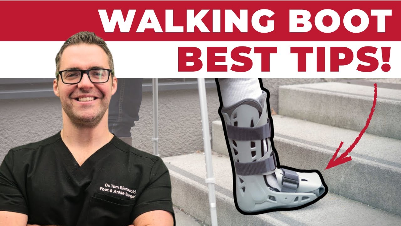
![Can You Walk on a Sprained Ankle? [Rolled Ankle Sprain Recovery Time]](https://www.michiganfootdoctors.com/wp-content/cache/flying-press/fad1e684e309d44cd86b7b5c8510c6a3.jpg)
Watch: Can You Walk on a Sprained Ankle? [Rolled Ankle Sprain Recovery Time] — MichiganFootDoctors YouTube
Medically reviewed by Dr. Tom Biernacki, DPM
Board-certified podiatric surgeon | Balance Foot & Ankle
Last reviewed: April 2026
You rolled your ankle, heard a crack (or maybe you didn’t), and now you’re staring at a rapidly swelling foot wondering: is this broken or just a bad sprain? This question comes up in our clinic constantly — and the honest answer is that you genuinely cannot tell just by looking at it or feeling it. Some of the worst fractures I’ve seen barely bruised. Some Grade 3 sprains that tore every ligament looked like disasters.
What I can do is walk you through exactly what distinguishes these injuries clinically, the evidence-based rules we use to decide who needs an X-ray, the specific fracture patterns you need to know about, and the very different treatment paths that follow. Getting this diagnosis right matters enormously for long-term ankle function.
The most important clinical decision with Sprained Ankle Vs Broken Ankle isn’t which treatment to start with — it’s identifying the correct subtype. That changes everything. Call (810) 206-1402.
The most important clinical decision with Sprained Ankle Vs Broken Ankle isn’t which treatment to start with — it’s identifying the correct subtype. That changes everything. Call (810) 206-1402.
Understanding the Anatomy
The ankle joint is formed by three bones: the tibia (inner lower leg), fibula (outer lower leg), and talus (the top bone of the foot). These are held together by multiple ligaments. When the ankle inverts (rolls outward, the most common mechanism), the lateral ligament complex is stressed: anterior talofibular ligament (ATFL), calcaneofibular ligament (CFL), and posterior talofibular ligament (PTFL). This is an ankle sprain.
When the force is greater than the ligaments can absorb, the force transfers to bone — and fractures occur. The most common fracture sites with ankle injury are: the distal fibula (lateral malleolus), the medial malleolus (inner ankle bump), the base of the 5th metatarsal (the bony bump on the outer mid-foot, often mistakenly called “the ankle”), and the navicular bone.
Key takeaway: An important anatomical fact: the base of the 5th metatarsal — the bony prominence on the outer side of your midfoot — is one of the most commonly fractured sites in ankle injuries. Many patients present believing they’ve sprained their ankle when they actually have an avulsion fracture or Jones fracture here.
The Ottawa Ankle Rules: The Clinical Standard
The Ottawa Ankle Rules are the most widely validated clinical decision tool for determining whether an ankle X-ray is needed after injury. Published in 1992 and validated in over 50 studies with a sensitivity approaching 99% for detecting fractures, these rules form the backbone of emergency and urgent care triage for ankle injuries.
X-ray is required if there is pain in the malleolar zone AND any of the following:
- Bone tenderness along the distal 6cm of the posterior edge of the fibula (outer ankle bone) or the tip of the lateral malleolus
- Bone tenderness along the distal 6cm of the posterior edge of the tibia or the tip of the medial malleolus (inner ankle)
- Inability to bear weight (take 4 steps) immediately after the injury AND in the emergency room
X-ray is required for the foot if there is pain in the midfoot zone AND any of the following:
- Bone tenderness at the base of the 5th metatarsal
- Bone tenderness at the navicular bone (top of the midfoot)
- Inability to bear weight immediately and in the emergency room
If NONE of these criteria are present, the probability of a clinically significant fracture is less than 2%, and X-rays can be safely deferred. This is why a skilled clinician can often avoid unnecessary radiation — but only by systematically applying these rules.
Warning: ⚠️ Go to urgent care or ER immediately if:
- Obvious deformity of the ankle or foot
- You cannot bear any weight at all
- Numbness or pins-and-needles in the foot
- The bone is pressing against the skin or the skin appears tented
- Severe pain that’s not improving with elevation and ice after 30 minutes
- Any crushing mechanism (car, heavy object) — high-energy fracture patterns
Symptoms: Sprain vs. Fracture Comparison
While overlap is substantial (both cause swelling, pain, and bruising), certain features shift the probability toward fracture or sprain. These are tendencies, not rules — any fracture can present mildly, and severe sprains can present dramatically.
More Likely a Sprain
- Pain concentrated over the soft tissue in front of and below the ankle bone (ligament territory), not over the bone itself
- Able to bear some weight immediately after injury, even if limping
- No specific point tenderness directly over bone
- Swelling develops over 12–24 hours rather than appearing within the first hour
- Previous similar injury to the same ankle (ligament laxity predisposes to sprains)
- Low-energy mechanism (simple rollover while walking or running)
More Likely a Fracture
- An audible or felt “crack” or “pop” at the time of injury (though this also occurs with ligament tears)
- Point tenderness directly over bone — palpating the fibula, tibia, 5th metatarsal base, or navicular produces exquisite localized pain
- Immediate inability to bear weight (within the first few minutes)
- Rapid swelling and bruising within the first 1–2 hours
- High-energy mechanism: jump landing, collision, significant fall
- Age > 55 (higher osteoporosis risk) or known bone density issues
- Tenderness specifically at the medial malleolus (inner ankle bone)
Common Ankle Fracture Types
Lateral Malleolus (Fibula) Fracture
The most common ankle fracture. A low fibula fracture at or below the ankle joint level (Weber A) is typically stable and treated with a walking boot for 4–6 weeks. Weber B fractures (at the joint level) and Weber C fractures (above the joint) involve more complex stability assessment and may require surgical fixation with plates and screws.
Bimalleolar and Trimalleolar Fractures
Fractures of both the lateral and medial malleolus (bimalleolar) or adding the posterior tibial rim (trimalleolar) represent unstable injuries that virtually always require surgical fixation to restore the ankle mortise alignment. These high-energy fractures cause significant swelling and require urgent orthopedic evaluation.
5th Metatarsal Fractures (Frequently Misdiagnosed as Ankle Sprain)
Two important fracture types occur at the 5th metatarsal after ankle inversion:
- Avulsion fracture (Dancer’s fracture): A piece of bone is pulled off by the peroneus brevis tendon during the inversion force. This is the most common 5th metatarsal fracture and heals well conservatively with a walking boot over 4–6 weeks.
- Jones fracture: A fracture at the junction of the metaphysis and diaphysis of the 5th metatarsal (about 1.5cm from the tip of the base). This zone has notoriously poor blood supply and high non-union rates. Jones fractures in athletes are often treated surgically (intramedullary screw) for faster, more reliable healing and quicker return to sport.
Treatment: Sprain vs. Fracture
Ankle Sprain Treatment
- Grade 1 (stretch, no tear): RICE protocol for 48–72 hours, functional rehabilitation starting within 1 week, return to sport in 1–2 weeks
- Grade 2 (partial tear): Protected weight bearing (lace-up brace or walking boot) for 1–2 weeks, physical therapy for proprioception and strength, return to sport in 3–6 weeks
- Grade 3 (complete tear): Boot for 2–4 weeks, comprehensive physical therapy for 6–12 weeks, possible surgical reconstruction for chronic instability not responding to rehab. Return to sport 6–12 weeks with functional bracing
Ankle Fracture Treatment
- Stable, non-displaced fractures (Weber A, isolated avulsion): Walking boot, 4–6 weeks non-weight bearing or protected weight bearing, followed by physical therapy
- Displaced or unstable fractures: Open reduction and internal fixation (ORIF) with plates and screws, 6–12 weeks protected weight bearing post-surgery, physical therapy for 3–6 months, return to full activity at 6–12 months
- Jones fracture in athletes: Often screw fixation for reliable healing and faster return to sport (8–12 weeks vs. 12–20 weeks conservative)
Frequently Asked Questions
Can you walk on a broken ankle?
Some ankle fractures — particularly stable, non-displaced fibula fractures below the joint line (Weber A) — allow limited weight bearing with a boot. Others, especially displaced fractures or those involving the ankle mortise, cannot be walked on safely. The ability to bear some weight does not rule out a fracture. This is why the Ottawa Ankle Rules specifically assess whether you can take 4 steps, not whether walking is completely comfortable.
How do I know if my ankle is fractured without an X-ray?
You cannot definitively rule out a fracture without imaging. However, the Ottawa Ankle Rules help estimate fracture probability: if you can bear weight (take 4 steps), and have no specific point tenderness directly over the fibula, tibia, 5th metatarsal base, or navicular, the probability of a significant fracture is below 2%. If any of these criteria are positive, you need an X-ray. When in doubt, imaging is always the right call.
How long does a sprained ankle take to heal vs. a broken ankle?
Grade 1 sprains typically heal in 1–2 weeks. Grade 2 in 3–6 weeks. Grade 3 in 6–12 weeks. Simple, stable ankle fractures in a boot heal in 6–8 weeks but require physical therapy for another 4–8 weeks. Complex fractures requiring surgery may take 6–12 months to full recovery. Both conditions have similar recovery windows for moderate injuries, which is another reason clinical examination alone can’t distinguish them.
What does a hairline fracture of the ankle feel like?
A stress fracture (hairline fracture) of the ankle or foot typically produces a dull aching pain that’s worse with activity and better with rest — different from the acute pain of a traumatic fracture. There is usually exquisite point tenderness directly over the fracture site. Stress fractures often don’t appear on plain X-rays for 2–4 weeks; MRI is the gold standard for early detection. Common locations in the foot include the metatarsals, navicular, and calcaneus.
Should I go to the ER or urgent care for an ankle injury?
Go to the ER if: there is obvious deformity, severe swelling preventing any weight bearing, the bone is near the skin surface, or you have numbness. Urgent care is appropriate for: suspected sprains, moderate pain with some ability to bear weight, no deformity, and stable vital signs. Our clinic also evaluates acute ankle injuries and can perform weight-bearing X-rays the same day. For athletes, early evaluation with a podiatrist or orthopedic sports medicine provider ensures the best management and fastest return to sport.
Sources
- Stiell IG, et al. Implementation of the Ottawa Ankle Rules. JAMA. 1994;271(11):827–832.
- van den Bekerom MP, et al. What is the evidence for rest, ice, compression, and elevation therapy in the treatment of ankle sprains in adults? J Athl Train. 2012;47(4):435–443.
- Clanton TO, McGarvey W. Athletic injuries to the soft tissues of the foot and ankle. In: Coughlin MJ, Mann RA, Saltzman CL, eds. Surgery of the Foot and Ankle. 8th ed. 2007.
- Mologne TS, et al. Early operative versus conservative management of fibular avulsion fractures. Am J Sports Med. 1997;25(3):372–375.
Ready to Get Relief?
Same-day appointments available in Howell & Bloomfield Township, MI
4.9★ | 1,123 Reviews | 3,000+ Surgeries
Or call: (810) 206-1402
When Shoes Aren’t Enough — Dr. Tom’s Top 9 Orthotics
About 30% of patients I see for foot pain need MORE than a great shoe — they need a structured insole. Below: my complete 2026 orthotic ranking with pros, cons, and the specific patient I’d give each one to.
Best All-Purpose Orthotic for Most Patients
Semi-rigid arch shell + dual-layer cushion + deep heel cup. The orthotic I’ve fitted to more patients than any other for 15 years. APMA-accepted. Trim-to-fit design works in athletic shoes, casual shoes, and most work boots.
✓ Pros
- Semi-rigid arch shell provides true biomechanical correction
- Deep heel cup centers the heel and reduces lateral instability
- Dual-layer cushion (top + bottom) lasts 9-12 months daily wear
- Available in 8 sizes for precise fit
- APMA-accepted and clinically validated
- APMA-accepted with superior cushioning versus rigid alternatives
✗ Cons
- Too thick for most dress shoes (use ProTech Slim instead)
- Some break-in period required (3-7 days for arch tolerance)
- Not enough correction for severe pes planus or rigid pes cavus
Dr. Tom’s Recommendation: If a patient has run-of-the-mill plantar fasciitis, mild flat feet, or arch fatigue, this is the first orthotic I try. Better value than most premium alternatives for 90% of patients, which is why it’s the first orthotic I reach for in the clinic. Sub-$50 typically.
Maximum Motion Control · Flat Feet & Severe Over-Pronation
PowerStep’s most aggressive stability orthotic. Adds a 2°-7° medial heel post on top of the standard PowerStep platform — designed specifically for flat-footed patients and severe pronators who need real corrective force.
✓ Pros
- 2°-7° medial heel post adds aggressive pronation control
- Same trusted PowerStep arch shell, more correction
- Built specifically for flat-foot biomechanics
- Excellent for posterior tibial tendon dysfunction (PTTD)
- Removable top cover for cleaning
✗ Cons
- Too aggressive for neutral-arch patients
- Needs longer break-in (10-14 days) due to stronger correction
- Adds 2-3 mm of stack height — won’t fit slim dress shoes
Dr. Tom’s Recommendation: When a patient comes in with significant flat feet AND symptoms (heel pain, arch pain, knee pain), the Original PowerStep isn’t aggressive enough. The Maxx is what gets prescribed. About 25% of my flat-footed patients end up here.
Low-Profile · Fits Dress Shoes & Narrow Casuals
3 mm slim profile with podiatrist-designed tri-planar arch technology. Engineered specifically to fit inside dress shoes, oxfords, loafers, and women’s flats without crowding the toe box. Vionic was founded by an Australian podiatrist.
✓ Pros
- 3 mm slim profile (vs 7-10 mm for standard orthotics)
- Tri-planar arch technology adds support without bulk
- Built-in deep heel cup despite slim design
- Fits dress shoes WITHOUT having to remove the factory insole
- Trim-to-fit · APMA-accepted
✗ Cons
- Less arch support than full-volume orthotics
- Top cover wears faster than thicker alternatives
- Not enough correction for severe foot deformities
Dr. Tom’s Recommendation: My default when a patient says ‘I need orthotics but I have to wear dress shoes for work.’ Slim enough to fit in oxfords and pumps without the heel sliding out. The single highest-impact change you can make for office workers with foot pain.
Built-In Metatarsal Pad · Morton’s Neuroma · Ball-of-Foot Pain
Standard Pinnacle orthotic with a built-in metatarsal pad positioned proximal to the metatarsal heads — the exact location that offloads neuromas and metatarsalgia. No need for separate met pads or pad placement guesswork.
✓ Pros
- Built-in met pad eliminates DIY pad placement errors
- Specifically designed for Morton’s neuroma + metatarsalgia
- Same trusted PowerStep arch + heel cup platform
- Top cover protects sensitive forefoot skin
- Faster relief than orthotics + add-on met pads
✗ Cons
- Met pad position is fixed (can’t fine-tune individual placement)
- Some patients with very small or very large feet need custom
- Slightly thicker than the standard Pinnacle
Dr. Tom’s Recommendation: If a patient has Morton’s neuroma, sesamoiditis, or generalized ball-of-foot pain (metatarsalgia), this saves a clinic visit and a prescription. The built-in pad placement is anatomically correct for 80% of feet. Way better than DIY met pads.
Adaptive Dynamic Arch · Athletic & Daily Wear
Currex’s flagship adaptive arch technology — the orthotic flexes with your gait instead of fighting it. Different stiffness zones along the length give you targeted support at the heel, midfoot, and forefoot. Available in three arch heights (low/medium/high).
✓ Pros
- Dynamic flex zones adapt to natural gait cycle
- Three arch heights ensure precise fit
- Lighter than rigid orthotics (no ‘heavy foot’ feel)
- Excellent for runners and athletic walkers
- European podiatric design (German engineering)
✗ Cons
- More expensive than PowerStep Original ($55-65 typically)
- Less aggressive correction than Pinnacle Maxx for severe cases
- Three arch heights means you must self-select correctly
Dr. Tom’s Recommendation: I started recommending Currex three years ago for runners who said PowerStep felt ‘too rigid.’ The dynamic flex zones respect natural gait. Best for active patients who walk 8K+ steps daily and don’t need maximum motion control.
Running-Specific · Heel Strike + Forefoot Strike Compatible
Currex’s purpose-built running orthotic. The midfoot flex zone is positioned for runner’s gait mechanics, with a flared heel cushion for heel strikers and a forefoot rocker for midfoot/forefoot strikers. Tested on 1000+ runners during product development.
✓ Pros
- Designed by German biomechanics lab specifically for runners
- Dynamic arch flexes with running gait (not static like PowerStep)
- Three arch heights (low/medium/high)
- Reduces overuse injury risk in mid-distance runners
- Lightweight (no impact on cadence)
✗ Cons
- Premium price ($60-75)
- Not aggressive enough for severe over-pronators (use Pinnacle Maxx)
- Runner-specific design = less ideal for daily walking shoes
Dr. Tom’s Recommendation: If a patient runs 20+ miles per week and has plantar fasciitis or shin splints, this is the orthotic I prescribe. The dynamic flex zones respect running biomechanics in a way that no rigid PowerStep can match. Pricier but worth it for serious runners.
Cavus Foot & High-Arch Patients
Polyurethane base with a deeper heel cup and higher arch profile than PowerStep — built for cavus (high-arched) feet that need maximum cushion and support. The 5-zone cushioning system addresses the unique pressure points of high-arch feet.
✓ Pros
- Deeper heel cup centers the heel for cavus foot stability
- Higher arch profile fills the void under high arches
- 5-zone cushioning addresses cavus foot pressure points
- Polyurethane base lasts 12+ months
- Available in Wide width
✗ Cons
- Too tall/aggressive for normal or low arches
- Won’t fit slim dress shoes
- Pricier than PowerStep Original
- Some patients find the arch height uncomfortable initially
Dr. Tom’s Recommendation: Cavus foot patients are often misdiagnosed and given low-arch orthotics — that makes everything worse. Spenco’s Total Support has the arch profile that high-arch feet actually need. About 15% of my patients have cavus feet; this is what they wear.
Cushion Layer · Standing All Day · Gel Pressure Relief
NOT a true biomechanical orthotic — this is a cushion insole. But for patients who want gel pressure relief instead of arch correction (or to add ON TOP of factory insoles in work boots), this is the best gel option on Amazon.
✓ Pros
- Genuine gel cushioning (not foam pretending to be gel)
- Targeted gel waves under heel and ball of foot
- Trim-to-fit · works in most shoe types
- Sub-$15 price (most affordable option in this list)
- Massaging texture is genuinely soothing
✗ Cons
- ZERO arch support — this is cushion only
- Won’t fix plantar fasciitis or flat-foot issues
- Compresses faster than PowerStep (4-6 months)
- Top cover wears through in high-mileage applications
Dr. Tom’s Recommendation: I recommend these to patients who tell me ‘I just want my feet to stop hurting at the end of my shift’ and who don’t have a biomechanical issue. Construction workers, factory workers, retail. Pure cushion does the job for them.
Tight-Fitting Shoes · Cycling Shoes · Hockey Skates
Tread Labs Pace insole with firm orthotic arch support for flat feet and plantar fasciitis relief. The replaceable top cover design makes it one of the most durable picks in this guide — backed by a million-mile guarantee and recommended for tight-fitting athletic footwear.
✓ Pros
- Firm orthotic arch support shell (podiatrist-grade)
- Slim profile fits tight athletic footwear
- Lasts 12+ months daily wear
- Excellent for cycling shoes specifically
- Built-in odor-control treatment
✗ Cons
- Premium price ($45-55)
- Less cushion than PowerStep equivalents
- Not as aggressive correction as Pinnacle Maxx for flat feet
- The signature ‘heel cup feel’ takes 1-2 weeks to adapt to
Dr. Tom’s Recommendation: If you’re a cyclist with foot numbness, hot spots, or knee pain — this is the orthotic. The stabilizer cap solves cycling-specific biomechanical issues that no other orthotic addresses. Worth the premium for athletes.
None of these solving your foot pain?
Some patients (about 30%) need custom-molded prescription orthotics. We make 3D-scanned custom orthotics in our Howell and Bloomfield Township offices — specifically built for your foot mechanics.
Schedule a Custom Orthotic Fitting →FSA/HSA eligible · Most insurance accepted · (810) 206-1402
What is Ankle sprain?
Ankle sprain is a common foot/ankle condition that affects mobility and quality of life. Understanding the underlying cause is the first step in successful treatment. Our podiatrists at Balance Foot & Ankle perform a hands-on biomechanical exam, review your activity history, and use diagnostic imaging when appropriate to identify the root cause—not just treat the symptom. Many patients have been told to “rest and ice” without a deeper diagnostic workup; our approach is different.
Symptoms and warning signs
Common signs of ankle sprain include pain that worsens with activity, morning stiffness, swelling, tenderness when palpated, and difficulty bearing weight. If you experience sudden severe pain, inability to walk, visible deformity, numbness or color change, contact our office the same day or visit urgent care—these can signal a more serious injury such as a fracture, tendon rupture, or vascular compromise. Diabetics with any foot wound should seek same-day care.
Conservative treatment options
Most cases of ankle sprain respond to non-surgical care: structured rest, supportive footwear changes, custom orthotics, targeted stretching and strengthening protocols, anti-inflammatory medications when medically appropriate, and in-office procedures such as ultrasound-guided injections. We also offer advanced therapies including MLS laser therapy, EPAT/shockwave, regenerative injections, and image-guided procedures. Treatment is sequenced from least invasive to most invasive, and we explain the rationale at every step.
When is surgery considered?
Surgery is reserved for cases that fail 3-6 months of well-structured conservative care, when there is structural pathology (severe deformity, complete tear, advanced arthritis), or when imaging shows damage that will not heal without intervention. Our surgeons have performed 3,000+ foot and ankle procedures and prioritize minimally-invasive techniques whenever appropriate. We discuss recovery timelines, return-to-activity milestones, and realistic outcome expectations before any procedure is scheduled.
Recovery timeline and prevention
Recovery from ankle sprain varies based on severity and chosen treatment path. Conservative cases often improve within 4-8 weeks with consistent adherence to the protocol. Post-procedural recovery may range from a few days (in-office procedures) to several months (reconstructive surgery). Long-term prevention involves footwear assessment, activity modification, structured strengthening, and regular check-ins with your podiatrist if you have a history of recurrence. We provide written home-exercise plans and digital follow-up support.
Ready to feel better?
Same-week appointments available in Howell and Bloomfield Township, Michigan.
Book Your VisitIn-Office Treatment at Balance Foot & Ankle
If home treatment isn’t providing relief for your sprained ankle vs broken ankle, our podiatry team at Balance Foot & Ankle can help with same-day evaluations and advanced in-office care.
American Academy of Orthopaedic Surgeons: Sprained vs Broken Ankle
Ready to Get Relief?
Same-day appointments available in Howell & Bloomfield Township, MI
4.9★ | 1,123 Reviews | 3,000+ Surgeries
Or call: (810) 206-1402

Dr. Tom Biernacki, DPM is a board-certified foot & ankle surgeon (ABFAS & ABPM) at Balance Foot & Ankle Specialists in Southeast Michigan. With over a decade of clinical experience, he specializes in heel pain, bunions, diabetic foot care, sports injuries, and minimally invasive surgery. Dr. Biernacki is a member of the APMA and ACFAS, and his patient education content on MichiganFootDoctors.com and YouTube has made him one of the most-followed foot & ankle educators on YouTube.
More questions patients ask
What is the difference between a sprained and broken ankle?
A sprained ankle involves injury to the ligaments — the connective tissue bands connecting bones. A broken ankle (fracture) involves a crack or break in one or more bones. Both can cause immediate pain, swelling, and difficulty walking, which is why physical examination alone cannot reliably distinguish them. X-rays are required to rule out fracture whenever there is bony tenderness, inability to bear weight, or significant mechanism of injury.
What are the signs that point toward a fracture rather than a sprain?
Signs favoring fracture include point tenderness directly over a bone (fibula, tibia, base of fifth metatarsal, or navicular), inability to take four steps at the time of injury and at evaluation (Ottawa Ankle Rules), bone-deep pain rather than soft-tissue pain, an audible crack rather than a pop at moment of injury, rapid severe swelling within minutes, and significant deformity or abnormal angulation at the ankle.
Can a broken ankle be treated like a sprain?
No — treating a fracture like a sprain can result in malunion (bones healing in incorrect position), chronic pain, ankle arthritis, and instability. Some fractures that appear stable on initial X-ray can displace with walking. Conversely, many patients with severe lateral ankle sprains are more painful than those with minor fractures. Accurate diagnosis with X-rays directs appropriate treatment — immobilization, non-weight-bearing, or surgical fixation when needed.
How long does it take to recover from a broken versus sprained ankle?
Grade I ankle sprains typically recover in 1–2 weeks; Grade II in 3–6 weeks; Grade III in 6–12 weeks. Non-displaced fibular fractures in a cast or boot require 4–8 weeks of protected weight-bearing. Bimalleolar or trimalleolar fractures requiring surgery take 3–4 months non-weight-bearing plus 6+ months of rehabilitation. A high ankle (syndesmotic) fracture-dislocation can take 6–12 months to return to full sport. See a podiatrist for any significant ankle injury.
Still have a question about coverage or cost? Book online and we will check your benefits before your visit: Book in Howell · Book in Bloomfield Township. Prefer to talk it through first? Call (810) 206-1402.