
Quick answer: Stress Fracture In Foot affects roughly 1 in 4 adults in our practice. Effective treatment starts with a targeted diagnosis, conservative-first treatment, and escalation only when needed. We treat this regularly at our Howell and Bloomfield Hills practices. Call (810) 206-1402.
✅ Medically reviewed by Dr. Tom Biernacki, DPM — Board-Certified Podiatrist · Last updated April 6, 2026
Last Updated: April 2026 | Reading Time: 11 min
This article is for informational purposes only and does not replace professional medical advice. Schedule an appointment for personalized care.
In This Article
- What Is a Stress Fracture?
- Common Foot Stress Fracture Locations
- Symptoms: How to Tell If You Have One
- Causes and Risk Factors
- How a Podiatrist Diagnoses a Stress Fracture
- Treatment: What Actually Heals a Stress Fracture
- Recovery Timeline: Week by Week
- Recommended Products for Recovery
- How to Prevent Stress Fractures
- FAQ
That nagging ache on top of your foot that appeared a week into your new running program? The one that fades when you sit down but returns the moment you push off again? There’s a good chance it’s a stress fracture — and ignoring it is one of the most common mistakes we see in our office. A stress fracture won’t show up on initial X-rays, doesn’t cause dramatic swelling, and can feel like “just a bruise.” That’s exactly why so many people walk on one for weeks before seeking help, turning a hairline crack into a complete fracture that requires far more aggressive treatment.
The most important clinical decision with Stress Fracture In Foot isn’t which treatment to start with — it’s identifying the correct subtype. That changes everything. Call (810) 206-1402.
What Is a Stress Fracture?
A stress fracture is a microscopic crack in bone caused by repetitive, submaximal force — meaning the force itself isn’t enough to break the bone in one blow, but over hundreds or thousands of cycles, the bone’s repair mechanism can’t keep up with the damage being inflicted. Think of bending a paperclip back and forth: no single bend breaks it, but eventually it snaps. That’s essentially what happens in bone when load exceeds the body’s ability to remodel.
Stress fractures are fundamentally different from acute fractures (which result from a single traumatic event like a fall or a dropped object). With a stress fracture, there’s usually no single moment of injury — instead, pain develops gradually over days to weeks and progressively worsens with continued activity. This gradual onset is the defining clinical feature and the reason many people delay seeking care.
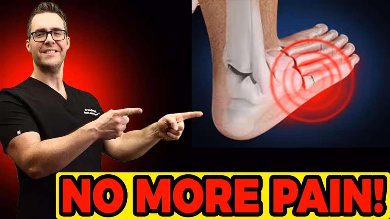
The foot is the most common location for stress fractures in the body because it absorbs the full force of every step. During running, each foot strike generates forces of 2–3 times body weight. Over a 5-mile run, each foot absorbs impact roughly 4,000 times. When bone remodeling can’t keep pace with that repetitive load — due to training errors, poor footwear, nutritional deficiencies, or underlying bone weakness — the result is a stress fracture.
Common Foot Stress Fracture Locations
Not all foot stress fractures are created equal. The location determines the severity, healing time, and treatment approach. Here’s a comparison of the most common sites:
| Location | Frequency | Risk Level | Typical Healing | Key Feature |
|---|---|---|---|---|
| 2nd/3rd Metatarsal | Most common (~50%) | Low | 6–8 weeks | Pain on top of midfoot; swelling over metatarsal shaft |
| 5th Metatarsal (Jones) | ~20% | High | 8–12 weeks (may need surgery) | Pain on outside of foot; poor blood supply = slow healing |
| Navicular | ~15% | Very High | 8–12 weeks non-weight-bearing | Vague pain on top of midfoot; often missed on X-ray |
| Calcaneus (Heel) | ~10% | Low | 6–8 weeks | Deep heel pain; often confused with plantar fasciitis |
| Sesamoids | ~5% | High | 8–12+ weeks | Pain under big toe joint; poor blood supply complicates healing |
The distinction between “low-risk” and “high-risk” stress fractures matters enormously. Low-risk fractures (2nd/3rd metatarsal, calcaneus) occur in areas with good blood supply and typically heal predictably with activity modification. High-risk fractures (5th metatarsal Jones fracture, navicular, sesamoids) occur in “watershed” areas with poor blood supply, are more likely to progress to complete fractures or nonunion, and frequently require more aggressive treatment including strict non-weight-bearing or surgery.
Symptoms: How to Tell If You Have a Stress Fracture
The classic stress fracture presentation follows a predictable pattern that distinguishes it from other foot injuries:
Activity-related pain with a crescendo pattern. The pain starts as mild discomfort during or after activity — easy to dismiss. Over the next 1–3 weeks, it appears earlier during activity, lasts longer afterward, and becomes more intense. Eventually, it occurs during normal walking and sometimes at rest. This progressive worsening over days to weeks is the signature symptom.
Pinpoint tenderness. Unlike soft tissue injuries that produce diffuse soreness, a stress fracture creates detailed tenderness at one specific spot on the bone. You can often place one finger directly on the painful point. Pressing on that spot reproduces the pain precisely.
Swelling. Mild to moderate swelling on top of the foot, directly over the affected bone. The swelling is usually more subtle than an acute fracture or sprain — you might notice a slight puffiness or that one shoe feels tighter than the other.
Pain that improves with rest. A stress fracture typically feels significantly better after sitting or sleeping and returns with weight-bearing activity. This rest-responsive pattern is a key differentiator. If your foot pain is constant regardless of activity, other diagnoses should be considered.
The “hop test.” A simple self-assessment: hop on the affected foot. If this produces sharp, localized pain at the suspected site, a stress fracture is likely. If hopping is painless, a stress fracture is less likely (though not ruled out).
Causes and Risk Factors
Stress fractures result from an imbalance between bone breakdown and bone repair. Several factors tip this balance toward injury:
Training errors (the #1 cause). Increasing mileage, intensity, or frequency too quickly — the classic “too much, too soon” mistake. The general guideline is the 10% rule: don’t increase weekly running volume by more than 10% per week. Runners who jump from 15 to 30 miles per week, or weekend warriors who go from sedentary to daily high-impact exercise, are prime candidates for stress fractures.
Poor footwear. Worn-out shoes lose their shock absorption capacity. Running shoes should generally be replaced every 300–500 miles. Flat, unsupportive shoes (ballet flats, flip-flops, minimalist shoes) provide inadequate force distribution across the foot bones, concentrating stress on the metatarsals.
Bone density issues. Osteopenia or osteoporosis weakens bone and lowers the threshold for stress fracture development. This is particularly relevant for postmenopausal women, women with the female athlete triad (disordered eating, menstrual irregularity, low bone density), and anyone on long-term corticosteroids.
Nutritional deficiencies. Inadequate calcium (below 1,000–1,300mg/day) and vitamin D (below 600–800 IU/day) impair bone remodeling. Low caloric intake relative to energy expenditure (relative energy deficiency in sport, or RED-S) is a major risk factor, particularly in endurance athletes and dancers.
Foot mechanics. High arches concentrate force on the metatarsals and heel. Flat feet shift excessive load to the navicular. Overpronation creates asymmetric stress distribution. Leg length discrepancies create uneven loading between feet. These biomechanical factors explain why some people get stress fractures at relatively modest activity levels.
Surface and activity type. Hard surfaces (concrete, asphalt) transmit more impact force than softer surfaces (grass, trails, tracks). High-impact activities (running, basketball, dance) carry significantly higher stress fracture risk than low-impact activities (cycling, swimming).
How a Podiatrist Diagnoses a Stress Fracture
Clinical diagnosis starts with a thorough history — when did the pain start, how has it progressed, what activities trigger it, and what recent changes in training or footwear occurred. The physical exam focuses on pinpoint palpation along each bone, the tuning fork test (vibration placed on the suspected bone reproduces pain), and provocative maneuvers like single-leg hopping.
X-rays are the first imaging step, but here’s the critical point that many patients don’t know: initial X-rays are normal in up to 70% of stress fractures. Stress fractures don’t become visible on X-ray until the bone starts healing and a callus forms — typically 2–3 weeks after symptom onset. A normal X-ray does not rule out a stress fracture. If clinical suspicion is high and the X-ray is negative, we either treat empirically or order advanced imaging.
MRI is the gold standard for stress fracture diagnosis. It can detect bone marrow edema (the earliest sign of stress reaction) within days of symptom onset — long before X-ray changes appear. MRI also grades the severity: a stress reaction (bone edema without a fracture line) is milder than a stress fracture (edema plus a visible fracture line), and treatment intensity may differ accordingly.
Bone scan is highly sensitive but less specific than MRI — it will light up at the site of increased bone turnover but can’t differentiate a stress fracture from infection, tumor, or other pathology. MRI has largely replaced bone scan as the advanced imaging study of choice for stress fractures.
Treatment: What Actually Heals a Stress Fracture
Stress fracture treatment follows one principle: reduce the mechanical load on the bone below the threshold that caused the fracture, and maintain that reduced load long enough for the bone to heal completely. The specific approach depends on the fracture location and risk level.
Low-Risk Stress Fractures (2nd/3rd Metatarsal, Calcaneus)
Relative rest — not complete immobilization. You can walk, but impact activities (running, jumping, prolonged standing) must stop. A stiff-soled shoe or walking boot distributes force across the foot and offloads the fracture site. Most patients can continue daily activities with this modification.
Walking boot. A CAM (controlled ankle motion) boot is the standard treatment for most metatarsal stress fractures. The rigid sole prevents the metatarsals from bending during push-off — which is the motion that stresses them most. Typical boot use is 4–6 weeks, with gradual transition back to regular shoes.
Activity modification. Replace running with cycling, swimming, or pool running. Maintain cardiovascular fitness without impact loading. This is the hardest part for athletes, but continuing to run on a stress fracture is the fastest way to convert a hairline crack into a complete break requiring surgery.
High-Risk Stress Fractures (5th Metatarsal, Navicular, Sesamoid)
Strict non-weight-bearing with crutches, knee scooter, or walking boot with no weight-bearing is often necessary for navicular and 5th metatarsal Jones fractures. These bones have tenuous blood supply, and any continued loading can prevent healing or cause the fracture to propagate.
Surgical fixation may be recommended for displaced fractures, Jones fractures in athletes who need reliable and timely return to sport, navicular fractures that show no healing after 6–8 weeks of conservative care, or any stress fracture that progresses to a complete fracture. The most common procedure is intramedullary screw fixation — a single screw is placed across the fracture line to compress and stabilize it.
Adjunctive Treatments
Bone stimulator. Pulsed electromagnetic field (PEMF) devices applied to the fracture site for 20–30 minutes daily have been shown to accelerate healing in delayed-union stress fractures. Insurance often covers bone stimulators for fractures that haven’t healed after 90 days.
Nutritional optimization. Ensure adequate calcium (1,000–1,300mg/day), vitamin D (1,000–2,000 IU/day during healing), and overall caloric intake. A sports medicine dietitian can be invaluable for athletes with stress fractures linked to energy deficiency.
Custom orthotics. Once the fracture has healed, custom orthotics address the biomechanical factors that contributed to the stress fracture — offloading the metatarsal heads, supporting the arch, correcting overpronation. This is a critical step in preventing recurrence.
Recovery Timeline: Week by Week
| Timeframe | What’s Happening | What You Can Do |
|---|---|---|
| Weeks 1–2 | Inflammatory phase; bone callus begins forming | Walking boot or stiff shoe; ice 15 min after activity; zero impact exercise; swimming/cycling OK |
| Weeks 3–4 | Soft callus forming; pain at rest resolving | Continue boot; may begin walking more normally; pool running OK; no land impact |
| Weeks 5–6 | Hard callus forming; most tenderness resolved | Transition out of boot into supportive shoe with orthotic; begin walking program |
| Weeks 7–8 | Bone remodeling; fracture line fading on X-ray | Gradual return to impact: walk/jog intervals; increase by 10% per week |
| Weeks 9–12 | Full remodeling; bone strength returning to baseline | Progressive return to full activity; address underlying risk factors (footwear, training load, nutrition) |
Important: The timeline above applies to low-risk metatarsal and calcaneal stress fractures. High-risk fractures (navicular, Jones, sesamoid) take 8–16 weeks and may require a longer non-weight-bearing period before transitioning to activity. Your podiatrist will use repeat imaging to confirm healing before clearing you for impact.
Recommended Products for Recovery
These are products we frequently recommend to patients recovering from foot stress fractures. They support healing, prevent recurrence, and support return to activity.
⭐ OUR #1 PICK
PowerStep Pinnacle Orthotic Insoles
The best over-the-counter orthotic for stress fracture recovery. The semi-rigid arch support redistributes pressure away from the metatarsal heads and provides the biomechanical control needed to prevent recurrence. We recommend these as the first step before considering custom orthotics — they work well for the majority of patients and cost a fraction of the price.
Best for: Metatarsal stress fractures, arch support during return to activity, plantar pressure redistribution
Hoka Bondi 9
Maximum cushioning for the transition out of a walking boot back into regular shoes. The thick EVA midsole absorbs significantly more impact than standard athletic shoes, reducing the repetitive force transmitted to healing bone. The meta-rocker geometry smooths the gait cycle and reduces peak pressure at push-off — exactly where metatarsal stress fractures are most vulnerable during recovery.
Best for: Transitioning out of walking boot, return-to-activity phase, runners with history of stress fractures
New Balance 990v6
The best everyday walking shoe for long-term stress fracture prevention. The FuelCell midsole provides excellent energy return without sacrificing stability, and the wide toe box prevents lateral compression of the metatarsals. This is the shoe we recommend for daily wear after healing — structured enough to support the foot, cushioned enough to absorb impact, and durable enough to maintain its protective properties for 500+ miles.
Best for: Long-term prevention, daily walking shoe, patients with history of recurrent stress fractures
How to Prevent Stress Fractures
Follow the 10% rule. Never increase your weekly running mileage, workout frequency, or exercise intensity by more than 10% per week. This gives bone time to adapt to increasing loads through the normal remodeling cycle.
Replace your shoes on schedule. Running shoes lose meaningful shock absorption after 300–500 miles. If you run 20 miles per week, that means replacing shoes every 4–6 months. Track your shoe mileage and replace before they feel worn out — the cushioning degrades before the tread shows it.
Cross-train. Alternate high-impact days with low-impact activities (cycling, swimming, elliptical). This maintains cardiovascular fitness while giving bones recovery time between impact sessions.
Optimize nutrition. Ensure you’re consuming adequate calcium (1,000–1,300mg/day from food and supplements), vitamin D (check your blood level; most people in Michigan are deficient), and total calories relative to energy expenditure. Chronic caloric deficit is a major stress fracture risk factor, especially in female endurance athletes.
Use orthotics if indicated. If you have biomechanical risk factors (flat feet, high arches, leg length discrepancy), over-the-counter or custom orthotics redistribute plantar pressure and reduce focal stress on vulnerable bones. This is particularly important after recovering from a first stress fracture — the recurrence rate without biomechanical correction is 20–30%.
Listen to your body. Stress fractures always give warning signals — a new ache that appears during activity and resolves with rest. If you develop persistent, localized foot pain that worsens over several days, stop the aggravating activity and get evaluated. Catching a stress reaction before it becomes a complete stress fracture dramatically simplifies treatment.
⚠️ Warning Signs — See a Doctor Immediately
- Sudden sharp pain with an audible pop or snap (possible complete fracture)
- Inability to bear weight on the affected foot
- Visible bruising or deformity on top of the foot
- Significant swelling that develops rapidly over hours
- Pain that doesn’t improve after 1–2 weeks of rest
- Numbness or tingling in the toes beyond the fracture site
- History of multiple stress fractures — this may indicate underlying bone density issues requiring workup
More Podiatrist-Recommended Stress Fracture Essentials
Max-Cushion Walking Shoe
Hoka Bondi 9 — maximum shock absorption during stress fracture recovery.
Foam Roller for Recovery
TriggerPoint foam roller — maintains lower-leg mobility during return to activity.
Supportive Insole
PowerStep Pinnacle — distributes impact evenly across the foot.
As an Amazon Associate, Balance Foot & Ankle earns from qualifying purchases. Product recommendations are based on clinical experience; prices and availability shown above update live from Amazon.
When to See a Podiatrist
Most foot stress fractures heal in 6-8 weeks of protected weight-bearing — but rushing back to activity can turn a hairline fracture into a full break. Balance Foot & Ankle confirms stress fractures on X-ray or MRI and guides your return-to-running protocol. Don’t guess — we’ll tell you the exact week you can start jogging again.
Call Balance Foot & Ankle: (810) 206-1402 · Book online · Offices in Howell & Bloomfield Hills
Frequently Asked Questions
Can I walk on a stress fracture?
It depends on the location and severity. Most metatarsal stress fractures allow walking in a supportive boot or stiff-soled shoe — you don’t need crutches. However, every step on an unprotected stress fracture risks propagating the crack. Walking in regular shoes, especially flexible or flat ones, is not recommended. Navicular and Jones fractures may require strict non-weight-bearing with crutches. Your podiatrist will determine the appropriate level of activity restriction based on imaging findings and fracture location.
How long until I can run again after a stress fracture?
For low-risk metatarsal stress fractures, most runners can begin a walk/jog program at 6–8 weeks, with full running volume restored by 10–12 weeks. High-risk fractures (navicular, Jones) take longer — typically 12–16 weeks before impact activity resumes. The return must be gradual: start with walk/jog intervals (1 minute jog, 2 minutes walk) and increase the jog intervals by 10% per week. Pain during or after running means you’re progressing too fast. Your podiatrist will use repeat X-rays or MRI to confirm adequate healing before clearing you for impact.
Will a stress fracture show up on X-ray?
Not initially, in most cases. Up to 70% of stress fractures are invisible on initial X-rays because the crack is too small to see. X-rays typically become positive 2–6 weeks after symptoms begin, once a healing callus forms around the fracture. This is why a normal X-ray does not rule out a stress fracture. If your doctor suspects a stress fracture despite a normal X-ray, MRI is the next step — it can detect bone edema within days of injury, making it far more sensitive for early diagnosis.
What’s the difference between a stress fracture and a stress reaction?
They exist on the same spectrum. A stress reaction is the earlier, milder stage — bone edema and microdamage without a visible fracture line on MRI. A stress fracture is the more advanced stage — bone edema plus a discrete fracture line. Clinically, a stress reaction typically produces milder symptoms and heals faster (4–6 weeks), while a stress fracture requires a full 6–8+ week recovery. The treatment principles are the same (reduce load, protect the bone), but the timeline and activity restrictions may be less aggressive for a stress reaction.
The Bottom Line
Stress fractures are overuse injuries that develop gradually — they always give warning signals before they become complete breaks. The key is recognizing the pattern (pain that worsens with activity and improves with rest, progressively worsening over days to weeks) and acting early. Most foot stress fractures heal in 6–8 weeks with a walking boot and activity modification. Don’t ignore persistent foot pain and don’t rely on a normal X-ray to rule out a stress fracture — MRI is far more sensitive for early diagnosis. Once healed, address the underlying cause (training errors, footwear, biomechanics, nutrition) to prevent recurrence.
Sources
- Nattiv A, Kennedy G, Barrack MT, et al. “Correlation of MRI grading of bone stress injuries with clinical risk factors and return to play: a 5-year prospective study in collegiate track and field athletes.” Am J Sports Med. 2013;41(8):1930-1941.
- Behrens SB, Deren ME, Matson A, et al. “Stress fractures of the pelvis and legs in athletes: a review.” Sports Health. 2013;5(2):165-174.
- Torg JS, Balduini FC, Zelko RR, et al. “Fractures of the base of the fifth metatarsal distal to the tuberosity: classification and guidelines for non-surgical and surgical management.” J Bone Joint Surg Am. 1984;66(2):209-214.
- Saxena A, Fullem B, Hannaford D. “Results of treatment of 22 navicular stress fractures and a new proposed radiographic classification system.” J Foot Ankle Surg. 2000;39(2):96-103.
- Fredericson M, Jennings F, Beaulieu C, Matheson GO. “Stress fractures in athletes.” Top Magn Reson Imaging. 2006;17(5):309-325.
Suspect a Stress Fracture?
Don’t wait for a hairline crack to become a complete break. Our podiatrists can diagnose stress fractures early with clinical evaluation and advanced imaging, and create a recovery plan that gets you back to activity safely.
Balance Foot & Ankle — Howell & Bloomfield Hills | (810) 206-1402
Suspect a Stress Fracture?
Our podiatrists use advanced imaging to diagnose stress fractures and provide expert treatment to ensure proper healing.
Clinical References
- Bica D, Sprouse RA, Armen J. Diagnosis and management of common foot fractures. Am Fam Physician. 2016;93(3):183-191.
- Pegrum J, Dixit V, Copelin II, Mayooran N. The pathophysiology, diagnosis, and management of foot stress fractures. Phys Sportsmed. 2014;42(4):87-99.
- Torg JS, Balduini FC, Zelko RR, Pavlov H, Peff TC, Das M. Fractures of the base of the fifth metatarsal distal to the tuberosity. J Bone Joint Surg Am. 1984;66(2):209-214.
Insurance Accepted
BCBS · Medicare · Aetna · Cigna · United Healthcare · HAP · Priority Health · Humana · View All →
👟 Dr. Tom Also Recommends
Podiatrist Recommended Shoes 2026: Dr. Tom’s Top Picks for Every Condition
The right footwear can make or break your recovery. Dr. Tom’s complete guide to the best shoes for plantar fasciitis, flat feet, neuropathy, bunions & more — with clinical picks for every foot type.
Howell Office
4330 E Grand River Ave
Howell, MI 48843
Get Directions →
Bloomfield Hills Office
43494 Woodward Ave, #208
Bloomfield Hills, MI 48302
Get Directions →
Your Board-Certified Podiatrists


Ready to Get Back on Your Feet?
Same-week appointments available at both locations.
Podiatrist-Recommended Products
🏆 Doctor Hoy’s Natural Pain Relief Gel — Top recommendation for reducing foot pain and inflammation naturally.
PowerStep Pinnacle Orthotic Insoles — Physician-grade arch support in an OTC package.
CURREX Support Insoles — Dynamic arch support in multiple profiles.
In-Office Treatment at Balance Foot & Ankle
When conservative care isn’t enough, Dr. Tom Biernacki and the team at Balance Foot & Ankle offer advanced, same-day options — including Foot & Ankle Fracture Repair Michigan at our Howell and Bloomfield Hills clinics.
Same-day appointments available. Call (810) 206-1402 or book online.
In-Office Treatment at Balance Foot & Ankle
If home treatment isn’t providing relief for your stress fractures, our podiatry team at Balance Foot & Ankle can help with same-day evaluations and advanced in-office care.
Same-day appointments available. (810) 206-1402
Frequently Asked Questions
When should I see a podiatrist?
If symptoms persist past 2 weeks, affect your normal activity, or are accompanied by red-flag symptoms (warmth, redness, swelling, inability to bear weight).
What does treatment cost?
Most diagnostic visits and conservative treatments are covered by Medicare and major insurers. Out-of-pocket costs vary by your specific plan.
How quickly can I get an appointment?
Most non-urgent cases see us within 5 business days. Urgent cases (sudden pain, possible fracture) typically same or next business day.
What is Stress fracture?
Stress fracture is a common foot/ankle condition that affects mobility and quality of life. Understanding the underlying cause is the first step in successful treatment. Our podiatrists at Balance Foot & Ankle perform a hands-on biomechanical exam, review your activity history, and use diagnostic imaging when appropriate to identify the root cause—not just treat the symptom. Many patients have been told to “rest and ice” without a deeper diagnostic workup; our approach is different.
Symptoms and warning signs
Common signs of stress fracture include pain that worsens with activity, morning stiffness, swelling, tenderness when palpated, and difficulty bearing weight. If you experience sudden severe pain, inability to walk, visible deformity, numbness or color change, contact our office the same day or visit urgent care—these can signal a more serious injury such as a fracture, tendon rupture, or vascular compromise. Diabetics with any foot wound should seek same-day care.
Conservative treatment options
Most cases of stress fracture respond to non-surgical care: structured rest, supportive footwear changes, custom orthotics, targeted stretching and strengthening protocols, anti-inflammatory medications when medically appropriate, and in-office procedures such as ultrasound-guided injections. We also offer advanced therapies including MLS laser therapy, EPAT/shockwave, regenerative injections, and image-guided procedures. Treatment is sequenced from least invasive to most invasive, and we explain the rationale at every step.
When is surgery considered?
Surgery is reserved for cases that fail 3-6 months of well-structured conservative care, when there is structural pathology (severe deformity, complete tear, advanced arthritis), or when imaging shows damage that will not heal without intervention. Our surgeons have performed 3,000+ foot and ankle procedures and prioritize minimally-invasive techniques whenever appropriate. We discuss recovery timelines, return-to-activity milestones, and realistic outcome expectations before any procedure is scheduled.
Recovery timeline and prevention
Recovery from stress fracture varies based on severity and chosen treatment path. Conservative cases often improve within 4-8 weeks with consistent adherence to the protocol. Post-procedural recovery may range from a few days (in-office procedures) to several months (reconstructive surgery). Long-term prevention involves footwear assessment, activity modification, structured strengthening, and regular check-ins with your podiatrist if you have a history of recurrence. We provide written home-exercise plans and digital follow-up support.
Ready to feel better?
Same-week appointments available in Howell and Bloomfield Hills, Michigan.
Same-Week Appointments in Howell & Bloomfield Hills
Three board-certified podiatric surgeons. 1,123+ five-star reviews. Most insurance accepted.
Dr. Tom Biernacki, DPM is a double board-certified podiatrist and foot & ankle surgeon at Balance Foot & Ankle Specialists in Southeast Michigan. With over a decade of clinical experience, he specializes in heel pain, bunions, diabetic foot care, sports injuries, and minimally invasive surgery. Dr. Biernacki is a member of the APMA and ACFAS, and his patient education content on MichiganFootDoctors.com and YouTube has reached over one million views.
Dr. Tom Biernacki, DPM is a board-certified foot & ankle surgeon (ABFAS & ABPM) at Balance Foot & Ankle Specialists in Southeast Michigan. With over a decade of clinical experience, he specializes in heel pain, bunions, diabetic foot care, sports injuries, and minimally invasive surgery. Dr. Biernacki is a member of the APMA and ACFAS, and his patient education content on MichiganFootDoctors.com and YouTube has made him one of the most-followed foot & ankle educators on YouTube.