
Medically reviewed by Dr. Tom Biernacki, DPM
Board-certified podiatric surgeon | Balance Foot & Ankle, Howell & Bloomfield Hills, MI
Last reviewed: May 2026
Balance Foot & Ankle offers same-day appointments for urgent foot and ankle conditions across Southeast Michigan — but the most important factor in outcomes isn’t getting seen quickly. Our podiatrists explain what to do in the first 24-48 hours before your appointment that most patients skip entirely. Call (810) 206-1402 — expert podiatric care across Michigan.

| Tendon | Location | Function | Classic Mechanism | Hallmark Symptom | Key Exam Finding |
|---|---|---|---|---|---|
| Achilles (non-insertional) | Posterior calf, 2–7cm above heel | Plantarflexion + push-off | Running load increase, tight gastroc | Morning posterior ankle stiffness, fusiform swelling | Arc sign with ankle movement |
| Achilles (insertional) | At calcaneal insertion | Plantarflexion | Haglund’s impingement, direct pressure | Posterior heel pain with shoe back counter | Pain at palpation of insertion |
| Posterior Tibial | Behind medial malleolus → navicular | Arch support, inversion | Overuse, overpronation, obesity | Medial ankle pain, arch fatigue | Painful single-leg heel rise |
| Peroneal Brevis | Behind lateral malleolus → 5th base | Eversion, ankle stability | Inversion sprain, chronic instability | Posterior lateral ankle, 5th MTB tenderness | Pain with resisted eversion |
| Extensor Hallucis Longus | Dorsal foot, big toe | Toe/ankle dorsiflexion | Tight shoe lacing, shoe pressure | Dorsal foot pain with tight laces | Pain with resisted dorsiflexion |
| Flexor Hallucis Longus | Posterior ankle → plantar big toe | Push-off power, big toe flexion | Ballet, gymnastics, aggressive push-off sports | Posterior ankle pain, triggering big toe | Silfverskiold + FHL trigger test |
| Tibialis Anterior | Anterior ankle → 1st metatarsal base | Ankle dorsiflexion | Spontaneous rupture age >50; traction injury | Anterior ankle pain, foot slap gait | Foot drop if complete rupture |
| Treatment | Phase | Mechanism | Duration | Applies To |
|---|---|---|---|---|
| Relative Rest (activity modification) | Acute — first 2 weeks | Reduce tensile load to allow healing | 2–6 weeks | All tendons |
| Ice (cryotherapy) | Acute | Reduce blood flow, local analgesia | 15–20 min, 3x daily | All — first 48–72 hours especially |
| NSAIDs (ibuprofen/naproxen) | Acute | COX inhibition — reduce prostaglandin inflammation | 7–14 days (avoid long-term for tendinosis) | Acute tendonitis phase |
| Eccentric Loading Protocol | Subacute → return to sport | Stimulate collagen remodeling, tendon strengthening | 12 weeks progressive | Achilles, peroneal, FHL, posterior tibial |
| Boot Immobilization | Acute-subacute (moderate-severe) | Complete rest to allow inflammatory phase resolution | 4–8 weeks | Moderate-severe tendonitis, partial tears |
| Custom Orthotics | All phases | Correct biomechanical driver (pronation, supination) | Long-term | PT, peroneal, Achilles insertional |
| PRP Injection | Chronic tendinosis — failed PT >12 weeks | Growth factors stimulate tendon collagen regeneration | 1–2 injections | Any tendon with chronic tendinopathy |
| Surgical Debridement | Chronic — failed all conservative | Remove degenerative tissue, stimulate healing response | 3–6 months recovery | Achilles, peroneal, FHL — severe tendinosis |

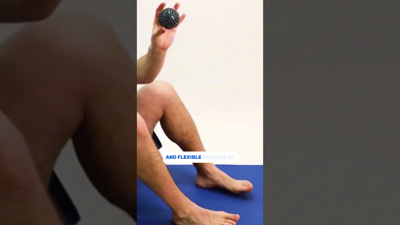
![Achilles Tendonitis & Back of Heel Pain [BEST Home Treatments 2024!]](https://www.michiganfootdoctors.com/wp-content/cache/flying-press/d645ea3f3d27024136b88a074b2b7894.jpg)
Watch: Achilles Tendonitis & Back of Heel Pain [BEST Home Treatments 2024!] — MichiganFootDoctors YouTube
Medically Reviewed | Dr. Tom Biernacki, DPM | Board-Certified Podiatric Surgeon | Balance Foot & Ankle, Michigan

Tendinopathy vs. Tendonitis: Why the Distinction Matters
The term “tendonitis” implies active inflammation — neutrophils, macrophages, the classical signs of redness, heat, and swelling from an acute inflammatory cascade. True acute tendonitis exists, typically in the first 24–72 hours after significant tendon overload. However, the chronic tendon pain that brings most patients to our office is not tendonitis. It is tendinosis — a degenerative, failed-healing response characterized by disorganized collagen fibers, neovascularization, and increased ground substance without meaningful inflammatory cell infiltrate.
This distinction matters because treatment principles differ. Tendinosis requires tendon loading through an appropriate progressive eccentric exercise protocol — loading the tendon to stimulate tenocyte activity and collagen remodeling. Rest alone allows pain to subside temporarily but doesn’t address the degenerative tissue. Prolonged anti-inflammatory medication use in tendinosis is not only ineffective but may be harmful — NSAIDs can impair tendon healing by blocking prostaglandin-mediated repair signaling.
Diagnostic musculoskeletal ultrasound distinguishes acute from chronic changes, measures tendon thickness, identifies partial-thickness tears, and guides injection therapy — all in-office at the initial evaluation.
Achilles Tendinopathy: Mid-Portion vs. Insertional
Mid-Portion Achilles Tendinopathy
Mid-portion Achilles tendinopathy (2–6cm proximal to the calcaneal insertion) is the most common tendon condition in runners, producing a characteristic fusiform palpable nodule within the tendon substance. The Alfredson eccentric heel-drop protocol — 3 sets of 15 repetitions of eccentric calf lowering on the edge of a step, both straight-leg and bent-knee, twice daily — is the most evidence-supported conservative treatment with 70–80% success rates at 12 weeks when properly executed. The protocol is painful initially — the guideline is “train through pain that is no worse than 5/10.” Patients who avoid pain during eccentric loading consistently fail to improve.
For recalcitrant mid-portion Achilles tendinopathy unresponsive to 12 weeks of eccentric training, extracorporeal shockwave therapy (ESWT) has Level I evidence supporting its efficacy. Ultrasound-guided PRP injection targeting the areas of neovascularization and tendinosis on ultrasound imaging represents an additional option for patients who fail ESWT.
Insertional Achilles Tendinopathy
Insertional Achilles tendinopathy at the calcaneal enthesis is a biologically and mechanically distinct entity from mid-portion disease. The compressive component — from the Haglund’s bony prominence and from the dorsal calcaneal surface directly loading the tendon insertion — means that pure eccentric loading (which loads the insertion compressively) may worsen rather than improve symptoms. Modified Alfredson protocol performed on flat ground rather than a step edge reduces compressive load. Retrocalcaneal bursitis is frequently concurrent and responds to ultrasound-guided corticosteroid injection directed into the bursa (not the tendon itself). Surgical calcaneal exostectomy with Achilles debridement and suture anchor reattachment produces excellent outcomes in properly selected patients failing conservative care.
Peroneal Tendinopathy
The peroneal tendons — peroneus brevis and peroneus longus — course posteriorly around the lateral malleolus within the peroneal groove, held in place by the superior peroneal retinaculum (SPR). Peroneus brevis tendinopathy and longitudinal split tears (most commonly at the fibular tip) produce lateral ankle and proximal lateral foot pain — often misdiagnosed as lateral ankle sprain. Peroneus longus tendinopathy produces lateral and plantar foot pain at the cuboid groove, frequently associated with cavovarus foot type that overloads the peroneus longus.
Dynamic ultrasound — imaging the peroneal tendons with active inversion and eversion — identifies subluxation of the tendons over the fibular tip (peroneal subluxation from SPR rupture) that is missed on static MRI. Appropriate conservative management (ASO ankle brace, physical therapy focusing on peroneal strengthening and proprioception) combined with correction of underlying biomechanical drivers (custom orthotics for cavovarus or flexible flatfoot) produces good outcomes in most cases. Operative tubularization of longitudinal split tears or SPR repair for subluxation is reserved for failed conservative care.
Posterior Tibial Tendinopathy
The posterior tibial tendon is the primary dynamic stabilizer of the medial arch — its failure produces the progressive adult acquired flatfoot deformity described in detail on our flatfoot page. Tendinopathy (early Stage I PTTD) presents as medial ankle pain just posterior to the medial malleolus with normal arch height and intact single-limb heel rise. Ultrasound shows tendon thickening, heterogeneous echotexture, and peritendinous fluid. Treatment at Stage I — before functional deficit develops — is immobilization in a CAM boot for 4–6 weeks, followed by Arizona AFO bracing and progressive physical therapy. Failure to diagnose and treat Stage I PTTD before progression to Stage II represents a significant missed opportunity for conservative management.
Flexor Hallucis Longus Tendinopathy
FHL tendinopathy is underdiagnosed and produces pain and crepitus along the medial ankle and plantar first ray — aggravated by push-off activities. It is the primary occupational injury of ballet dancers (“dancer’s tendinopathy”) but also affects recreational runners and athletes who push off forcefully. Hallmark: pain with resisted great toe plantarflexion and passive dorsiflexion. Ultrasound identifies tenosynovitis and tendinosis within the FHL tunnel posterior to the medial malleolus. Conservative treatment with activity modification, FHL eccentric strengthening, and ultrasound-guided corticosteroid injection into the tendon sheath resolves most cases.
Dr. Tom's Product Recommendations
Theraband CLX Resistance Band Set
⭐ Highly Rated
The Theraband CLX is the gold standard for Achilles eccentric rehabilitation, peroneal strengthening, and posterior tibial tendon strengthening exercises. The loop design allows independent or bilateral use and provides progressive resistance for systematic tendon loading protocols.
Dr. Tom says: “Dr. Biernacki gave me a specific eccentric Achilles protocol using the Theraband. After 8 weeks of consistent twice-daily sessions, my Achilles tendinopathy has improved more than anything else I tried.”
Achilles eccentric protocol, peroneal strengthening, posterior tibial tendon rehab
Protocol-dependent — must follow the correct exercise prescription for your specific tendon
Disclosure: We earn a commission at no extra cost to you.
Hoka Bondi 8 – Maximum Cushion Running Shoe
⭐ Highly Rated
Maximum cushion with extended heel bevel and rocker geometry reduces Achilles tendon load at initial contact — the primary shoe recommendation for Achilles mid-portion and insertional tendinopathy. The 4mm heel-to-toe drop combined with rocker geometry unloads the Achilles insertion significantly.
Dr. Tom says: “My Achilles tendinopathy dramatically improved after switching to the Bondi 8. The rocker bottom really does reduce the heel cord strain I could feel in my old neutral trainers.”
Achilles tendinopathy, plantar fasciitis, maximum impact reduction
Not ideal for peroneal tendinopathy where lateral stability is needed
Disclosure: We earn a commission at no extra cost to you.
ASO Ankle Stabilizer Brace
⭐ Highly Rated
The ASO provides figure-8 lateral stability that protects peroneal tendons during activity and prevents the inversion events that aggravate peroneal tendinopathy. Essential for peroneal tendon rehabilitation and prevention of subluxation during return to sport.
Dr. Tom says: “After my peroneal tendon split tear diagnosis, Dr. Biernacki had me in the ASO for 6 weeks during rehab. The lateral support was exactly what I needed to keep training without aggravating it.”
Peroneal tendinopathy, lateral ankle instability, peroneal tendon rehab
Not specifically designed for Achilles or posterior tibial tendon conditions
Disclosure: We earn a commission at no extra cost to you.
✅ Pros / Benefits
- Diagnostic ultrasound differentiates tendinosis from partial tear from complete rupture — critical for correct treatment
- Tendon-specific rehabilitation protocols — Alfredson eccentric for Achilles, peroneal strengthening for peroneal tendinopathy
- Ultrasound-guided injections target the bursa or tendon sheath precisely — avoiding intratendinous injection
- Shockwave therapy (ESWT) for recalcitrant Achilles tendinopathy in-office
- Biomechanical correction addressing footwear and custom orthotics as root cause
❌ Cons / Risks
- Tendinopathy rehabilitation requires consistent effort over 12+ weeks — there is no shortcut to tendon remodeling
- Partial tendon tears may require MRI for complete characterization if ultrasound findings are equivocal
- Some tendon conditions (complete rupture, failed conservative care with structural tear) ultimately require surgery
Dr. Tom Biernacki’s Recommendation
The most important thing I tell patients with tendon pain is this: rest alone doesn’t heal tendinosis. It just makes you feel better temporarily. The tendon needs progressive loading to stimulate proper collagen remodeling. The Alfredson eccentric protocol for Achilles tendinopathy is the best-studied tendon rehab protocol in medicine — it’s painful for the first few weeks, it requires consistency, and it genuinely works when done correctly. We teach it properly, monitor your progress with ultrasound, and adjust the approach if you’re not responding. That’s the difference between a generic ‘stretch and ice’ handout and actual tendon rehabilitation.
— Dr. Tom Biernacki, DPM | Board-Certified Podiatric Surgeon | Balance Foot & Ankle
Frequently Asked Questions
What is the difference between Achilles tendinopathy and an Achilles tear?
Achilles tendinopathy is degeneration of the tendon substance — thickening, disorganized collagen, neovascularization — without disruption of tendon continuity. A partial tear involves disruption of some but not all tendon fibers. A complete rupture means total loss of tendon continuity, typically producing a positive Thompson test (squeezing the calf doesn’t produce foot plantarflexion) and a palpable defect in the tendon. Diagnostic ultrasound differentiates these with high accuracy in-office. The distinction is critical because complete ruptures require either surgical repair or functional bracing in plantarflexion — not eccentric exercise.
How long does Achilles tendinopathy take to heal?
The Alfredson eccentric protocol typically produces meaningful improvement in 70–80% of mid-portion Achilles tendinopathy cases at 12 weeks. However, full pain resolution and tendon normalization on ultrasound may take 6–12 months. Insertional tendinopathy tends to respond more slowly than mid-portion disease. The critical variable is consistency — patients who complete the eccentric protocol twice daily without skipping sessions reliably improve faster than those who do it occasionally.
Should I get a cortisone injection for my tendon pain?
It depends on location. Corticosteroid injection into the retrocalcaneal bursa (not the Achilles tendon itself) is appropriate for retrocalcaneal bursitis concurrent with insertional Achilles tendinopathy. Injection into the FHL tendon sheath is appropriate for FHL tenosynovitis. However, direct intratendinous corticosteroid injection into the Achilles tendon is generally contraindicated — it has been associated with tendon rupture in multiple studies. We use ultrasound guidance for all tendon-adjacent injections to ensure precise placement and avoid intratendinous injection.
Can peroneal tendon problems be confused with an ankle sprain?
Yes — lateral ankle pain from peroneal tendinopathy or a peroneal tendon split tear is commonly misdiagnosed as ankle sprain. The key distinction: ankle sprain pain typically resolves in 3–6 weeks with appropriate rehabilitation; peroneal tendon pain that persists or worsens beyond 6 weeks suggests tendon pathology rather than simple ligament sprain. Clinical examination (tenderness posterior to the lateral malleolus rather than anterior to it, pain with resisted eversion) and ultrasound differentiate these. Treating peroneal tendinopathy as a simple sprain leads to months of ineffective management.
Michigan Foot Pain? See Dr. Biernacki In Person
4.9★ rated | 1,123 Reviews | 3,000+ Surgeries
Same-week appointments · Howell & Bloomfield Hills
📞 (810) 206-1402 Book Online →What does treatment cost?
Most diagnostic visits and conservative treatments are covered by Medicare and major insurers. Out-of-pocket costs vary by your specific plan.
How quickly can I get an appointment?
Most non-urgent cases see us within 5 business days. Urgent cases (sudden pain, possible fracture) typically same or next business day.
Related Conditions
In-Office Treatment at Balance Foot & Ankle
If home treatment isn’t providing relief for your foot and ankle issues, our podiatry team at Balance Foot & Ankle can help with same-day evaluations and advanced in-office care.
American Podiatric Medical Association: Find a Podiatrist
Ready to Get Relief?
Same-day appointments available in Howell & Bloomfield Hills, MI
4.9★ | 1,123 Reviews | 3,000+ Surgeries
Or call: (810) 206-1402

Dr. Tom Biernacki, DPM is a board-certified foot & ankle surgeon (ABFAS & ABPM) at Balance Foot & Ankle Specialists in Southeast Michigan. With over a decade of clinical experience, he specializes in heel pain, bunions, diabetic foot care, sports injuries, and minimally invasive surgery. Dr. Biernacki is a member of the APMA and ACFAS, and his patient education content on MichiganFootDoctors.com and YouTube has made him one of the most-followed foot & ankle educators on YouTube.