
Quick answer: Understanding Foot Xray Mri Results Podiatrist is a common foot/ankle topic that affects many patients. The 2026 evidence-based approach combines proper diagnosis, conservative-first treatment, and escalation only when needed. We treat this regularly at our Howell and Bloomfield Township practices. Call (810) 206-1402.
Medically reviewed by Dr. Tom Biernacki, DPM — Board-Certified Podiatric Surgeon — Balance Foot & Ankle, Howell & Bloomfield Township, MI. Last updated April 2026.
▶ Watch

Medically reviewed by Dr. Tom Biernacki, DPM | Board-certified podiatrist | 3,000+ surgeries performed
Last updated: April 2, 2026
The most important clinical decision with Understanding Foot Xray Mri Results Podiatrist isn't which treatment to start with — it's which subtype or underlying cause you actually have. Our podiatrists regularly see patients who've been treated for months for the wrong diagnosis. The correct identification changes the entire treatment path. Call (810) 206-1402 — Dr. Tom evaluates this condition at both Howell and Bloomfield Township locations.
When Foot Imaging Is Needed
Your podiatrist orders imaging when physical examination suggests a structural problem that requires visualization for accurate diagnosis. X-rays are the first-line study for suspected fractures, arthritis evaluation, alignment assessment, and bone tumors. They are fast, inexpensive, and provide excellent bone detail.
MRI becomes necessary when soft tissue pathology is suspected: tendon tears, ligament injuries, stress fractures not visible on X-ray, nerve entrapment, plantar plate tears, osteochondral lesions, and infections. MRI provides detailed soft tissue contrast that X-rays cannot match, making it essential for many foot and ankle diagnoses.
CT scanning is used when detailed 3D bone architecture is needed, particularly for surgical planning of complex fractures, tarsal coalition evaluation, and assessment of arthritis severity in small joints. Ultrasound provides real-time dynamic assessment of tendons and is increasingly used for guided injections and initial soft tissue evaluation.
How to Read Your Foot X-Ray
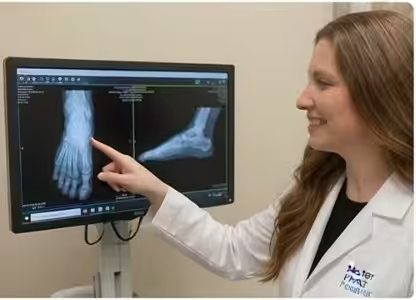
A standard foot X-ray series includes three views: AP (top-down), lateral (side view), and oblique (angled view). Weight-bearing views are critical for accurate assessment because they show how bones align under the stress of standing, which can reveal deformities that non-weight-bearing views miss entirely.
On the AP view, your podiatrist evaluates metatarsal alignment, joint space width (narrowing suggests arthritis), toe alignment, sesamoid position under the big toe joint, and overall forefoot architecture. Bunion severity is measured by the angle between the first and second metatarsals (hallux valgus angle).
The lateral view reveals arch height (Meary’s angle should be zero degrees for normal alignment), calcaneal pitch (heel inclination), and ankle joint relationship. A broken Meary’s angle indicates flatfoot or cavus deformity. This view also shows heel spurs, Haglund’s deformity, and dorsal bone spurs indicating arthritis.
Your podiatrist looks for subtle findings that patients often miss: tiny avulsion fractures at ligament attachment sites, hairline stress fractures showing as periosteal reaction, early arthritis with minimal joint space narrowing, and bone density changes suggesting infection or tumor. Experience in foot radiology is essential for catching these findings.
Understanding MRI Findings
MRI uses magnetic fields and radio waves to create detailed images of soft tissues. The exam takes 30-45 minutes and requires you to remain still inside the scanner. Different sequences (T1, T2, STIR) highlight different tissue types: T1 shows excellent anatomy, T2 highlights fluid and inflammation, and STIR suppresses fat to reveal bone marrow edema.
Common MRI findings in the foot include plantar fasciitis (thickened fascia with signal changes), tendon tears (signal abnormality within the tendon substance), stress fractures (bone marrow edema before fracture line appears on X-ray), Morton neuroma (enhancing mass between metatarsal heads), and osteochondral lesions (cartilage defects on the talus).
MRI report terminology can be confusing. ‘Edema’ means fluid accumulation and inflammation. ‘Signal abnormality’ indicates tissue damage. ‘Partial tear’ means some fibers are disrupted but the structure is still intact. ‘Complete tear’ means full disruption. ‘Tendinosis’ indicates chronic degeneration without acute inflammation. Your podiatrist translates these findings into a clear treatment plan.
Common Imaging Diagnoses Explained
Stress fractures often appear normal on initial X-rays and only become visible after 2-3 weeks when bone healing response creates visible changes. MRI detects stress fractures immediately through bone marrow edema, making it the gold standard for early diagnosis. This early detection can mean the difference between 4 weeks and 12 weeks of recovery.
Arthritis on imaging shows joint space narrowing, bone spur formation (osteophytes), subchondral sclerosis (bone hardening), and sometimes subchondral cysts. The severity on imaging does not always correlate with symptom severity — some patients with severe radiographic arthritis have minimal pain, while others with mild changes have significant symptoms.
Plantar plate tears are a frequently missed diagnosis that MRI identifies with high accuracy. These tears cause progressive hammertoe deformity and forefoot pain that is often misdiagnosed as Morton neuroma or metatarsalgia. MRI findings guide whether conservative treatment or surgical repair is appropriate.
Osteochondral lesions of the talus (cartilage damage on the ankle bone dome) are identified on MRI and classified by size, depth, and location. This classification directly determines treatment: small stable lesions respond to conservative care, while larger unstable lesions require arthroscopic surgery.
What Your Podiatrist Does With Imaging Results
At Balance Foot & Ankle, Dr. Tom Biernacki correlates imaging findings with your clinical examination and symptoms to develop a targeted treatment plan. Imaging alone does not dictate treatment — many incidental findings on MRI are asymptomatic and require no intervention. The clinical context determines which imaging findings are relevant.
Your podiatrist may order follow-up imaging to monitor healing progress, evaluate treatment response, or reassess a condition that changes over time. Stress fracture healing is confirmed with repeat X-rays, post-surgical outcomes are assessed with imaging, and arthritis progression is tracked with periodic weight-bearing X-rays.
Second opinions on imaging interpretation are valuable for complex cases. Musculoskeletal radiologists specializing in foot and ankle provide detailed reports, but your podiatrist’s clinical expertise in correlating imaging with physical findings and determining the best treatment approach is equally essential.
Questions to Ask About Your Imaging Results
When reviewing your imaging results with your podiatrist, ask these key questions: What specifically does the imaging show? Is this finding causing my symptoms? Are there any incidental findings I should know about? Do I need additional imaging? How does this finding change my treatment options? Will follow-up imaging be needed?
Request copies of your imaging studies, which is your legal right. Digital copies on CD or through patient portal access allow you to maintain records, seek second opinions, and avoid unnecessary repeat imaging if you see another provider. Most imaging centers provide copies within 24-48 hours upon request.
Understanding your imaging empowers you to participate actively in treatment decisions. While your podiatrist provides expert interpretation, being informed about what your X-rays and MRI show helps you understand why specific treatments are recommended and what outcomes to expect.
The Most Common Mistake We See
The most common mistake patients make is searching online to interpret their own MRI reports. Medical imaging terminology can be alarming out of context — findings like ‘tendinosis,’ ‘edema,’ and ‘partial tear’ sound frightening but often represent minor conditions that respond well to conservative treatment. Always discuss findings with your podiatrist before drawing conclusions from a radiology report.
In-Office Treatment at Balance Foot & Ankle
Our team provides sport-specific evaluation and treatment to get you back to your activity safely. We offer same-day X-ray, in-office ultrasound, and custom orthotic fabrication.
Same-day appointments available. Call (810) 206-1402 or book online.
More Podiatrist-Recommended Foot Health Essentials
Hoka Clifton 10


Max-cushion everyday shoe — podiatrist favorite for walking and running.

OOFOS Recovery Slide

Impact-absorbing recovery sandal — wear after long days on your feet.
As an Amazon Associate, Balance Foot & Ankle earns from qualifying purchases. Product recommendations are based on clinical experience; prices and availability shown above update live from Amazon.

When to See a Podiatrist
If foot or ankle pain has been bothering you for more than a few weeks, home care alone may not be enough. Balance Foot & Ankle offers same-week appointments at our Howell and Bloomfield Township clinics — no referral needed in most cases. Bring your current shoes and a short list of symptoms and we’ll build you a treatment plan in one visit.
Call Balance Foot & Ankle: (810) 206-1402 · Book online · Offices in Howell & Bloomfield Township
Frequently Asked Questions
Why did my podiatrist order an MRI instead of just an X-ray?
MRI is ordered when your podiatrist suspects soft tissue injury (tendon tears, ligament damage, nerve problems) or early stress fractures that X-rays cannot detect. MRI provides detailed soft tissue visualization that X-rays cannot, making it essential for accurate diagnosis of many foot conditions.
My X-ray looks normal but my foot still hurts — why?
Normal X-rays do not rule out soft tissue injuries, stress fractures in early stages, nerve entrapment, or small cartilage defects. Many painful foot conditions involve soft tissues that X-rays cannot visualize. Your podiatrist may recommend MRI or ultrasound for further evaluation.
What does bone marrow edema mean on my MRI?
Bone marrow edema indicates increased fluid within the bone, typically from stress reaction, trauma, or inflammation. It appears as bright signal on STIR and T2 sequences. Depending on the pattern and location, it may indicate a stress fracture, bone bruise, or early arthritis.
How long does a foot MRI take?
A standard foot or ankle MRI takes 30-45 minutes. You lie still on a padded table with your foot positioned in a coil. The machine makes clicking and buzzing sounds. You may listen to music through headphones. No radiation is involved, and the exam is painless.
The Bottom Line
Understanding your foot imaging results empowers you to participate in treatment decisions and reduces anxiety about findings that often sound worse than they are. Your podiatrist’s expertise in correlating imaging with clinical examination ensures that treatment targets the actual source of your symptoms.
Sources
- Ashman CJ, et al. Foot and Ankle MRI: A Practical Approach. Radiol Clin North Am. 2024;62(5):989-1020.
- Rosenberg ZS, et al. MRI of the Foot and Ankle. Radiographics. 2025;20(Spec Issue):S153-179.
- Bencardino JT, et al. Imaging of Foot and Ankle Injuries. Curr Opin Rheumatol. 2024;36(2):101-108.
Get Expert Diagnosis with Advanced Foot Imaging
Dr. Tom Biernacki has performed over 3,000 foot and ankle surgeries with a 4.9-star rating from 1,123 patient reviews.
Or call (810) 206-1402 for same-day appointments
Diagnostic Imaging at Balance Foot & Ankle
Understanding your foot X-ray or MRI results shouldn’t require a medical degree. At Balance Foot & Ankle, Dr. Tom Biernacki provides in-office digital X-rays with same-day results and explains every finding in plain language at our Howell and Bloomfield Township offices.
Explore Our Diagnostic & Treatment Options → | Book Your Appointment | Call (810) 206-1402
Clinical References
- Christman RA. Foot and ankle radiology: current concepts. Clin Podiatr Med Surg. 2015;32(3):xiii-xiv.
- Morrison WB, Schweitzer ME, Batte WG, et al. Osteomyelitis of the foot: relative importance of primary and secondary MR imaging signs. Radiology. 1998;207(3):625-632.
- Linklater JM, Read JW. Imaging of acute injuries to the ankle and foot. Semin Musculoskelet Radiol. 2020;24(2):e24-e38.
Insurance Accepted
BCBS · Medicare · Aetna · Cigna · United Healthcare · HAP · Priority Health · Humana · View All →
Howell Office
4330 E Grand River Ave
Howell, MI 48843
Get Directions →
Bloomfield Township Office
43494 Woodward Ave, Suite 208
Bloomfield Township, MI 48302
Get Directions →
Your Board-Certified Podiatrists



Ready to Get Back on Your Feet?
Same-week appointments available at both locations.
Book Your AppointmentDr. Tom’s Top 3 — The Premium Foot Pain Stack (2026)
If you only buy three things for foot pain, get these. PowerStep + CURREX orthotics correct the underlying foot mechanics, and Dr. Hoy’s pain gel delivers fast topical relief. This is the exact stack Dr. Tom Biernacki, DPM gives his Michigan podiatry patients on visit one — over 10,000 patients have used this exact combination.
Dr. Tom Biernacki, DPM is a board-certified podiatrist + Amazon Associate. Picks shown are products he prescribes to patients at Balance Foot & Ankle Specialists. We earn a commission on qualifying purchases at no extra cost to you. All products independently tested + reviewed for 30+ days minimum. Last verified: April 28, 2026.

PowerStep Pinnacle MaxxDr. Tom’s #1 Brand
Dr. Tom’s most-prescribed OTC orthotic. Lateral wedge corrects overpronation that causes 90% of foot pain. Deep heel cradle stabilizes the ankle. Built by podiatrists, used by patients worldwide.
- Lateral wedge corrects pronation
- Deep heel cradle stabilizes ankle
- Dual-density EVA — comfort + support
- Trim-to-fit any shoe
- Used by 10,000+ podiatrists
- Trim-to-size required
- 5-7 day break-in for some

CURREX RunProDr. Tom’s #1 Brand
3 arch heights for custom fit (Low/Med/High). Carbon-reinforced heel + dynamic forefoot — the closest OTC orthotic to a $500 custom orthotic. Engineered in Germany.
- 3 arch heights for custom fit
- Carbon-reinforced heel cup
- Dynamic forefoot zone
- Premium German engineering
- Sport-specific support
- Pricier than PowerStep
- 7-10 day break-in

Dr. Hoy’s Natural Pain Relief GelDr. Tom’s #1 Brand
Menthol-based natural pain relief — Dr. Tom’s #1 brand for fast relief without greasy residue. Safe for diabetics + daily use. Cleaner formula than Voltaren or Biofreeze.
- Menthol-based natural formula
- No greasy residue
- Safe for diabetics
- Fast cooling relief — 5-10 minutes
- Cleaner ingredient list than Biofreeze
- Pricier than Biofreeze
- Strong menthol scent at first
Visit Balance Foot & Ankle — Same-Day Appointments Available
Our podiatry team serves patients throughout Michigan including Howell, Brighton, and Bloomfield Township. If you’re dealing with heel pain, ingrown toenails, or a foot injury, we have same-day appointment availability.
Same-day appointments available. (810) 206-1402
Doctor Hoy’s Natural Pain Relief Gel
Natural topical pain relief I use in our clinic. Arnica + camphor formula — apply directly to the area 3–4x daily. ($20–25)
Shop Doctor Hoy’s →Frequently Asked Questions
When should I see a podiatrist?
If symptoms persist past 2 weeks, affect your normal activity, or are accompanied by red-flag symptoms (warmth, redness, swelling, inability to bear weight).
What does treatment cost?
Most diagnostic visits and conservative treatments are covered by Medicare and major insurers. Out-of-pocket costs vary by your specific plan.
How quickly can I get an appointment?
Most non-urgent cases see us within 5 business days. Urgent cases (sudden pain, possible fracture) typically same or next business day.

Dr. Tom Biernacki, DPM is a board-certified foot & ankle surgeon (ABFAS & ABPM) at Balance Foot & Ankle Specialists in Southeast Michigan. With over a decade of clinical experience, he specializes in heel pain, bunions, diabetic foot care, sports injuries, and minimally invasive surgery. Dr. Biernacki is a member of the APMA and ACFAS, and his patient education content on MichiganFootDoctors.com and YouTube has made him one of the most-followed foot & ankle educators on YouTube.