

Quick answer: Yellow soles are most often from thickened skin (calluses), dryness, or harmless pigment from foods like carrots — usually not a medical concern. Less commonly, yellowing reflects jaundice, anemia, or circulation issues; if it comes with other symptoms or won’t resolve with exfoliation and moisturizing, see a doctor.
Ready to be seen? Book a real appointment time online, 24/7 — Howell or Bloomfield Township. Same-week openings are usually available.
Medically reviewed by Dr. Tom Biernacki, DPM
Board-certified podiatric surgeon | Balance Foot & Ankle
Last reviewed: May 2026
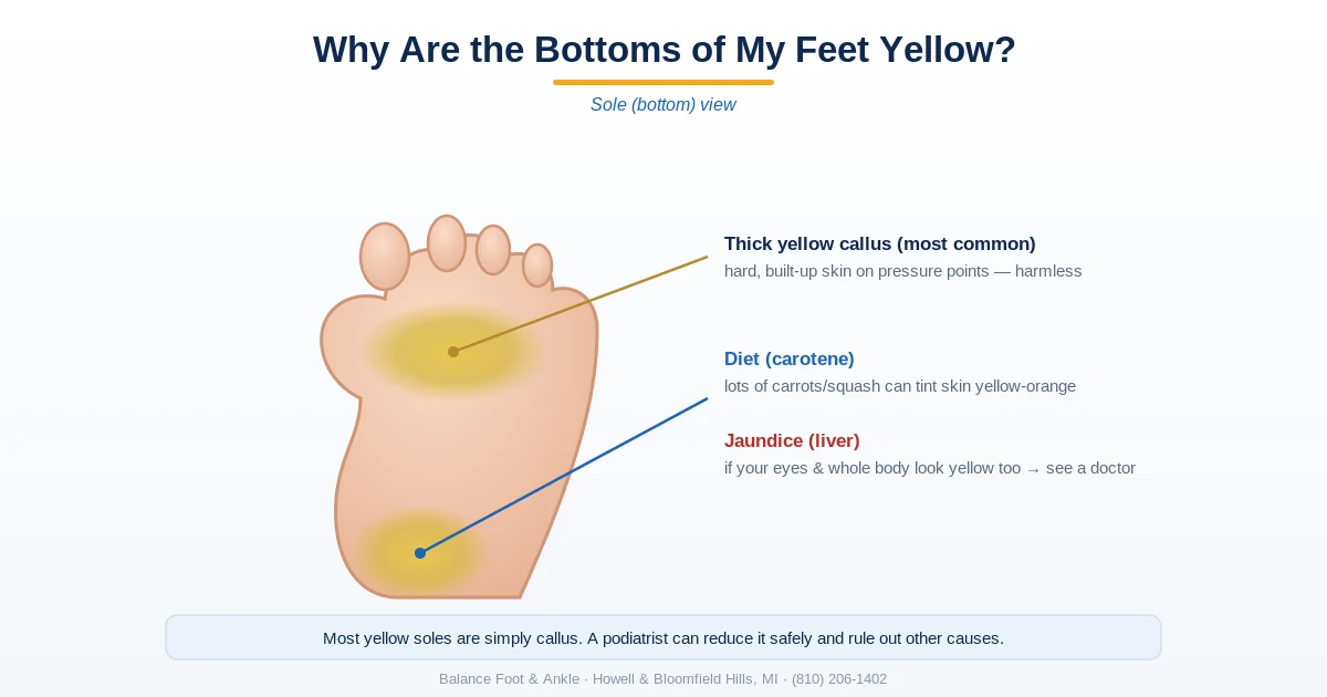
The most important clinical decision with Why Are The Bottom Of My Feet Yellow isn’t which treatment to start with — it’s identifying the correct subtype. That changes everything. Call (810) 206-1402 or book online.
Table of Contents
- How to Tell the Difference at a Glance
- Cause 1: Callus (Most Common)
- Cause 2: Carotenemia (Diet-Related)
- Cause 3: Moccasin Tinea Pedis
- Cause 4: Jaundice (Medical Emergency)
- Cause 5: Vascular / Ischemic Changes
- Cause 6: Xanthomas (Cholesterol Deposits)
- How We Evaluate Yellow Feet
- Warning Signs
- Frequently Asked Questions
When you notice the bottoms of your feet turning yellow, the range of possible causes runs from completely benign (too many carrots in your diet) to requiring urgent medical attention (liver disease). The good news is that most cases have an obvious benign explanation. The key is knowing the one red flag that changes everything: whether your eyes are yellow too.
How to Tell the Difference at a Glance
| Cause | How It Looks | Key Clues | Concern Level |
|---|---|---|---|
| Callus | Thick yellow patches on pressure points | Hard and rough over heels and balls of the feet | Harmless; easily treated |
| Carotenemia | Orange-yellow soles and palms | Whites of the eyes stay white; linked to a beta-carotene-rich diet | Harmless; diet-related |
| Moccasin tinea pedis (fungus) | Yellowish, dry, scaly sole | Itching, peeling, and spreading | Treatable with antifungals |
| Jaundice | Yellow skin and yellow eyes | Whole-body yellowing, often with dark urine | See a doctor promptly |
| Vascular / ischemic changes | Yellow with cool, pale, or shiny skin | Poor circulation, pain, or slow-healing wounds | Needs medical evaluation |
| Xanthomas | Firm yellow bumps or plaques | Cholesterol deposits under the skin | See a doctor to check cholesterol |
Before reading through every possible cause, use this two-question filter that handles the majority of cases. First: where exactly is the yellow? If it is on the thick-skinned pressure areas of the sole — the heel, ball, and lateral border — it is almost certainly callus. If it covers the entire sole evenly, including the arch, carotenemia or tinea pedis is more likely. Second: are the whites of your eyes (sclerae) yellow? If yes, stop reading this page and go to urgent care today — that is jaundice, not a foot problem.
The color tone also gives information. Ivory or pale yellow on pressure areas = callus. Orange-yellow on the entire sole and palms = carotenemia. Diffuse fine scale with mild yellow-tan discoloration across the whole sole = possible moccasin tinea pedis. Shiny, pale yellow, thin skin on the foot dorsum = vascular insufficiency. Yellow nodular deposits over tendons = xanthomas.
Key takeaway: The single most important distinguishing test: look at the whites of your eyes (sclerae). Yellow sclerae = jaundice = liver, bile duct, or blood disorder requiring urgent medical evaluation. Normal white sclerae + yellow feet/palms = almost certainly carotenemia or callus, which are benign.
Not sure which one you’re looking at?
Callus and carotenemia are the harmless two on this list. Jaundice and reduced circulation are not — and by eye they are the hardest of the six to tell apart from the rest. It is worth having a look in person if the yellow is spreading, if it is on one foot and not the other, or if it comes with numbness, swelling, or pain.
Howell — 4330 E Grand River Ave, Howell MI 48843 · Bloomfield Township — 43494 Woodward Ave #208, Bloomfield Township MI 48302
Cause 1: Callus (The Most Common Cause)
A callus is a diffuse area of thickened, hardened skin that develops in response to repetitive pressure or friction. On the plantar surface, calluses most commonly form over the heel, the ball of the foot, and the lateral forefoot edge. As the stratum corneum (outermost skin layer) thickens, it loses its normal translucency and turns pale yellow or ivory. This is the most common reason the bottom of your feet looks yellow — it is simply dead skin cells compressed into a dense, discolored layer.
Calluses are not dangerous but can cause discomfort when they become very thick, especially in people with diabetes where they increase plantar pressure and ulcer risk. Treatment is mechanical: pumice stone after soaking, keratolytic creams (urea 20–40%, salicylic acid 6–17%), and correcting the underlying pressure cause with orthotics or wider shoes. In our clinic, we use an electric nail drill to debride thick plantar calluses safely and effectively in a single visit. Regular debridement is part of diabetic foot care because callus buildup is independently associated with plantar ulcer formation.
Cause 2: Carotenemia (Diet-Related Orange-Yellow Skin)
Carotenemia is a benign condition caused by excess beta-carotene in the blood, which deposits in the skin and turns it orange-yellow. It most prominently affects areas of thick, fatty skin: the soles of the feet and the palms of the hands. The face may also be affected, particularly the nasolabial folds and forehead, but — crucially — the sclerae (whites of the eyes) remain white. This is the definitive distinguishing feature from jaundice.
Foods that cause carotenemia in large quantities: carrots, sweet potatoes, squash (especially butternut and acorn), pumpkin, cantaloupe, green smoothies with large amounts of kale or spinach, and carrot-based juices. The condition is especially common in people who have recently adopted plant-based diets or green juice protocols. Beta-carotene is fat-soluble and builds up in subcutaneous fat, which is why soles and palms (which have thick fat layers) are disproportionately affected.
Key takeaway: Carotenemia is harmless but does not resolve quickly — beta-carotene is fat-soluble and stored in subcutaneous fat. After stopping high-carotene foods (carrots, sweet potatoes, squash, green juice), the skin color normalizes over 2–6 weeks. No treatment is needed.
Cause 3: Moccasin-Type Tinea Pedis
The moccasin type of tinea pedis (athlete’s foot) covers the entire sole, heel, and sides of the foot with a diffuse fine powdery scale. Unlike the interdigital type (between the toes) which presents clearly as redness and maceration, the moccasin type is subtle — it looks like very dry skin with a slight yellowish-tan discoloration. Many patients with this presentation do not realize they have a fungal infection because there are no obvious blisters, and the itch may be mild.
The causative organism is almost always Trichophyton rubrum. The infection can be present for years without treatment because it is not recognized as a fungal problem. A KOH preparation (microscopic exam of a skin scraping with potassium hydroxide) reveals fungal hyphae and confirms the diagnosis. Treatment is topical terbinafine 1% cream applied twice daily for 4 weeks extended beyond the visible rash border, or oral terbinafine for severe or recalcitrant cases.
Key takeaway: Moccasin-type tinea pedis spreads diffuse fine scale across the entire sole and arch, producing a subtle yellow-tan discoloration that is frequently mistaken for dry skin or callus. If the soles itch and have fine powdery scale, try terbinafine cream for 4 weeks before assuming it is callus.
Cause 4: Jaundice
Jaundice (icterus) is yellowing of the skin, sclerae, and mucous membranes caused by elevated bilirubin — a breakdown product of hemoglobin that accumulates when the liver cannot clear it properly. Jaundice is never a benign finding. The causes include hepatitis (viral, alcoholic, autoimmune), bile duct obstruction (gallstones, cancer), hemolytic anemia (accelerated red blood cell destruction), liver cirrhosis, and pancreatic cancer. Neonatal jaundice (in newborns) is common and usually managed with phototherapy.
The key distinguishing feature from carotenemia: jaundice turns the sclerae yellow; carotenemia does not. Bilirubin has a high affinity for the elastic tissue in the sclerae, which is why jaundice is detected there first. If you look in the mirror and the whites of your eyes have a yellow tint, that is not dietary. A podiatric exam is not the right next step — same-day urgent care or emergency room evaluation for liver function tests, bilirubin, and imaging is.
Cause 5: Vascular and Ischemic Changes
In patients with peripheral arterial disease (PAD), reduced blood flow to the foot produces a characteristic skin appearance: the skin becomes shiny, atrophic (thin), and pale to yellowish, with loss of hair, delayed capillary refill, and diminished pedal pulses. The yellowing in this context is not a pigment issue — it is the color of poorly perfused, thin skin seen over underlying fat and tendon. The toenails often become thick and yellow separately due to slow nail growth from reduced blood supply.
This presentation is most common in older patients with atherosclerosis risk factors (smoking, diabetes, hypertension, hyperlipidemia). PAD-related skin changes combined with rest pain (foot pain when lying down, relieved by dangling the foot over the bed), non-healing wounds, or absent pedal pulses indicate possible critical limb ischemia requiring vascular surgery consultation. This is not a condition for conservative podiatric management alone — vascular input is essential.
Key takeaway: Yellow skin in someone with diabetes or peripheral arterial disease is never cosmetic until proven otherwise. Yellowish, shiny, atrophic skin on the dorsum of the foot can indicate critical ischemia — poor blood flow that puts tissue at risk. A vascular exam is warranted.
Cause 6: Xanthomas (Cholesterol Deposits)
Xanthomas are yellow deposits of cholesterol-laden macrophages in the skin and tendons. On the feet, the most common type is tendinous xanthoma, appearing as firm yellow nodular swellings over the Achilles tendon or the extensor tendons on the foot dorsum. Xanthelasma appears on the eyelids. Eruptive xanthomas appear as clusters of small yellow papules and are associated with severe hypertriglyceridemia. Xanthomas are markers of lipid disorders — familial hypercholesterolemia (FH), Type III hyperlipoproteinemia, or secondary dyslipidemia from diabetes or hypothyroidism. Their presence warrants a fasting lipid panel and cardiac risk assessment.
How We Evaluate Yellow Feet
In our clinic, a systematic approach to yellow-colored feet takes about 10 minutes. We check the sclerae immediately — if yellow, the patient is directed to urgent medical care before we proceed. If sclerae are normal, we assess the distribution pattern (pressure areas only vs. entire sole vs. diffuse foot), the color tone (ivory vs. orange vs. scale-covered vs. shiny atrophic), and the texture (hardened and rough = callus; scaly and slightly itchy = tinea; smooth and thin = vascular).
For suspected tinea pedis, a KOH preparation confirms the diagnosis at the same visit. For suspected callus, we measure the callus thickness and assess the underlying pressure distribution with a plantar pressure scan. For suspected carotenemia, a diet history is usually sufficient; serum beta-carotene can be measured if confirmation is needed. For suspected vascular changes, we perform ankle-brachial index (ABI) measurement and palpate pedal pulses. Xanthomas trigger lipid panel ordering and rheumatology or endocrinology referral as appropriate.
⚠️ Seek same-day evaluation if your yellow feet are accompanied by
- Yellowing of the whites of your eyes (sclerae) — this is jaundice, a medical emergency
- Dark urine and pale stools alongside yellow skin
- Sudden onset of yellow skin combined with fatigue, abdominal pain, or nausea
- Yellow skin with leg or foot ulcers that are not healing
- Foot pain at rest or at night combined with skin color changes (critical limb ischemia)
- Yellow skin developing rapidly over days rather than gradually over weeks
Frequently Asked Questions
Is it normal for the bottom of your feet to be yellow?
Mild ivory-yellow discoloration of the heel and ball of the foot from callus is very common and considered a normal response to pressure and activity. An orange-yellow color on both soles and palms in someone who eats large amounts of carrots, sweet potatoes, or green juice is also benign carotenemia. What is not normal: yellow color extending to the sclerae (jaundice), rapidly developing yellow skin, yellow skin with pain at rest or non-healing wounds, or yellow skin with associated systemic symptoms (fatigue, nausea, dark urine). Those presentations require prompt medical evaluation.
Can liver problems cause yellow feet?
Yes — jaundice from liver disease causes yellow skin throughout the body, including the soles. The liver is responsible for processing bilirubin (a breakdown product of red blood cells). When liver function is impaired by hepatitis, cirrhosis, bile duct obstruction, or other conditions, bilirubin accumulates in the blood and deposits in the skin. The most reliable sign that yellow feet are due to liver disease rather than a benign cause is yellow sclerae (whites of the eyes). If your eyes look normal, liver-based jaundice is very unlikely to be causing the foot discoloration.
How do I get rid of yellow calluses on the bottom of my feet?
Soak feet in warm water for 10–15 minutes to soften the callus, then use a pumice stone or foot file to gently reduce the thickened skin. Apply a urea 20–40% cream or salicylic acid lotion to the area after soaking and cover with socks overnight to maximize penetration. Repeat nightly until the callus is at normal skin thickness. Address the underlying pressure cause — shoes with inadequate cushioning, improper fit, or biomechanical abnormalities — or the callus will return. If the callus is painful, very thick, or you have diabetes, professional debridement by a podiatrist is safer than at-home treatment.
My feet and hands are orange — is it carotenemia or something serious?
Orange-yellow color on both the hands and feet (particularly the palms and soles) in someone with a high-carotene diet is almost certainly carotenemia. It is a benign, cosmetic condition with no health consequences. Check your diet for large amounts of carrots, sweet potatoes, squash, or green juices, and check that your sclerae are white. If you reduce carotene intake and your eyes are white, you can observe for improvement over 2–6 weeks without any other workup. If the orange color is accompanied by yellow eyes, fatigue, or other symptoms, see a physician the same day for liver function tests.
The Bottom Line
Yellow discoloration on the bottom of the feet most commonly comes from one of three benign causes: callus buildup over pressure areas, carotenemia from excess dietary beta-carotene, or moccasin-type tinea pedis. The one finding that changes the entire picture is yellow sclerae — if the whites of your eyes are also yellow, that is jaundice requiring urgent medical evaluation, not a podiatry visit. For the benign causes, the treatments are straightforward: mechanical callus debridement, dietary carotene reduction, or antifungal cream. When yellow skin is accompanied by vascular symptoms, non-healing wounds, or systemic illness, a more thorough evaluation is warranted.
Sources
- Maharshak N, Shapiro J, Trau H. Carotenoderma — a review of the current literature. Int J Dermatol. 2003;42(3):178-181.
- Hay RJ. Tinea pedis: evidence-based treatment. Curr Opin Infect Dis. 2015;28(2):139-142.
- Frykberg RG, et al. Diabetic foot disorders: a clinical practice guideline. J Foot Ankle Surg. 2006;45(5 Suppl):S1-S66.
- Goldberg A, et al. Peripheral arterial disease: a clinical review for the internist. Am J Med. 2023;136(4):337-345.
Ready to Get Relief?
Same-day appointments available in Howell & Bloomfield Township, MI
4.9★ | 1,123 Reviews | 3,000+ Surgeries
Or call: (810) 206-1402
📋 Dr. Tom Biernacki, DPM, FACFAS answers:
The bottom of the feet turning yellow is most often due to simple callus formation — thickened skin over pressure points that takes on a yellowish or ivory color. This is benign and very common, especially in people who stand for long hours or wear ill-fitting shoes. The second most common cause is carotenemia — high beta-carotene from eating large amounts of carrots, sweet potatoes, squash, or taking supplements. Carotenemia preferentially deposits in the palms and soles and is completely harmless; it resolves when intake normalizes. The cause that must not be missed is jaundice: if the yellow color is accompanied by yellowing of the whites of your eyes, dark urine, or pale stools, see a doctor the same day — this indicates liver or bile duct disease. A quick rule: if only your feet are yellow and your eyes are white, it’s almost certainly callus or diet-related; if your eyes are also yellow, it’s a medical emergency.
Related foot-skin guides
More on skin color and texture changes on the feet:
Yellow soles that won’t fade?
Persistent yellow feet can mean thick callus, fungus, or a circulation issue worth checking. In Michigan? Our podiatrists can find the cause and treat it. Same-week visits in Howell & Bloomfield Township.
📅 Book an Appointment 📞 (810) 206-1402★ 4.9 from 1,123+ Google reviews · Same-week visits · We verify your insurance free — most visits are just a copay.
Book an appointment with a podiatrist
Yellow soles are most often thickened callus doing exactly what callus does, but the same colour can accompany circulatory and metabolic changes worth identifying. If you are diabetic, or the change came on quickly, it deserves a look rather than a pumice stone. Balance Foot & Ankle sees patients at two offices — Howell in Livingston County, and Bloomfield Township in Oakland County.
Call (810) 206-1402 or use the form below and our team will find you an appointment time.

Dr. Tom Biernacki, DPM is a board-certified foot & ankle surgeon (ABFAS & ABPM) at Balance Foot & Ankle Specialists in Southeast Michigan. With over a decade of clinical experience, he specializes in heel pain, bunions, diabetic foot care, sports injuries, and minimally invasive surgery. Dr. Biernacki is a member of the APMA and ACFAS, and his patient education content on MichiganFootDoctors.com and YouTube has made him one of the most-followed foot & ankle educators on YouTube.
More questions patients ask
Are yellow feet a sign of liver disease?
Yellow skin on the feet can be a sign of jaundice, which may indicate liver disease, hepatitis, or bile duct obstruction. Unlike callus-related yellowing (which is localized to pressure areas) or carotenemia (orange-yellow), jaundice typically also affects the whites of the eyes and causes generalized yellow skin.
How do I get rid of yellow feet?
For callus-related yellowing: regular pumice stone exfoliation, moisturizing, and cushioned footwear. For carotenemia: reduce beta-carotene intake (carrots, sweet potato). For jaundice: see your doctor immediately. A podiatrist can confirm whether the yellowing is callus-based and treat it appropriately.
Can yellow feet indicate liver problems?
Yellow discoloration of the feet can be a sign of jaundice (icterus) from liver disease, but distinguishing true jaundice from other causes is important: True jaundice: the yellow color involves not just the feet but the entire skin, and critically the whites of the eyes (sclera) turn yellow; it is caused by elevated bilirubin from liver disease (hepatitis, cirrhosis), bile duct obstruction, or hemolytic anemia; other symptoms accompany it (dark urine, light-colored stools, fatigue, abdominal pain). Carotenemia (excess dietary carotene): affects palms and soles most prominently but NOT the sclera — sclera remain white; it is harmless and resolves with dietary modification. If your feet are yellow and your eye whites are also yellow, seek medical evaluation promptly. Balance Foot & Ankle coordinates appropriate medical referrals when systemic conditions are suspected — call (810) 206-1402.
Do calluses cause yellow coloring on the bottom of the foot?
Yes — calluses are the most common cause of yellow discoloration on the plantar surface of the feet. The mechanism: keratin (the structural protein of skin, hair, and nails) has a naturally yellowish color; when skin thickens from repetitive pressure and friction, the concentrated layers of keratin create a visibly yellow tint; calluses under the metatarsal heads (ball of foot), the heel, and over bony prominences are often distinctly yellow or yellowish-white. The color intensifies with callus thickness. Professional debridement by a podiatrist removes the yellow hyperkeratotic tissue. Addressing the underlying pressure cause (custom orthotics, footwear modification) prevents recurrence. At Balance Foot & Ankle, callus removal is a routine part of foot care visits — call (810) 206-1402.
What is carotenemia and does it cause yellow feet?
Carotenemia is a benign condition causing yellowish skin discoloration from elevated blood carotene levels. It develops from excessive consumption of beta-carotene-rich foods (carrots, sweet potatoes, pumpkin, winter squash, leafy greens) or from impaired carotene metabolism. Characteristics: yellow color is most prominent on palms, soles, and nasolabial folds (where carotene deposits in fat-rich skin), the sclera (whites of eyes) remain white (critical distinction from jaundice), there are no other symptoms, and it is completely harmless. It resolves spontaneously over weeks to months when dietary carotene is reduced. Common in infants fed high-carotene puréed vegetables. If in doubt whether yellow skin is from carotenemia or jaundice, a simple blood test (bilirubin vs. serum carotene level) distinguishes them. Balance Foot & Ankle in Howell and Bloomfield Township — call (810) 206-1402.
When should yellow feet be evaluated by a doctor?
Seek evaluation for yellow foot discoloration when: the yellow color involves the sclera (whites of eyes) — possible jaundice requiring urgent liver evaluation; the discoloration appeared suddenly without dietary changes; it is accompanied by dark urine or light stools; there is itching throughout the body; yellow skin is accompanied by other systemic symptoms (fatigue, abdominal pain, weight loss); or the discoloration is not explained by calluses or dietary carotene. Yellow toenails (without foot skin yellowing) most commonly indicate fungal nail infection. Callus-related yellowness on the sole with no systemic symptoms does not require urgent evaluation. At Balance Foot & Ankle in Howell (4330 E Grand River) and Bloomfield Township (43494 Woodward Ave #208), we evaluate the full range of foot skin changes and coordinate appropriate referrals — call (810) 206-1402.
What causes yellow skin on the bottom of the feet?
The most common cause is callus — thick, yellowish-tan skin that builds up over pressure points. Other causes include carotenemia (excess beta-carotene from carrots, sweet potatoes, or supplements), jaundice (bilirubin buildup from liver or gallbladder disease, which also yellows the eyes), athlete's foot with secondary hyperkeratosis, and in diabetic patients, glycation of the skin. Yellow spots rather than diffuse discoloration suggest focal callus, a corn, or a wart in early stages.
When is yellow skin on the feet a sign of liver disease?
Jaundice-related yellowing of the feet is accompanied by yellowing of the whites of the eyes (scleral icterus), dark urine, pale stools, and fatigue. If you have yellow feet AND any of these other signs, see a doctor urgently — it indicates the liver cannot process bilirubin. Callus and carotenemia cause yellow skin on the feet only, without eye involvement.
How do you get rid of yellow calluses on the bottom of your feet?
Callus removal involves mechanical debridement (a pumice stone after soaking, or professional paring by a podiatrist), followed by daily moisturizer with urea 20–40% to prevent regrowth. Most importantly, the pressure cause must be addressed — custom orthotics redistribute load away from callus-forming sites. A podiatrist can debride even severe calluses painlessly in a single visit.
Still have a question about coverage or cost? Call (810) 206-1402 and we will check your benefits before you come in — or book online: Book in Howell · Book in Bloomfield Township