

Medically reviewed by Dr. Tom Biernacki, DPM
Board-certified podiatric surgeon | Balance Foot & Ankle, Howell & Bloomfield Township, MI
Last reviewed: May 2026
Medically reviewed by Dr. Tom Biernacki, DPM
Board-certified podiatric surgeon | Balance Foot & Ankle
Last reviewed: April 2026
Most people know they ‘sprained their ankle’ — but few know which specific ligaments were damaged, which matters enormously for recovery. An anterior talofibular ligament sprain heals differently than a syndesmotic injury. The location, severity, and specific structures involved determine whether you need a brace, a boot, physical therapy, or surgery. Let’s break down the ankle ligament anatomy and what problems occur in each area.
Ankle Ligament Anatomy
The ankle joint relies on three groups of ligaments for stability. Each group protects the ankle against different directions of excessive movement:
Lateral (Outer) Ankle Ligaments
The three ligaments on the outside of the ankle are injured in 85% of ankle sprains — the classic ‘rolling inward’ mechanism:
- Anterior talofibular ligament (ATFL) — runs from the fibula forward to the talus; the most commonly sprained ankle ligament; injured when the foot rolls inward with the toes pointing down
- Calcaneofibular ligament (CFL) — runs from the fibula downward to the heel bone; injured in more severe lateral sprains alongside the ATFL
- Posterior talofibular ligament (PTFL) — runs from the fibula backward to the talus; the strongest lateral ligament; rarely injured except in complete dislocations
Medial (Inner) Ankle Ligaments — Deltoid Complex
The deltoid ligament is a strong, fan-shaped complex on the inside of the ankle. It resists outward rolling (eversion) and provides critical medial stability. It’s divided into superficial and deep layers. Isolated deltoid sprains account for only about 5% of ankle sprains but are common in athletes with repeated forced eversion.
Syndesmotic (High Ankle) Ligaments
The syndesmosis holds the tibia and fibula together just above the ankle joint. The anterior inferior tibiofibular ligament (AITFL) is most commonly injured in high ankle sprains. This injury mechanism — external rotation of the foot while the leg is fixed — is common in football linemen and skiers. High ankle sprains take significantly longer to heal than lateral sprains (8–12+ weeks vs. 4–6 weeks).
How Ankle Ligament Injuries Are Classified
- Grade 1: Microscopic tearing — ligament is stretched but intact. Mild pain, minimal swelling, no instability. 1–2 weeks to return to activity.
- Grade 2: Partial tear — moderate damage with some loss of ligament continuity. Notable pain, swelling, bruising. Some instability. 4–6 weeks to return to sport.
- Grade 3: Complete rupture — total loss of ligament continuity. Severe swelling, bruising, significant instability. 8–12 weeks; may require surgical repair.
Key takeaway: The Ottawa Ankle Rules: a validated clinical decision tool used worldwide. If there’s bone tenderness at the posterior tip of the fibula, the tip of the medial malleolus, or the base of the 5th metatarsal — or if the patient can’t bear weight immediately after injury and in our clinic — we order X-rays to rule out fractures. These rules have 99% sensitivity for clinically significant fractures.
Diagnosing Ankle Ligament Injuries
Diagnosis starts with the mechanism of injury (rolling inward vs. outward, rotation) and precise physical exam — palpating each ligament, performing stability tests (anterior drawer, talar tilt), and assessing the syndesmosis with the squeeze test and external rotation stress test.
- X-rays — rule out fractures (fibula, 5th metatarsal, talar dome)
- Stress X-rays — assess ligament integrity under controlled force
- MRI — gold standard for visualizing ligament tears, detecting talar dome lesions, and evaluating chronic instability
- Diagnostic ultrasound — real-time imaging of ligament continuity; can be performed during examination
Treatment of Ankle Ligament Injuries
Acute Phase (0–72 Hours)
- RICE protocol: rest, ice (15–20 min every 2 hours), compression wrap, elevation
- NSAIDs for pain and swelling management
- Crutches if weight-bearing is painful (Grade 2–3)
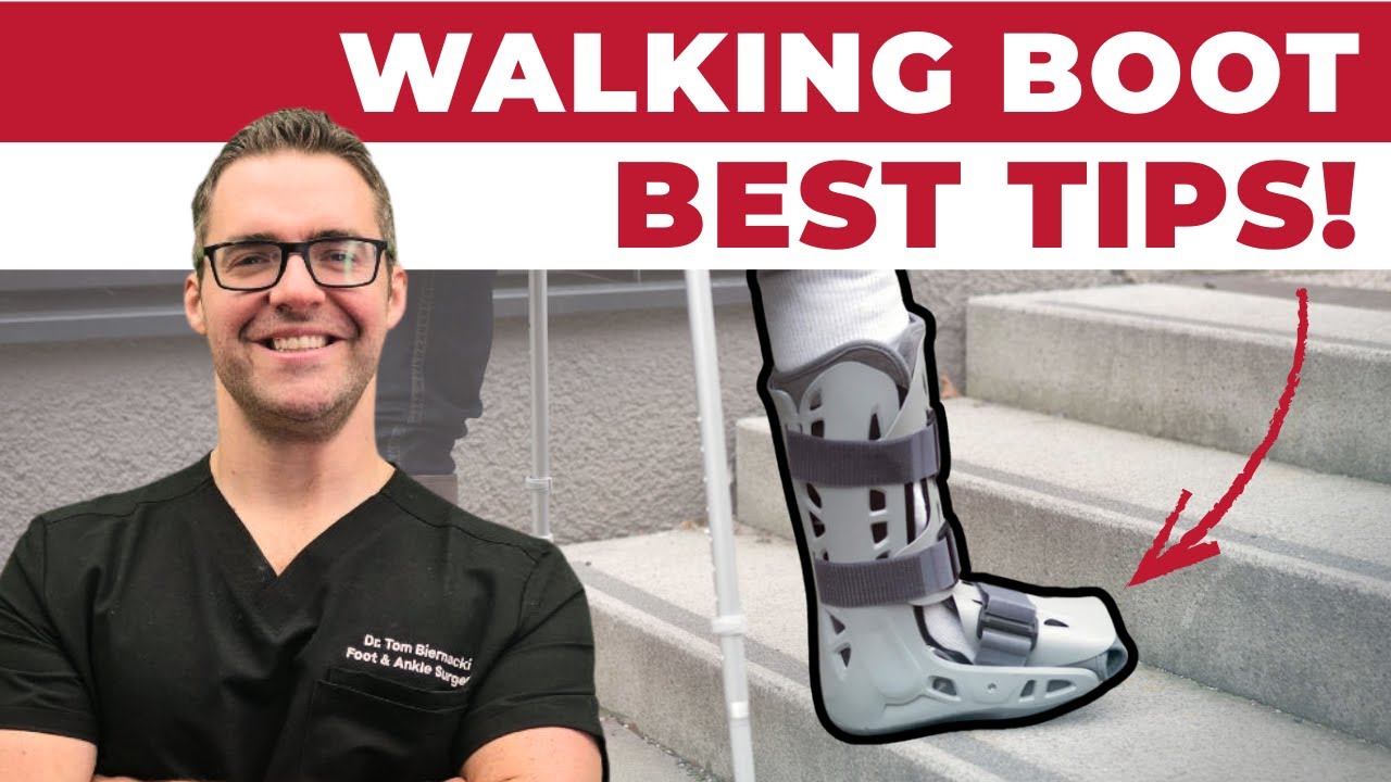
- Lace-up ankle brace for Grade 1–2 sprains; walking boot for Grade 2–3
Rehabilitation Phase
- Range of motion exercises starting when acute swelling subsides
- Strengthening of peroneal muscles (primary dynamic stabilizers of the lateral ankle)
- Proprioception/balance training — essential to prevent recurrence; the joint’s positional sense is disrupted by ligament injury
- Sport-specific training before return to athletics
Chronic Instability and Surgery
If ankle instability persists despite 3–6 months of rehabilitation, chronic lateral ankle instability (CLAI) may be diagnosed. Surgical options include the Broström-Gould repair — the gold standard lateral ligament reconstruction, tightening the ATFL and CFL and reinforcing them with the extensor retinaculum. Excellent long-term results.
Frequently Asked Questions
How long does an ankle ligament take to heal?
ATFL Grade 1 sprains: 1–2 weeks. Grade 2 lateral sprains: 4–6 weeks. Grade 3 complete tears: 8–12 weeks. High ankle (syndesmotic) sprains: 8–16 weeks. These timelines assume appropriate rehabilitation — skipping physical therapy dramatically increases recurrence and the risk of chronic instability.
Can ankle ligaments heal without surgery?
Yes — the vast majority of ankle ligament injuries, including complete Grade 3 tears, heal successfully with conservative care and physical therapy. Surgery is reserved for cases with persistent instability after 6+ months of rehab, or for syndesmotic injuries where the tibia and fibula relationship is significantly disrupted.
What is chronic ankle instability?
Chronic ankle instability (CAI) develops when a previously sprained ankle never fully regains its mechanical and neuromuscular stability. Patients describe the ankle ‘giving way,’ repeated sprains, and reduced confidence in activities. About 40% of acute lateral ankle sprains lead to CAI if rehabilitation is inadequate — proprioception training is the single most important preventive measure.
Does an ankle brace prevent re-injury?
Wearing an ankle brace for 6–12 months after a significant sprain reduces re-injury risk by 50–60%. The brace compensates for the ligament laxity while the soft tissue heals and neuromuscular training is completed. Lace-up braces (like the ASO) offer better evidence-based support than sleeve-type braces for preventing ankle sprains.
Dr. Tom’s Recovery Kit
Menthol + arnica + magnesium for post-injury and post-surgical soreness. Used in our clinic — apply 3-4x daily.
View on Amazon →
FTC Disclosure: As an Amazon Associate and Foundation Wellness affiliate, we earn from qualifying purchases. Dr. Biernacki only recommends products used in our clinic or personally vetted.
Ready to Get Relief?
Book online 24/7 in Howell & Bloomfield Township, MI
4.9★ | 1,200+ Reviews | 3,000+ Surgeries
Or call: (810) 206-1402
Sources
- Doherty C, et al. Treatment and prevention of acute and recurrent ankle sprain: an overview of systematic reviews with meta-analysis. Br J Sports Med. 2017.
- Vuurberg G, et al. Diagnosis, treatment and prevention of ankle sprains: update of an evidence-based clinical guideline. Br J Sports Med. 2018.
- Gribble PA, et al. Evidence review for the 2016 International Ankle Consortium consensus statement on the prevalence, impact and long-term consequences of lateral ankle sprains. Br J Sports Med. 2016.
- American Orthopaedic Foot & Ankle Society. Ankle Sprain. aofas.org. 2025.
In-Office Treatment at Balance Foot & Ankle
If home treatment isn’t providing relief for your ankle ligaments, our podiatry team at Balance Foot & Ankle can help with expert evaluations and advanced in-office care.
AAOS OrthoInfo: Ankle Ligament Anatomy
Ready to Get Relief?
Book online 24/7 in Howell & Bloomfield Township, MI
4.9★ | 1,200+ Reviews | 3,000+ Surgeries
Or call: (810) 206-1402

Dr. Tom Biernacki, DPM is a board-certified foot & ankle surgeon (ABFAS & ABPM) at Balance Foot & Ankle Specialists in Southeast Michigan. With over a decade of clinical experience, he specializes in heel pain, bunions, diabetic foot care, sports injuries, and minimally invasive surgery. Dr. Biernacki is a member of the APMA and ACFAS, and his patient education content on MichiganFootDoctors.com and YouTube has made him one of the most-followed foot & ankle educators on YouTube.