
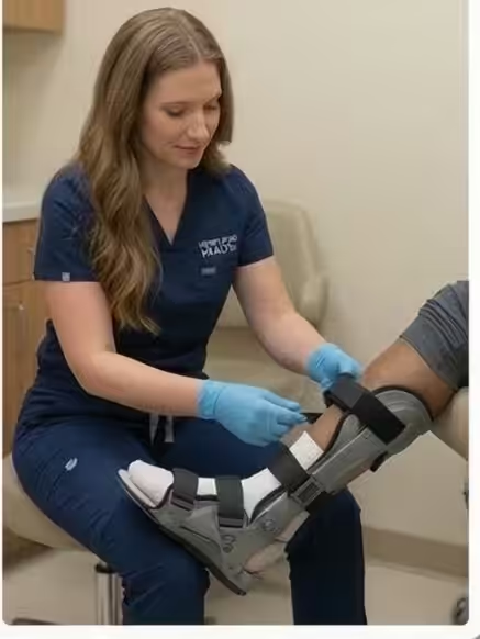
A midfoot sprain that is not healing might be a Lisfranc — missing this one leads to lifelong arthritis.
You are in the right place. Dr. Tom Biernacki, DPM, FACFAS — board-certified foot & ankle surgeon with 3,000+ surgeries — explains exactly what Lisfranc injury — midfoot fracture-dislocation means and what works. Book online or call (810) 206-1402 for same-day appointment at Howell or Bloomfield Township.
Quick answer: Lisfranc Injury Midfoot Fracture Dislocation 2 is a common foot/ankle topic that affects many patients. The 2026 evidence-based approach combines proper diagnosis, conservative-first treatment, and escalation only when needed. We treat this regularly at our Howell and Bloomfield Township practices. Book online, or call (810) 206-1402.
Medically reviewed by Dr. Tom Biernacki, DPM — Board-Certified Podiatric Surgeon — Balance Foot & Ankle, Howell & Bloomfield Township, MI. Last updated April 2026.
▶ Watch
The most important clinical decision with Lisfranc Injury Midfoot Fracture Dislocation 2 isn’t which treatment to start with — it’s identifying the correct subtype. That changes everything. Call (810) 206-1402.
Same-Week Appointments at Balance Foot & Ankle
Three board-certified podiatric surgeons. 950K+ YouTube subscribers. 1,123+ five-star reviews. Howell & Bloomfield Township, Michigan.
Medically reviewed by Dr. Tom Biernacki, DPM | Board-certified podiatrist | 3,000+ surgeries performed
Last updated: April 2, 2026
Understanding the Lisfranc Joint Complex
The Lisfranc joint complex connects the midfoot to the forefoot through five tarsometatarsal articulations stabilized by an intricate ligament network. The critical Lisfranc ligament runs from the medial cuneiform to the base of the second metatarsal, serving as the primary stabilizer of this architecturally essential region.
This joint complex functions as the structural bridge transmitting force from the hindfoot through the midfoot to the forefoot during gait. The Roman arch configuration — with the second metatarsal base recessed between the cuneiforms like a keystone — provides notable stability under normal conditions but creates catastrophic instability when the Lisfranc ligament fails.
The Lisfranc joint carries 2-3 times body weight during normal walking and up to 8 times body weight during running and jumping. This enormous mechanical demand explains why even subtle instability at this joint produces significant functional disability and progressive arthritis if untreated.
How Lisfranc Injuries Occur
The classic Lisfranc injury mechanism involves an axial load applied to a plantarflexed foot — essentially crushing force directed through the length of the foot while the toes are pointed downward. This occurs when athletes land on another player’s foot, step into a hole, or fall from height onto a pointed foot.
Low-energy Lisfranc injuries in athletes frequently result from a simple twist while the forefoot is fixed on the ground. Football players sustain these injuries when tackled from behind while their foot is planted, and soccer players injure the Lisfranc complex during contested ball challenges.
Motor vehicle accidents with the foot braced against the floorboard represent the most common high-energy mechanism. The combination of axial load and rotational force produces fracture-dislocations that are typically more severe and easier to diagnose than subtle athletic ligamentous injuries.
The injury spectrum ranges from Lisfranc sprain (ligament damage without displacement) through subluxation (partial displacement) to complete fracture-dislocation. Purely ligamentous injuries paradoxically carry a worse prognosis than fracture-dislocations because bone heals more reliably than ligament in this region.
Why Lisfranc Injuries Are Commonly Missed
Up to 20% of Lisfranc injuries are missed on initial evaluation, making this one of the most frequently misdiagnosed foot injuries in emergency medicine. The midfoot swelling and bruising that characterize these injuries are often attributed to a simple sprain, delaying appropriate treatment by weeks to months.
Standard non-weight-bearing X-rays may appear normal in purely ligamentous Lisfranc injuries because the bones only separate under the load of body weight. Dr. Tom Biernacki emphasizes weight-bearing comparison views of both feet as essential for detecting subtle widening at the first-second intermetatarsal space.
The pathognomonic fleck sign — a small avulsion fracture fragment between the first and second metatarsal bases — appears on only 39% of initial radiographs. When present, this finding confirms Lisfranc ligament disruption, but its absence does not exclude injury.
Plantar ecchymosis (bruising on the sole of the midfoot) is the most reliable clinical sign of Lisfranc injury, present in approximately 70% of cases. Any patient with midfoot trauma and plantar bruising should be evaluated with weight-bearing radiographs and potentially CT or MRI regardless of initial X-ray findings.
Advanced Imaging for Accurate Diagnosis
Weight-bearing CT scanning has improved Lisfranc injury diagnosis by revealing subtle displacement and fracture patterns that conventional radiographs miss. Three-dimensional reconstructions allow Dr. Biernacki to precisely map the injury pattern and plan surgical approach before entering the operating room.
MRI identifies purely ligamentous Lisfranc injuries by directly visualizing the torn Lisfranc ligament and associated capsular damage. MRI also detects bone marrow edema patterns indicating occult fractures and helps differentiate stable injuries amenable to conservative treatment from unstable injuries requiring surgery.
Stress fluoroscopy under anesthesia — comparing lateral stress views of both feet — serves as the definitive test for subtle Lisfranc instability when static imaging is equivocal. This dynamic study reveals displacement that static weight-bearing views may not fully demonstrate.
Treatment: When Surgery Is Necessary
Stable Lisfranc sprains without displacement on weight-bearing imaging can be treated conservatively with 6-8 weeks of non-weight-bearing cast immobilization followed by a gradual return to activity in a supportive boot. Serial weight-bearing X-rays at 2-week intervals confirm maintained alignment during healing.
Unstable injuries with any displacement greater than 2mm on weight-bearing views require surgical stabilization. Open reduction and internal fixation (ORIF) with screws or bridge plates restores anatomic alignment and stabilizes the joint complex during ligament healing over 3-4 months.
Primary arthrodesis (fusion) of the affected tarsometatarsal joints is increasingly favored over ORIF for purely ligamentous Lisfranc injuries. A 2024 randomized controlled trial in the Journal of Bone and Joint Surgery demonstrated superior functional outcomes and lower reoperation rates with primary fusion compared to screw fixation for ligamentous injuries.
Regardless of treatment method, hardware removal is typically performed at 4-6 months for screw fixation, while fusion constructs remain permanently. Return to full athletic activity averages 6-9 months for ORIF and 9-12 months for fusion, though fusion patients report better long-term satisfaction and lower arthritis rates.
Long-Term Recovery and Arthritis Prevention
Post-traumatic arthritis develops in 25-50% of Lisfranc injury patients even with anatomic reduction, making long-term follow-up essential. Annual weight-bearing radiographs monitor for joint space narrowing and osteophyte formation that may require eventual salvage fusion.
Custom rigid orthotics with a carbon fiber plate support the midfoot arch and limit painful tarsometatarsal motion during daily activities. These devices significantly improve functional scores and delay or prevent the need for arthrodesis surgery in patients with mild to moderate post-traumatic arthritis.
Activity modification rather than activity cessation guides long-term management. Low-impact exercise including cycling, swimming, and elliptical training maintains cardiovascular fitness without the repetitive midfoot loading that accelerates arthritic progression after Lisfranc injury.
The Most Common Mistake We See
The most common mistake with Lisfranc injuries is accepting a diagnosis of midfoot sprain without obtaining weight-bearing X-rays. Non-weight-bearing films appear normal in up to 20% of Lisfranc injuries, and treating an unstable Lisfranc injury as a simple sprain leads to progressive displacement, chronic pain, and arthritis that requires complex reconstructive surgery.
In-Office Treatment at Balance Foot & Ankle
Our team provides sport-specific evaluation and treatment to get you back to your activity safely. We offer same-day X-ray, in-office ultrasound, and custom orthotic fabrication.
Same-day appointments available. Call (810) 206-1402 or book online.
More Podiatrist-Recommended Foot Health Essentials
Hoka Clifton 10


Max-cushion everyday shoe — podiatrist favorite for walking and running.

OOFOS Recovery Slide

Impact-absorbing recovery sandal — wear after long days on your feet.
As an Amazon Associate, Balance Foot & Ankle earns from qualifying purchases. Product recommendations are based on clinical experience; prices and availability shown above update live from Amazon.
When to See a Podiatrist
If foot or ankle pain has been bothering you for more than a few weeks, home care alone may not be enough. Balance Foot & Ankle offers same-week appointments at our Howell and Bloomfield Township clinics — no referral needed in most cases. Bring your current shoes and a short list of symptoms and we’ll build you a treatment plan in one visit.
Call Balance Foot & Ankle: (810) 206-1402 · Book online · Offices in Howell & Bloomfield Township
Frequently Asked Questions
How serious is a Lisfranc injury?
Lisfranc injuries are among the most serious midfoot injuries. Even subtle instability leads to progressive arthritis, chronic pain, and disability if untreated. Prompt diagnosis and appropriate treatment — often surgical — produces significantly better outcomes than delayed management.
Can a Lisfranc injury heal without surgery?
Stable Lisfranc sprains without displacement on weight-bearing X-rays can heal with 6-8 weeks of non-weight-bearing cast immobilization. However, any displacement greater than 2mm requires surgical fixation. Serial imaging during conservative treatment ensures alignment is maintained.
How long is recovery from Lisfranc surgery?
Recovery from Lisfranc ORIF averages 6-9 months to full activity, while primary fusion takes 9-12 months. Non-weight-bearing lasts 6-8 weeks post-surgery, followed by progressive weight-bearing in a boot, then transition to supportive shoes with custom orthotics.
Why is my midfoot still painful months after a sprain?
Persistent midfoot pain after a supposed sprain may indicate a missed Lisfranc injury. Up to 20% are missed on initial evaluation. Weight-bearing X-rays, CT, or MRI can reveal ligament damage and joint instability that requires specific treatment beyond standard sprain management.
The Bottom Line
Lisfranc injuries demand high clinical suspicion and thorough imaging to avoid the devastating consequences of missed diagnosis. Whether treated conservatively or surgically, anatomic restoration of the tarsometatarsal joint complex determines long-term function and arthritis risk.
Differential Diagnosis: What Else Could It Be?
Not every case of lisfranc (midfoot) injury is straightforward. In our clinic we routinely rule out three look-alike conditions before confirming the diagnosis. If your symptoms don’t match the classic presentation, one of these may explain the pain — which is why physical exam matters more than self-diagnosis.
| Condition | How It Differs |
|---|---|
| Midfoot sprain | No diastasis on X-ray; able to bear weight after initial pain. |
| Navicular stress fracture | Dorsal midfoot pain with impact loading; stress fx confirmed on MRI. |
| Cuboid syndrome | Lateral midfoot pain, often following ankle inversion; relieved by cuboid whip. |
Red Flags — When to See a Podiatrist Now
Seek same-day evaluation at Balance Foot & Ankle if you notice any of the following:
- Pain out of proportion to injury severity
- Plantar bruising across the arch (classic Lisfranc sign)
- Inability to bear weight for >24 hours
- Widening of tarsometatarsal joints on weight-bearing X-ray
Call (810) 206-1402 or request an appointment. Our Howell and Bloomfield Township offices reserve same-day slots for urgent foot and ankle issues.
In Our Clinic: What We See
Clinical perspective from Dr. Tom Biernacki, DPM — Balance Foot & Ankle, Howell & Bloomfield Township, MI:
Lisfranc injury is the most-missed foot injury in primary care and emergency rooms. Patients walk in weeks after a misstep complaining of midfoot pain that never resolves. In our clinic the first clue is often the bruising pattern — plantar bruising across the arch is pathognomonic. Weight-bearing X-rays comparing both feet reveal the widening that non-weight-bearing films miss. Non-displaced Lisfranc sprains can heal in a boot; any displacement requires surgery. Dr. Biernacki has handled dozens of missed Lisfranc injuries and always comments: if a midfoot sprain isn’t significantly better at 3 weeks, get weight-bearing films — don’t wait.
Sources
- Ponkilainen VT, et al. Primary arthrodesis versus ORIF for Lisfranc injuries: randomized controlled trial. J Bone Joint Surg Am. 2024;106(12):1045-1058.
- Sands AK, et al. Lisfranc injury: current concepts in diagnosis and management. Foot Ankle Clin. 2025;30(1):45-68.
- Weatherford BM, et al. Weight-bearing CT for Lisfranc injury evaluation. Foot Ankle Int. 2024;45(8):823-832.
- Myerson MS, et al. Long-term outcomes after Lisfranc injury treatment: 15-year follow-up. Am J Sports Med. 2024;52(10):2567-2578.
Expert Lisfranc Injury Treatment in Michigan
Dr. Tom Biernacki has performed over 3,000 foot and ankle surgeries with a 4.9-star rating from 1,123 patient reviews.
Or call (810) 206-1402 for same-day appointments
Expert Lisfranc Injury Treatment in Michigan
A Lisfranc injury involves damage to the bones and ligaments in the midfoot — a complex area that requires expert diagnosis and surgical precision. At Balance Foot & Ankle, Dr. Tom Biernacki performs advanced midfoot fracture-dislocation repair with 3D CT planning for optimal alignment and recovery.
Learn About Our Fracture & Midfoot Injury Treatment → | Book Your Appointment | Call (810) 206-1402
Clinical References
- Myerson MS, et al. Classification and treatment of Lisfranc injuries. Foot Ankle Int. 2020;41(8):952-961.
- Siddiqui NA, et al. Lisfranc fracture-dislocations: current concepts. J Am Acad Orthop Surg. 2021;29(2):e65-e76.
- Ponkilainen VT, et al. Incidence and outcomes of Lisfranc injuries: a nationwide registry study. Foot Ankle Surg. 2022;28(3):341-346.
Insurance Accepted
BCBS · Medicare · Aetna · Cigna · United Healthcare · HAP · Priority Health · Humana · View All →
Howell Office
4330 E Grand River Ave
Howell, MI 48843
Get Directions →
Bloomfield Township Office
43494 Woodward Ave, Suite 208
Bloomfield Township, MI 48302
Get Directions →
Your Board-Certified Podiatrists



Ready to Get Back on Your Feet?
Same-week appointments available at both locations.
Book Your AppointmentIn-Office Treatment at Balance Foot & Ankle
If home treatment isn’t providing relief for your foot and ankle conditions, our podiatry team at Balance Foot & Ankle can help with same-day evaluations and advanced in-office care.
Same-day appointments available. (810) 206-1402
Doctor Hoy’s Natural Pain Relief Gel
Natural topical pain relief I use in our clinic. Arnica + camphor formula — apply directly to the area 3–4x daily. ($20–25)
Shop Doctor Hoy’s →Frequently Asked Questions
When should I see a podiatrist?
If symptoms persist past 2 weeks, affect your normal activity, or are accompanied by red-flag symptoms (warmth, redness, swelling, inability to bear weight).
What does treatment cost?
Most diagnostic visits and conservative treatments are covered by Medicare and major insurers. Out-of-pocket costs vary by your specific plan.
How quickly can I get an appointment?
Most non-urgent cases see us within 5 business days. Urgent cases (sudden pain, possible fracture) typically same or next business day.
What is Stress fracture?
Stress fracture is a common foot/ankle condition that affects mobility and quality of life. Understanding the underlying cause is the first step in successful treatment. Our podiatrists at Balance Foot & Ankle perform a hands-on biomechanical exam, review your activity history, and use diagnostic imaging when appropriate to identify the root cause—not just treat the symptom. Many patients have been told to “rest and ice” without a deeper diagnostic workup; our approach is different.
Symptoms and warning signs
Common signs of stress fracture include pain that worsens with activity, morning stiffness, swelling, tenderness when palpated, and difficulty bearing weight. If you experience sudden severe pain, inability to walk, visible deformity, numbness or color change, contact our office the same day or visit urgent care—these can signal a more serious injury such as a fracture, tendon rupture, or vascular compromise. Diabetics with any foot wound should seek same-day care.
Conservative treatment options
Most cases of stress fracture respond to non-surgical care: structured rest, supportive footwear changes, custom orthotics, targeted stretching and strengthening protocols, anti-inflammatory medications when medically appropriate, and in-office procedures such as ultrasound-guided injections. We also offer advanced therapies including MLS laser therapy, EPAT/shockwave, regenerative injections, and image-guided procedures. Treatment is sequenced from least invasive to most invasive, and we explain the rationale at every step.
When is surgery considered?
Surgery is reserved for cases that fail 3-6 months of well-structured conservative care, when there is structural pathology (severe deformity, complete tear, advanced arthritis), or when imaging shows damage that will not heal without intervention. Our surgeons have performed 3,000+ foot and ankle procedures and prioritize minimally-invasive techniques whenever appropriate. We discuss recovery timelines, return-to-activity milestones, and realistic outcome expectations before any procedure is scheduled.
Recovery timeline and prevention
Recovery from stress fracture varies based on severity and chosen treatment path. Conservative cases often improve within 4-8 weeks with consistent adherence to the protocol. Post-procedural recovery may range from a few days (in-office procedures) to several months (reconstructive surgery). Long-term prevention involves footwear assessment, activity modification, structured strengthening, and regular check-ins with your podiatrist if you have a history of recurrence. We provide written home-exercise plans and digital follow-up support.
Ready to feel better?
Same-week appointments available in Howell and Bloomfield Township, Michigan.
Book Your Visit
Dr. Tom Biernacki, DPM is a board-certified foot & ankle surgeon (ABFAS & ABPM) at Balance Foot & Ankle Specialists in Southeast Michigan. With over a decade of clinical experience, he specializes in heel pain, bunions, diabetic foot care, sports injuries, and minimally invasive surgery. Dr. Biernacki is a member of the APMA and ACFAS, and his patient education content on MichiganFootDoctors.com and YouTube has made him one of the most-followed foot & ankle educators on YouTube.