


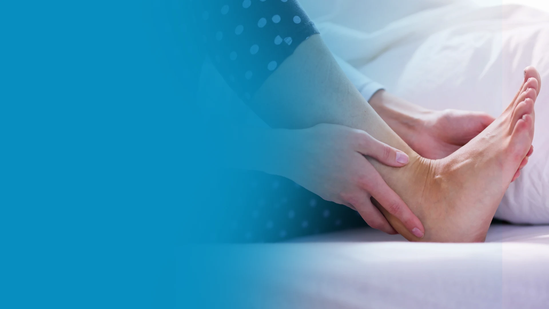
Achilles tendon reconstruction with allograft augmentation is for large tendon defects from neglected ruptures or chronic tears — restoring function patients had given up on.
You’re in the right place. Dr. Tom Biernacki, DPM, FACFAS — board-certified foot & ankle surgeon with 3,000+ surgeries — explains exactly what Achilles tendon reconstruction means and what works. Call (810) 206-1402 for same-day appointment at Howell or Bloomfield Hills.
Quick answer: Achilles Tendon Reconstruction Allograft Augmentation Large Defects is a common foot/ankle topic that affects many patients. The 2026 evidence-based approach combines proper diagnosis, conservative-first treatment, and escalation only when needed. We treat this regularly at our Howell and Bloomfield Hills practices. Call (810) 206-1402.
Medically reviewed by Dr. Tom Biernacki, DPM — Board-Certified Podiatric Surgeon — Balance Foot & Ankle, Howell & Bloomfield Hills, MI. Last updated April 2026.
Medically Reviewed by Dr. Tom Biernacki, DPM — Board-Certified Podiatrist, Balance Foot & Ankle Specialists, Michigan. Last updated April 2026.
Quick Answer
Achilles tendon reconstruction is a surgical procedure to repair a severely damaged, ruptured, or chronically degenerated Achilles tendon that cannot heal with conservative treatment alone. Techniques range from direct repair for acute ruptures to tendon transfer procedures (FHL transfer) for chronic cases with significant tissue loss. Recovery takes 4-9 months depending on the technique, with structured rehabilitation essential for regaining strength and returning to activity.
Medical Review
Medically reviewed by Dr. Tom Biernacki, DPM — Board-certified podiatrist and foot/ankle surgeon at Balance Foot & Ankle, Southeast Michigan. Dr. Biernacki performs Achilles tendon repair and reconstruction for acute ruptures, chronic tears, and degenerative tendinopathy.
Affiliate disclosure: This page contains affiliate links to products we recommend. We may earn a small commission at no extra cost to you. We only recommend products used in our clinical practice.
Table of Contents
- What Is Achilles Tendon Reconstruction?
- When Is Reconstruction Necessary?
- Acute Achilles Rupture Repair
- Chronic Achilles Tendon Reconstruction
- FHL Tendon Transfer Technique
- V-Y Advancement and Turndown Flaps
- Allograft and Synthetic Augmentation
- Surgical vs. Non-Surgical Treatment
- Preparing for Surgery
- Recovery Timeline
- Rehabilitation Protocol
- Post-Recovery Orthotic Support
- Pain Management After Surgery
- Compression for Swelling Control
- Most Common Mistake
- Warning Signs of Complications
- Long-Term Outcomes and Expectations
- Video Guide
- Frequently Asked Questions
- Sources
- Book an Appointment
What Is Achilles Tendon Reconstruction?
Achilles tendon reconstruction encompasses a range of surgical procedures designed to restore the structural integrity and function of a severely damaged Achilles tendon. The Achilles is the strongest and largest tendon in the human body, connecting the powerful gastrocnemius and soleus calf muscles to the calcaneus (heel bone). It must withstand forces of up to 8-12 times body weight during running and jumping, making it both essential for mobility and vulnerable to catastrophic failure.
The term “reconstruction” distinguishes more complex procedures from simple primary repair. While a fresh, clean Achilles rupture in a healthy tendon can often be sutured back together (primary repair), chronic tears, neglected ruptures, and tendons with extensive degenerative damage may have a gap that cannot be closed by direct suturing. Reconstruction bridges this gap using the patient’s own tissue (autograft), donor tissue (allograft), or synthetic materials.
Achilles reconstruction is among the most technically demanding procedures in foot and ankle surgery. The surgeon must restore not only the structural continuity of the tendon but also its mechanical properties — appropriate tension, length, and the ability to transmit the powerful forces generated by the calf muscles. A tendon reconstructed too loosely will not generate adequate push-off power. A tendon repaired too tightly will limit ankle flexibility and predispose to re-rupture.
When Is Achilles Reconstruction Necessary?
Not every Achilles tendon problem requires surgery. Mild to moderate Achilles tendinopathy typically responds well to eccentric strengthening exercises, physical therapy, and activity modification. However, several clinical scenarios warrant surgical reconstruction rather than continued conservative management.
Acute complete Achilles rupture is the most common indication for surgical intervention, particularly in active adults under 60 who want to return to sport. While non-surgical treatment with functional bracing and early rehabilitation has shown comparable outcomes in some studies, surgical repair reduces the re-rupture rate from approximately 12% (non-surgical) to 4% (surgical) and produces slightly better plantarflexion strength — an important consideration for athletes.
Chronic Achilles rupture — defined as a rupture diagnosed more than 4-6 weeks after the initial injury — presents a greater surgical challenge because the torn tendon ends retract and scar tissue fills the gap. The longer the delay between rupture and treatment, the larger the gap and the more complex the reconstruction required. Neglected ruptures identified months after the initial injury may require tendon transfer procedures to restore function.
Failed conservative treatment for insertional Achilles tendinopathy is another indication. When 6-12 months of comprehensive conservative management (eccentric exercises, orthotics, physical therapy, possibly shockwave therapy or PRP) fails to resolve symptoms, surgical debridement of the degenerated tendon with reattachment to the calcaneus (often using suture anchors) may be necessary. If more than 50% of the tendon substance is degenerated, reconstruction with augmentation may be required.
Acute Achilles Rupture Repair
Acute Achilles rupture repair performed within 2-4 weeks of injury offers the best surgical outcomes because the tendon ends have not yet retracted significantly and the tissue quality remains adequate for strong suture fixation. The procedure is typically performed through a medial or posterolateral incision approximately 8-12cm long, centered over the rupture site.
The surgeon identifies the ruptured tendon ends, debrids any frayed or necrotic tissue, and reapproximates the ends using a core suture technique (Krackow or Bunnell locking stitch) combined with an epitenon repair. The core suture provides the structural strength of the repair while the epitenon suture restores the smooth gliding surface and adds additional strength. Modern suture materials and techniques achieve repair strengths sufficient to begin early functional rehabilitation.
Minimally invasive and percutaneous techniques have gained popularity for acute ruptures, using smaller incisions and specialized instruments to pass sutures through the tendon ends. These techniques reduce wound complications — a significant concern with the traditional open approach, where wound healing problems occur in 5-15% of cases. However, minimally invasive repair carries a slightly higher risk of sural nerve injury (5-10%) compared to open repair (3-5%).
Chronic Achilles Tendon Reconstruction
Chronic Achilles ruptures present a fundamentally different surgical challenge than acute injuries. The tendon ends retract, the gap fills with disorganized scar tissue, the calf muscles shorten and develop fatty infiltration, and the overall quality of the remaining tendon deteriorates. The size of the gap between the retracted tendon ends largely determines the reconstruction technique required.
Gaps less than 2cm can sometimes be managed with extensive mobilization of the tendon ends and direct repair with augmentation. Gaps of 2-5cm typically require a V-Y lengthening of the proximal tendon or a turndown flap to bridge the defect. Gaps greater than 5cm usually necessitate a tendon transfer — most commonly the flexor hallucis longus (FHL) — to provide both structural bridging and a new motor unit for plantarflexion power.
The quality of the remaining Achilles tendon substance influences surgical planning significantly. Tendon with extensive mucoid degeneration, calcification, or xanthomatous changes may be too compromised to hold sutures reliably, requiring more aggressive reconstruction techniques even for smaller gaps. MRI evaluation before surgery helps characterize the tendon quality, gap size, and degree of calf muscle atrophy to guide surgical planning.
FHL Tendon Transfer: The Gold Standard for Large Defects
Flexor hallucis longus (FHL) tendon transfer has become the most widely used technique for reconstructing chronic Achilles defects greater than 5cm. The FHL muscle is harvested through the same posterior incision used for Achilles exposure and transferred to the calcaneus to function as a supplementary plantarflexor alongside the remaining Achilles tissue.
The FHL is ideally suited for this role for several reasons. It lies immediately adjacent to the Achilles tendon, making harvest straightforward. Its muscle belly extends further distally than other calf muscles, providing vascularized tissue that fills the reconstruction gap. The FHL fires in synchrony with the gastrocnemius-soleus complex during the gait cycle, meaning it is already neurologically programmed for the plantarflexion function it assumes. The functional deficit from sacrificing the FHL — reduced great toe flexion strength — is generally well tolerated and rarely noticed by patients in daily activities.
The FHL tendon is secured to the calcaneus through a bone tunnel or with a suture anchor system (interference screw fixation). The remaining Achilles tissue is then sutured over the FHL transfer to create a reinforced construct. This combination of tendon transfer with host tissue augmentation produces consistently good functional outcomes, with 85-90% of patients reporting satisfaction and ability to return to recreational activities.
V-Y Advancement and Turndown Flaps
For moderate gaps (2-5cm), V-Y advancement of the gastrocnemius aponeurosis provides additional length to the proximal tendon stump, allowing direct repair without tendon transfer. The surgeon makes a V-shaped incision in the gastrocnemius aponeurosis proximal to the rupture, advances the tendon distally to close the gap, and closes the aponeurosis in a Y configuration. This technique effectively lengthens the musculotendinous unit by 2-3cm while preserving the continuity of the native Achilles tendon.
Turndown flaps involve rotating a strip of the proximal tendon stump 180 degrees on its base and suturing it into the distal stump to bridge the gap. This technique uses autologous tissue with the same mechanical properties as the native tendon, potentially providing superior strength compared to synthetic augmentation. However, turndown flaps weaken the proximal tendon, and the flap may not be viable if the proximal tissue quality is poor.
These techniques are often combined — for example, a V-Y advancement with FHL transfer augmentation provides both length and additional motor unit for larger defects. The choice of technique depends on the gap size, tissue quality, patient activity demands, and surgeon experience. Your surgeon will discuss the planned approach based on your preoperative imaging and may adjust the technique intraoperatively based on findings at the time of surgery.
Allograft and Synthetic Augmentation
When the native tendon tissue is insufficient for reconstruction, allograft (donor tissue) or synthetic materials can augment the repair. Acellular dermal matrix (ADM) products — processed human or animal tissue scaffolds — provide a biological framework that the patient’s cells can infiltrate and remodel over time. These products are particularly useful for wrapping and reinforcing a primary repair that has marginal tissue quality.
Fresh-frozen Achilles tendon allograft can replace large segments of missing tendon, providing immediate structural support. The allograft gradually incorporates as the patient’s cells migrate into the scaffold and lay down new collagen. However, allograft tissue takes longer to fully incorporate than autograft and carries a small risk of disease transmission and immune rejection, although modern processing techniques have made these risks extremely low.
Synthetic scaffolds and suture tape augmentation provide immediate mechanical reinforcement that protects the biological repair during the early healing phase when the tissue is weakest. These devices share the load with the healing tendon, allowing earlier mobilization and potentially accelerating rehabilitation. However, long-term outcomes data for synthetic devices is still limited compared to established biological techniques.
Surgical vs. Non-Surgical Treatment for Achilles Rupture
The debate between surgical and non-surgical management of acute Achilles ruptures has evolved significantly with improvements in functional rehabilitation protocols. Modern non-surgical treatment — using early functional bracing with progressive weight bearing and early motion rather than prolonged casting — has narrowed the outcomes gap between surgical and conservative approaches.
Surgical repair consistently shows a lower re-rupture rate (2-5% vs. 8-12% non-surgical) and slightly better plantarflexion strength at long-term follow-up. Non-surgical treatment avoids surgical wound complications (5-15% with open surgery), anesthesia risks, and the costs of surgery. Patient-reported functional outcomes are remarkably similar between groups when both receive structured rehabilitation.
We generally recommend surgical repair for active patients under 60 who plan to return to sport, patients with large tendon gaps on ultrasound, and athletes who require maximum plantarflexion power. Non-surgical treatment may be preferred for patients over 60, those with significant medical comorbidities that increase surgical risk, patients with minimal gap on ultrasound, and individuals who are primarily sedentary. Each case requires individualized decision-making based on the patient’s goals, health status, and imaging findings.
Preparing for Achilles Reconstruction Surgery
Optimal preparation improves surgical outcomes and reduces complication risk. Stop smoking at least 4 weeks before surgery — nicotine impairs wound healing and increases the risk of infection and tendon healing failure. Optimize nutrition with a high-protein diet and ensure adequate vitamin C, zinc, and vitamin D levels. If diabetic, work with your primary care physician to achieve the best possible glucose control before surgery.
Prepare your home for the recovery period: arrange bedroom on the main floor if possible, install shower grab bars, stock up on groceries, arrange help with cooking and household tasks for the first 2 weeks, and acquire necessary mobility aids (crutches, knee scooter, or hands-free crutch) before surgery day. Set up a comfortable recovery station with leg elevation pillows, entertainment, phone charger, and medications within easy reach.
Recovery Timeline After Achilles Reconstruction
Weeks 1-2: Non-weight bearing in a posterior splint with the ankle in slight plantarflexion. Strict elevation above heart level. Pain management with prescribed medications and ice therapy. First post-operative visit for wound check and dressing change at 10-14 days.
Weeks 2-6: Transition to a walking boot with heel lifts. Non-weight bearing continues. Sutures removed at 2-3 weeks. Gentle ankle range-of-motion exercises begin at 4 weeks (surgeon-directed). Heel lifts are gradually reduced over this period.
Weeks 6-12: Progressive weight bearing begins in the walking boot based on healing progress. Formal physical therapy initiated with focus on gentle stretching, isometric strengthening, and gait training. Transition from boot to supportive shoes with a heel lift at approximately 10-12 weeks.
Months 3-6: Progressive strengthening — concentric and eccentric calf exercises. Walking for exercise begins. Swimming and cycling for cardiovascular fitness. Gradual return to normal daily activities without restrictions. Continue physical therapy 2-3 times per week.
Months 6-12: Sport-specific rehabilitation for athletes. Walk-to-jog progression begins at approximately 5-6 months. Full return to running by 6-8 months. Return to competitive sport with jumping and pivoting by 9-12 months. The tendon continues to remodel and strengthen for up to 18 months post-surgery.
Rehabilitation Protocol Highlights
Successful rehabilitation after Achilles reconstruction follows a progressive loading protocol that respects the biology of tendon healing while challenging the tissue enough to stimulate adaptation. The key principle is “progressive overload within the tissue’s capacity” — gradually increasing the demands on the healing tendon without exceeding its structural tolerance.
Isometric exercises (holding positions without movement) begin first because they load the tendon without elongating it, reducing the risk of disrupting the repair. Double-leg calf raises progress to single-leg calf raises as strength improves. Eccentric exercises (lowering the heel below a step) are introduced around 12 weeks because they stimulate organized collagen remodeling — the most important adaptation for long-term tendon health.
Proprioceptive training begins as soon as full weight bearing is achieved. Balance exercises, wobble board training, and dynamic stability drills retrain the neuromuscular system to control ankle position during functional activities. This proprioceptive retraining is essential for preventing re-injury and restoring confidence in the ankle’s stability during sport-specific movements.
Post-Recovery Orthotic Support
After completing rehabilitation from Achilles reconstruction, long-term orthotic support helps protect the healed tendon and optimize foot biomechanics. We recommend PowerStep Pinnacle Insoles for ongoing daily use after Achilles reconstruction. The cushioned heel absorbs impact forces that would otherwise be transmitted through the healed tendon, while the arch support maintains proper foot alignment that reduces the strain on the calf-Achilles complex during walking and activity.
A temporary heel lift (6-12mm) in both shoes may be recommended for 3-6 months after transitioning out of the boot. This slight elevation reduces the ankle dorsiflexion required during walking, protecting the repair from excessive stretching during the remodeling phase. The heel lift is gradually reduced and eventually removed as the tendon gains flexibility and strength.
Pain Management After Surgery
We recommend Doctor Hoy’s Natural Pain Relief Gel for managing residual discomfort during the later rehabilitation phase (after the incision is fully healed, typically 4-6 weeks post-surgery). Apply to the skin around the Achilles tendon before and after physical therapy sessions for cooling relief that supports your rehabilitation effort without the systemic effects of oral medications.
Compression for Swelling Control
We recommend DASS Compression Socks once cleared by your surgeon (typically 4-6 weeks post-op after wound healing). Graduated compression reduces the persistent ankle and calf swelling that accompanies Achilles reconstruction recovery and supports venous return during the months when activity is gradually increasing.
Most Common Mistake: Skipping Physical Therapy
The biggest mistake after Achilles reconstruction is treating the surgery as the cure rather than the beginning of recovery. Surgery restores structural continuity, but it is the structured rehabilitation that follows that determines your functional outcome. Patients who skip or abbreviate physical therapy consistently achieve worse strength recovery, reduced ankle flexibility, and higher rates of re-injury compared to those who complete the full 4-6 month rehabilitation protocol. Your surgeon repairs the tendon. Physical therapy rebuilds its function. Both are essential.
Warning Signs of Complications
- Sudden sharp pain and loss of push-off strength — possible re-rupture requiring urgent evaluation
- Wound drainage persisting beyond 5 days — may indicate wound healing problem or superficial infection
- Increasing redness and warmth around the incision — early infection requiring antibiotics
- Calf swelling with warmth and tenderness — possible deep vein thrombosis needing emergency assessment
- Progressive numbness on the outer foot — sural nerve irritation from the surgical approach
- Inability to achieve neutral ankle position by 8 weeks — concerning for adhesion or repair tightness
- Persistent significant pain at 6 weeks despite following the recovery protocol — warrants imaging evaluation
Long-Term Outcomes and Realistic Expectations
Long-term outcomes after Achilles reconstruction are generally favorable, with 85-95% of patients reporting good to excellent results. Most patients return to walking without limitation and can perform daily activities without pain. Plantarflexion strength typically recovers to 75-85% of the uninjured side — a deficit that is functionally significant only during explosive athletic activities.
Return to sport varies by sport type and level. Approximately 80% of recreational athletes return to their pre-injury sport, while competitive athletes return at lower rates (60-75%) — though this partly reflects the psychological barrier of trusting the repaired tendon during high-intensity activity. Persistent ankle stiffness and reduced calf power are the most common long-term complaints, typically manageable with ongoing stretching and strengthening exercises.
Re-rupture after surgical repair occurs in approximately 2-5% of cases, with the highest risk during the 3-6 month window when patients are returning to activity. Adherence to the rehabilitation timeline and gradual return to sport significantly reduces this risk. Long-term, the repaired Achilles tendon never fully returns to pre-injury tissue quality, making lifelong attention to calf flexibility, appropriate footwear, and gradual activity progression important for tendon health.
Video: Achilles Tendon Treatment Guide
Watch Dr. Biernacki discuss Achilles tendon conditions and treatment approaches, from conservative management to surgical reconstruction.
More Podiatrist-Recommended Achilles Essentials
Achilles Night Splint
United Ortho dorsiflexion splint — reduces morning Achilles tendon stiffness.
Cushioned Running Shoe

![Achilles Tendonitis & Back of Heel Pain [BEST Home Treatments 2024!]](https://www.michiganfootdoctors.com/wp-content/cache/flying-press/d645ea3f3d27024136b88a074b2b7894.jpg)
Watch: Achilles Tendonitis & Back of Heel Pain [BEST Home Treatments 2024!] — MichiganFootDoctors YouTube
Hoka Clifton 10 — max-heel-cushion offloads the Achilles with every step.
Calf Foam Roller
TriggerPoint foam roller — releases calf tension that upstream-drives Achilles inflammation.
As an Amazon Associate, Balance Foot & Ankle earns from qualifying purchases. Product recommendations are based on clinical experience; prices and availability shown above update live from Amazon.
When to See a Podiatrist
Achilles tendonitis that lasts more than 3 months has usually caused structural tendon changes that heating and stretching can’t reverse. Balance Foot & Ankle offers shockwave therapy and ultrasound-guided PRP for chronic Achilles pain — both treatments rebuild tendon tissue without surgery. If you’ve been icing, stretching, and modifying activity without improvement, it’s time for an in-office evaluation.
Call Balance Foot & Ankle: (810) 206-1402 · Book online · Offices in Howell & Bloomfield Hills
Frequently Asked Questions
How long does Achilles reconstruction surgery take?
The surgery itself typically takes 60-90 minutes for a primary repair and 90-120 minutes for complex reconstruction with tendon transfer (FHL transfer) or augmentation. The procedure is performed under regional anesthesia (ankle block or popliteal nerve block) or general anesthesia. You will spend 1-2 hours in the recovery room and most patients go home the same day.
Will I be able to run again after Achilles reconstruction?
Most patients can return to running 6-8 months after Achilles reconstruction, starting with a gradual walk-to-jog progression. Approximately 80% of recreational athletes return to running at their pre-injury level. Competitive athletes may take 9-12 months for full return. The key is completing the full rehabilitation protocol and achieving adequate calf strength before starting impact activities.
What is the difference between Achilles repair and reconstruction?
Achilles repair involves suturing the torn tendon ends directly back together, typically possible within 2-4 weeks of an acute rupture. Reconstruction is needed when the tendon gap is too large for direct repair (usually chronic or neglected ruptures) and involves bridging the defect using tendon transfers, tissue flaps, or graft materials. Reconstruction is a more complex procedure with a longer recovery.
How long before I can bear weight after Achilles surgery?
Weight-bearing timelines vary by procedure and surgeon preference. Most protocols begin partial weight bearing in a boot at 4-6 weeks and progress to full weight bearing by 8-10 weeks. Some accelerated protocols allow earlier weight bearing with protective bracing. Your surgeon will advance weight bearing based on the repair technique used and the quality of healing observed at follow-up visits.
What are the risks of Achilles reconstruction surgery?
Risks include wound healing complications (5-15%, higher in open techniques), infection (1-3%), sural nerve injury causing lateral foot numbness (3-10%), re-rupture (2-5%), deep vein thrombosis (1-2%), persistent weakness, and stiffness. Smoking, diabetes, and steroid use significantly increase complication rates. Most complications are manageable and do not prevent a good long-term outcome.
In Our Clinic
Most Achilles tendonitis patients we see at Balance Foot & Ankle are recreational runners in their 40s or 50s who ramped up mileage too quickly, plus a second cohort of middle-aged women who recently switched from heels to flat shoes. The first question we ask is whether the pain is at the insertion on the heel bone versus 2–6 cm up the mid-substance — the treatment ladder is genuinely different. Eccentric heel-drops, heel lifts, and a soft-strike gait retraining pass resolve ~80 % of cases. The ones who aren’t improving by week 8 usually have an unrecognized Haglund’s deformity or insertional calcific tendinosis that needs imaging.
Sources
- Maffulli N, et al. “Achilles tendon rupture management: techniques, outcomes, and complications.” EFORT Open Reviews. 2021;6(11):1011-1019.
- Metz R, et al. “Acute Achilles Tendon Rupture: Minimally Invasive Surgery Versus Non-Operative Treatment.” Journal of Bone and Joint Surgery. 2008;90(9):1876-1883.
- Hahn F, et al. “Augmented Repair of Acute Achilles Tendon Ruptures.” Foot and Ankle Clinics. 2019;24(3):441-457.
- Den Hartog BD. “Flexor Hallucis Longus Transfer for Chronic Achilles Tendonosis.” Foot and Ankle International. 2003;24(3):233-237.
- Olsson N, et al. “Stable Surgical Repair With Accelerated Rehabilitation Versus Nonsurgical Treatment for Acute Achilles Tendon Ruptures.” American Journal of Sports Medicine. 2013;41(12):2867-2876.
Achilles Tendon Problem? Get Expert Surgical Care
Dr. Biernacki at Balance Foot & Ankle performs Achilles tendon repair and reconstruction using modern techniques for optimal outcomes. From initial evaluation to full rehabilitation, we provide comprehensive Achilles care.
Related Articles
- Achilles Tendinitis Treatment Guide
- Ankle Surgery Options
- Foot Surgery Recovery Timeline
- Sports Podiatrist Near Me
- Podiatrist Recommended Foot Care Products
When to Consider Achilles Tendon Reconstruction
If you have a large Achilles tendon defect or a chronic rupture that hasn’t healed, reconstruction with allograft tissue can restore function. At Balance Foot & Ankle, Dr. Biernacki performs Achilles reconstruction at our Howell and Bloomfield Hills offices.
Learn About Our Achilles Treatment Options | Book Your Appointment | Call (810) 206-1402
Clinical References
- Den Hartog BD. “Surgical strategies: delayed diagnosis or neglected Achilles tendon ruptures.” Foot & Ankle International. 2008;29(4):456-463.
- Maffulli N, Via AG, Oliva F. “Chronic Achilles tendon rupture.” Open Orthopaedics Journal. 2017;11:660-669.
- Elias I, Besser M, Nazarian LN, Raikin SM. “Reconstruction for missed or neglected Achilles tendon rupture with V-Y lengthening and flexor hallucis longus tendon transfer through one incision.” Foot & Ankle International. 2007;28(12):1238-1248.
Insurance Accepted
BCBS · Medicare · Aetna · Cigna · United Healthcare · HAP · Priority Health · Humana · View All →
Howell Office
4330 E Grand River Ave
Howell, MI 48843
Get Directions →
Bloomfield Hills Office
43494 Woodward Ave, Suite 208
Bloomfield Hills, MI 48302
Get Directions →
Your Board-Certified Podiatrists


Ready to Get Back on Your Feet?
Same-week appointments available at both locations.
Book Your AppointmentWhen Shoes Aren’t Enough — Dr. Tom’s Top 9 Orthotics
About 30% of patients I see for foot pain need MORE than a great shoe — they need a structured insole. Below: my complete 2026 orthotic ranking with pros, cons, and the specific patient I’d give each one to.
★ DR. TOM’S COMPLETE 2026 ORTHOTIC RANKING
9 Best Prefab Orthotics by Use Case
PowerStep, Currex, Spenco, Vionic, and Superfeet — every orthotic I’ve fitted to thousands of patients across both Michigan offices. Each card includes pros, cons, and the specific patient I’d give it to. Real Amazon ratings, review counts, and prices below.
Best All-Purpose Orthotic for Most Patients
Semi-rigid arch shell + dual-layer cushion + deep heel cup. The orthotic I’ve fitted to more patients than any other for 15 years. APMA-accepted. Trim-to-fit design works in athletic shoes, casual shoes, and most work boots.

✓ Pros
- Semi-rigid arch shell provides true biomechanical correction
- Deep heel cup centers the heel and reduces lateral instability
- Dual-layer cushion (top + bottom) lasts 9-12 months daily wear
- Available in 8 sizes for precise fit
- APMA-accepted and clinically validated
- Lower price than Superfeet Green for equivalent function
✗ Cons
- Too thick for most dress shoes (use ProTech Slim instead)
- Some break-in period required (3-7 days for arch tolerance)
- Not enough correction for severe pes planus or rigid pes cavus
Dr. Tom’s Recommendation: If a patient has run-of-the-mill plantar fasciitis, mild flat feet, or arch fatigue, this is the first orthotic I try. Better value than Superfeet for 90% of patients, which is why I swapped it into our clinic kits three years ago. Sub-$50 typically.
Maximum Motion Control · Flat Feet & Severe Over-Pronation
PowerStep’s most aggressive stability orthotic. Adds a 2°-7° medial heel post on top of the standard PowerStep platform — designed specifically for flat-footed patients and severe pronators who need real corrective force.

✓ Pros
- 2°-7° medial heel post adds aggressive pronation control
- Same trusted PowerStep arch shell, more correction
- Built specifically for flat-foot biomechanics
- Excellent for posterior tibial tendon dysfunction (PTTD)
- Removable top cover for cleaning
✗ Cons
- Too aggressive for neutral-arch patients
- Needs longer break-in (10-14 days) due to stronger correction
- Adds 2-3 mm of stack height — won’t fit slim dress shoes
Dr. Tom’s Recommendation: When a patient comes in with significant flat feet AND symptoms (heel pain, arch pain, knee pain), the Original PowerStep isn’t aggressive enough. The Maxx is what gets prescribed. About 25% of my flat-footed patients end up here.
Low-Profile · Fits Dress Shoes & Narrow Casuals
3 mm slim profile with podiatrist-designed tri-planar arch technology. Engineered specifically to fit inside dress shoes, oxfords, loafers, and women’s flats without crowding the toe box. Vionic was founded by an Australian podiatrist.
✓ Pros
- 3 mm slim profile (vs 7-10 mm for standard orthotics)
- Tri-planar arch technology adds support without bulk
- Built-in deep heel cup despite slim design
- Fits dress shoes WITHOUT having to remove the factory insole
- Trim-to-fit · APMA-accepted
✗ Cons
- Less arch support than full-volume orthotics
- Top cover wears faster than thicker alternatives
- Not enough correction for severe foot deformities
Dr. Tom’s Recommendation: My default when a patient says ‘I need orthotics but I have to wear dress shoes for work.’ Slim enough to fit in oxfords and pumps without the heel sliding out. The single highest-impact change you can make for office workers with foot pain.
Built-In Metatarsal Pad · Morton’s Neuroma · Ball-of-Foot Pain
Standard Pinnacle orthotic with a built-in metatarsal pad positioned proximal to the metatarsal heads — the exact location that offloads neuromas and metatarsalgia. No need for separate met pads or pad placement guesswork.

✓ Pros
- Built-in met pad eliminates DIY pad placement errors
- Specifically designed for Morton’s neuroma + metatarsalgia
- Same trusted PowerStep arch + heel cup platform
- Top cover protects sensitive forefoot skin
- Faster relief than orthotics + add-on met pads
✗ Cons
- Met pad position is fixed (can’t fine-tune individual placement)
- Some patients with very small or very large feet need custom
- Slightly thicker than the standard Pinnacle
Dr. Tom’s Recommendation: If a patient has Morton’s neuroma, sesamoiditis, or generalized ball-of-foot pain (metatarsalgia), this saves a clinic visit and a prescription. The built-in pad placement is anatomically correct for 80% of feet. Way better than DIY met pads.
Adaptive Dynamic Arch · Athletic & Daily Wear
Currex’s flagship adaptive arch technology — the orthotic flexes with your gait instead of fighting it. Different stiffness zones along the length give you targeted support at the heel, midfoot, and forefoot. Available in three arch heights (low/medium/high).

✓ Pros
- Dynamic flex zones adapt to natural gait cycle
- Three arch heights ensure precise fit
- Lighter than rigid orthotics (no ‘heavy foot’ feel)
- Excellent for runners and athletic walkers
- European podiatric design (German engineering)
✗ Cons
- More expensive than PowerStep Original ($55-65 typically)
- Less aggressive correction than Pinnacle Maxx for severe cases
- Three arch heights means you must self-select correctly
Dr. Tom’s Recommendation: I started recommending Currex three years ago for runners who said PowerStep felt ‘too rigid.’ The dynamic flex zones respect natural gait. Best for active patients who walk 8K+ steps daily and don’t need maximum motion control.
Running-Specific · Heel Strike + Forefoot Strike Compatible
Currex’s purpose-built running orthotic. The midfoot flex zone is positioned for runner’s gait mechanics, with a flared heel cushion for heel strikers and a forefoot rocker for midfoot/forefoot strikers. Tested on 1000+ runners during product development.

✓ Pros
- Designed by German biomechanics lab specifically for runners
- Dynamic arch flexes with running gait (not static like PowerStep)
- Three arch heights (low/medium/high)
- Reduces overuse injury risk in mid-distance runners
- Lightweight (no impact on cadence)
✗ Cons
- Premium price ($60-75)
- Not aggressive enough for severe over-pronators (use Pinnacle Maxx)
- Runner-specific design = less ideal for daily walking shoes
Dr. Tom’s Recommendation: If a patient runs 20+ miles per week and has plantar fasciitis or shin splints, this is the orthotic I prescribe. The dynamic flex zones respect running biomechanics in a way that no rigid PowerStep can match. Pricier but worth it for serious runners.
Cavus Foot & High-Arch Patients
Polyurethane base with a deeper heel cup and higher arch profile than PowerStep — built for cavus (high-arched) feet that need maximum cushion and support. The 5-zone cushioning system addresses the unique pressure points of high-arch feet.

✓ Pros
- Deeper heel cup centers the heel for cavus foot stability
- Higher arch profile fills the void under high arches
- 5-zone cushioning addresses cavus foot pressure points
- Polyurethane base lasts 12+ months
- Available in Wide width
✗ Cons
- Too tall/aggressive for normal or low arches
- Won’t fit slim dress shoes
- Pricier than PowerStep Original
- Some patients find the arch height uncomfortable initially
Dr. Tom’s Recommendation: Cavus foot patients are often misdiagnosed and given low-arch orthotics — that makes everything worse. Spenco’s Total Support has the arch profile that high-arch feet actually need. About 15% of my patients have cavus feet; this is what they wear.
Cushion Layer · Standing All Day · Gel Pressure Relief
NOT a true biomechanical orthotic — this is a cushion insole. But for patients who want gel pressure relief instead of arch correction (or to add ON TOP of factory insoles in work boots), this is the best gel option on Amazon.

✓ Pros
- Genuine gel cushioning (not foam pretending to be gel)
- Targeted gel waves under heel and ball of foot
- Trim-to-fit · works in most shoe types
- Sub-$15 price (most affordable option in this list)
- Massaging texture is genuinely soothing
✗ Cons
- ZERO arch support — this is cushion only
- Won’t fix plantar fasciitis or flat-foot issues
- Compresses faster than PowerStep (4-6 months)
- Top cover wears through in high-mileage applications
Dr. Tom’s Recommendation: I recommend these to patients who tell me ‘I just want my feet to stop hurting at the end of my shift’ and who don’t have a biomechanical issue. Construction workers, factory workers, retail. Pure cushion does the job for them.
Tight-Fitting Shoes · Cycling Shoes · Hockey Skates
Superfeet’s slim version of their famous Green insole. The trademark stabilizer cap is preserved but the overall thickness is reduced — works in cycling shoes, hockey skates, ski boots, and other tight-fitting footwear that the standard Superfeet Green can’t fit into.

✓ Pros
- Stabilizer cap centers the heel (Superfeet’s signature feature)
- Slim profile fits tight athletic footwear
- Lasts 12+ months daily wear
- Excellent for cycling shoes specifically
- Built-in odor-control treatment
✗ Cons
- Premium price ($45-55)
- Less cushion than PowerStep equivalents
- Not as aggressive correction as Pinnacle Maxx for flat feet
- The signature ‘heel cup feel’ takes 1-2 weeks to adapt to
Dr. Tom’s Recommendation: If you’re a cyclist with foot numbness, hot spots, or knee pain — this is the orthotic. The stabilizer cap solves cycling-specific biomechanical issues that no other orthotic addresses. Worth the premium for athletes.
None of these solving your foot pain?
Some patients (about 30%) need custom-molded prescription orthotics. We make 3D-scanned custom orthotics in our Howell and Bloomfield Hills offices — specifically built for your foot mechanics.
Schedule a Custom Orthotic Fitting →FSA/HSA eligible · Most insurance accepted · (810) 206-1402
Frequently Asked Questions
When should I see a podiatrist?
If symptoms persist past 2 weeks, affect your normal activity, or are accompanied by red-flag symptoms (warmth, redness, swelling, inability to bear weight).
What does treatment cost?
Most diagnostic visits and conservative treatments are covered by Medicare and major insurers. Out-of-pocket costs vary by your specific plan.
How quickly can I get an appointment?
Most non-urgent cases see us within 5 business days. Urgent cases (sudden pain, possible fracture) typically same or next business day.
What is Achilles tendon?
Achilles tendon is a common foot/ankle condition that affects mobility and quality of life. Understanding the underlying cause is the first step in successful treatment. Our podiatrists at Balance Foot & Ankle perform a hands-on biomechanical exam, review your activity history, and use diagnostic imaging when appropriate to identify the root cause—not just treat the symptom. Many patients have been told to “rest and ice” without a deeper diagnostic workup; our approach is different.
Symptoms and warning signs
Common signs of Achilles tendon include pain that worsens with activity, morning stiffness, swelling, tenderness when palpated, and difficulty bearing weight. If you experience sudden severe pain, inability to walk, visible deformity, numbness or color change, contact our office the same day or visit urgent care—these can signal a more serious injury such as a fracture, tendon rupture, or vascular compromise. Diabetics with any foot wound should seek same-day care.
Conservative treatment options
Most cases of Achilles tendon respond to non-surgical care: structured rest, supportive footwear changes, custom orthotics, targeted stretching and strengthening protocols, anti-inflammatory medications when medically appropriate, and in-office procedures such as ultrasound-guided injections. We also offer advanced therapies including MLS laser therapy, EPAT/shockwave, regenerative injections, and image-guided procedures. Treatment is sequenced from least invasive to most invasive, and we explain the rationale at every step.
When is surgery considered?
Surgery is reserved for cases that fail 3-6 months of well-structured conservative care, when there is structural pathology (severe deformity, complete tear, advanced arthritis), or when imaging shows damage that will not heal without intervention. Our surgeons have performed 3,000+ foot and ankle procedures and prioritize minimally-invasive techniques whenever appropriate. We discuss recovery timelines, return-to-activity milestones, and realistic outcome expectations before any procedure is scheduled.
Recovery timeline and prevention
Recovery from Achilles tendon varies based on severity and chosen treatment path. Conservative cases often improve within 4-8 weeks with consistent adherence to the protocol. Post-procedural recovery may range from a few days (in-office procedures) to several months (reconstructive surgery). Long-term prevention involves footwear assessment, activity modification, structured strengthening, and regular check-ins with your podiatrist if you have a history of recurrence. We provide written home-exercise plans and digital follow-up support.
Ready to feel better?
Same-week appointments available in Howell and Bloomfield Hills, Michigan.
Book Your VisitOrthoInfo – AAOS: Achilles Tendinitis
In-Office Treatment at Balance Foot & Ankle
If home treatment isn’t providing relief for your Achilles tendon conditions, our podiatry team at Balance Foot & Ankle can help with same-day evaluations and advanced in-office care.
Same-day appointments available. (810) 206-1402
Get Expert Care at Balance Foot & Ankle
Same-week appointments at our Howell and Bloomfield Hills offices. Board-certified podiatric surgeons. Most insurance accepted.
Dr. Tom Biernacki, DPM is a board-certified foot & ankle surgeon (ABFAS & ABPM) at Balance Foot & Ankle Specialists in Southeast Michigan. With over a decade of clinical experience, he specializes in heel pain, bunions, diabetic foot care, sports injuries, and minimally invasive surgery. Dr. Biernacki is a member of the APMA and ACFAS, and his patient education content on MichiganFootDoctors.com and YouTube has made him one of the most-followed foot & ankle educators on YouTube.